244
Background: Our previous investigations demonstrated that citrus red mite (CRM) antigen could cause IgE-mediated bronchoconstriction in exposed farmers working on citrus farms.
Objective: This study was performed to confirm IgE-binding components and major allergens within the CRM antigens. Methods: Ten subjects who had been diagnosed as having CRM-induced asthma were enrolled. Serum-specific IgE anti-bodies to CRM antigens were measured by using an ELISA. To identify IgE-binding components and major allergens, SDS-PAGE, 2-dimensional SDS-PAGE, IgE-immunoblot analysis, and amino acid sequencing of major allergens were performed. Results: All the asthmatic subjects had high specific IgE anti-bodies to CRMs. Twelve percent SDS-PAGE analysis showed more than 10 protein bands ranging from 6 to 64 kd. SDS-PAGE and IgE-immunoblot analysis with each individual serum showed 5 IgE-binding components (11, 24, 35, 40, and 64 kd), with 2 (24 and 35 kd) of them bound in more than 50% of the study subjects. Two-dimensional PAGE and IgE-immunoblot analysis demonstrated that the major allergen at 24 kd had 2 bands with different isoelectric points of 4.75 and 5.1. Thirty-five kilodaltons had one band with an isoelectric point of 4.75. All amino acid sequencing of the 2 major aller-gens was performed, which was not homologous with any pre-viously characterized allergens.
Conclusion: Five IgE-binding components and 2 major aller-gens (24 and 35 kd) were identified within the CRM antigen. The N-terminal amino acid sequence of the 2 major allergens (24 and 35 kd) was determined. (J Allergy Clin Immunol 2001;107:244-8.)
Key words: Citrus red mite, IgE-binding component, major aller-gen, amino acid sequencing
There have only been a few reports of occupational
asthma to Tetranychidae species in other parts of the
world.
1-3Previous studies have revealed that citrus red
mite (CRM; Panonychus citri) is the most prevalent
inhalant allergen in farmers working on citrus farms in
Korea,
4whereas pyroglyphid mites (Dermatophagoides
species) are the most important inhalant allergens in
other parts of this country.
5The previous reports on
CRM-induced asthma suggested that IgE-mediated
response is a major pathogenic mechanism responsible
for the development of CRM-induced asthma.
4,6To the
best of our knowledge, there has been no published data
dealing with IgE-binding components or identification of
major allergens within the CRM antigen. In this study, to
confirm IgE-binding components and to identify major
allergens within the CRM antigen, SDS-PAGE,
2-dimen-sional PAGE, IgE-immunoblot analysis, and amino acid
sequencing of the major allergens were applied by using
the sera of patients with CRM-induced asthma.
METHODS
Subjects
All the subjects had complained of aggravation of their lower respiratory symptoms when working on citrus farms; their mean occupational duration was 20 years, ranging from 10 to 30 years. They all showed either reversible airway obstruction after inhalation of bronchodilator, airway hyperresponsiveness to methacholine, or both. Their bronchial sensitizations were confirmed by means of specific bronchial challenge with CRM extract (Table I).
Airway reversibility and methacholine
bronchial provocation testing
FEV1was measured with a computerized spirometer (Multi-spiro-SX), and the highest value of triplicate FEV1was adopted. Subjects with FEV1lower than 70% of predicted value inhaled 2 breaths of short-acting β2-agonist, and airway obstruction was assumed to be reversible if FEV1was at least 15% higher than before inhalation. The methacholine bronchial provocation test was done according to the method of Chai et al.7Airway hyperrespon-siveness to methacholine was defined if the provocative concentra-tion of methacholine that caused a 20% fall in FEV1was less than 25 mg/mL.
of citrus red mite in sera of patients
with citrus red mite–induced asthma
Hee-Yeon Kim, MD,aHae-Sim Park, MD,aYoon-Keun Kim, MD,bJee-Woong Son, MD,bHyun-Ah Kim, MS,aJung-Hee Suh, MS,aDong-Ho Nahm, MD,aSang-Heon Cho, MD,bKyung-Up Min, MD,band You-Young Kim, MDbSuwon and Seoul, Korea
From athe Department of Allergy and Clinical Immunology, Ajou University School of Medicine, Suwon; and bthe Department of Medicine, Seoul National University, Seoul.
Hee-Yeon-Kim is currently affiliated with Nowon Eulgi Hospital, and Jee-Woong Son is currently affiliated with Hallym University School of Med-icine.
Supported by a research grant (HMP-98-M-1-0001) from the Ministry of Health and Welfare, Republic of Korea.
Received for publication June 20, 2000; revised September 25, 2000; accept-ed for publication September 29, 2000.
Reprint requests: Hae-Sim Park, MD, Associate Professor, Department of Allergy and Clinical Immunology, Ajou University School of Medicine, Paldalgu Wonchondong San-5, Suwon, Korea, 442-749.
Copyright © 2001 by Mosby, Inc. 0091-6749/2001 $35.00 + 0 1/81/112123
doi:10.1067/mai.2001.112123
Abbreviations used CRM: Citrus red mite HDM: House dust mite
IEF: Isoelectric focusing RT: Room temperature TBST: TRIS-buffered saline
Preparation of CRM extract
CRM extract was obtained from the Citrus Research Center on Cheju Island, Korea. They were extracted into PBS (pH 7.5, 1:5 wt/vol) at 4°C overnight, followed by centrifugation at 5000 rpm. The supernatant was passed through a syringe filter and used for a bronchoprovocation test at a concentration of 1:10 wt/vol. For skin prick testing, the 1:5 wt/vol extract was mixed with an equal amount of sterile glycerin. The supernatant was dialyzed (cut-off molecular weight, 6000 d) against 4 L of PBS at 4°C for 48 hours and lyophilized at –70°C. The protein concentration, as determined by using the Bradford method, was 3.52 mg/mL.
Allergy skin prick testing
Skin prick tests with 11 commercially available inhalant aller-gens (Dermatophagoides pteronyssinus, Dermatophagoides fari-nae, Aspergillus species, Alternaria species, tree pollen mixture I and II, grass pollen mixture, mugwort and ragweed pollen, animal epithelium, and cockroach; Allergopharma Co), CRM extract (1:10 wt/vol), and histamine (1 mg/mL) were performed on the volar side of both forearms. After 15 minutes, the mean diameter of a wheal formed by the allergen was compared with that formed by hista-mine. If the former was the same or larger than the latter (A/H ratio,
≥1), it was considered positive.
Measurement of total serum IgE
The total serum IgE level was measured with a DPC kit, accord-ing to the manufacturer’s directions.
ELISA for specific IgE antibody to CRM
antigen
The presence of specific IgE antibody to CRM antigen was determined by using an ELISA according to the method described previously.4,6Microtitre plates (Dynatech) were first coated with 100 µL/well CRM (1 µg/well) extracts, and left at 4°C overnight. Each well was washed 3 times with 0.05% PBS, and the remaining binding sites were blocked by incubation with 350 µL of 3% BSA-PBS for 1 hour at room temperature (RT). The wells were then incu-bated for 3 hours at RT with 50 µL of either the patient or control sera (undiluted) from 30 patients showing negative skin prick test responses to common inhalant allergens, as well as to CRMs. After washing 3 times with PBS, 50 µL of the 1:500 vol/vol biotin-labeled goat anti-human IgE antibody (Vector Co) was added to the wells
and incubated for 1.5 hours at RT. The wells were then washed 3 times with PBS and incubated with 1:1000 vol/vol streptavidin-peroxidase (Sigma Co) for 30 minutes at RT before another wash-ing step, which was followed by incubation with 100 µL o-phenylenediamine dihydrochloride for 10 minutes at RT. The posi-tive cut-off value was decided as the mean ± 2-fold SD of the absorbance value of the controls.
SDS-PAGE and IgE-immunoblot analysis
SDS-PAGE and immunoblot analysis were done under reducing conditions according to the methods described previously.8,9CRM antigen was mixed with sample buffer (0.5 mol/L TRIS-HCl [pH 6.8] of 1 mL, glycerol of 1.6 mL, 10% [wt/vol] SDS of 1.6 mL, 0.5% 2-mercaptoethanol of 0.4 mL, and distilled water of 3 mL), which was then adjusted to 1 mg/mL and heated in boiling water for 5 min-utes. Ten microliters of standard marker (Novex) and antigen solu-tion were added to a Novex precast TRIS-glycine gel (12%) for the separation of antigens. Electrophoresis was performed with Novex TABLE I.Clinical characteristics of patients with CRM-induced occupational asthma
Patient Symptom FEV1† Skin response‡ Total IgE Specific
No. Sex Age (y) Latency (y) severity* (% predicted) Rhinitis (mm) (IU/mL) IgE§
1 M 30 7 IV 29 + 10 (6) 1140 2.42 2 F 57 10 IV 37 + 5.5 (4) 112 0.91 3 M 56 15 IV 56 + 3 (2) 446 2.31 4 F 55 16 IV 49 + 5 (5) 156 0.60 5 M 66 20 IV 59 – 4 (3) 245 1.91 6 F 59 15 IV 58 + 3.5 (3.5) 1580 1.96 7 M 33 13 III 83 + 5 (4) 970 1.02 8 M 38 9 III 95 + 7 (4) 465 1.69 9 F 45 8 II 98 + 7 (6) 320 0.63 10 F 44 15 I 103 + 8 (6) 1340 2.07
*Symptom severity: II, mild persistent; III, moderate persistent; and IV, severe. †Percent predicted value of FEV
1at initial visit.
‡Skin response is presented as mean wheal diameter formed by CRM; wheal diameter formed by histamine is presented in parentheses. §Absorbance value to represent serum-specific IgE level by ELISA.
FIG 1.Protein staining of 12% SDS-PAGE of CRM extract. MW,
Mini-cell for 90 minutes at 125 V. The gel was fixed and stained with Coomassie brilliant blue. For immunoblot analysis, transfer of the proteins onto nitrocellulose membrane (pore size 0.45 µm; Bio-rad Labs) was performed on a Novex Western transfer apparatus in a transfer buffer (25 mmol/L TRIS base, 192 mmol/L glycine in 20% methanol) at 200 mA for 90 minutes. The blotted nitrocellulose membrane was blocked by 5% BSA in TRIS-buffered saline (TBST) for 1 hour. The patient and control sera were diluted 1:1 vol/vol with 3% BSA-TBST. Each membrane was then incubated with patient and control sera overnight at RT and washed with TBST. Affinity-purified biotinylated goat anti-human IgE antibody (Vector Labs) was diluted 1:500 with TBST and used as a secondary antibody solu-tion. After washing with TBST, streptavidin-alkaline phosphatase (Sigma Chemical Co) was diluted 1:1000 with TBST and used to incubate the membrane for 30 minutes and then detected with a 5-bromo-4-chloro-3 indoly phosphate/nitro blue tetrazolium system (BCIP/NBT system, Sigma Co) as a substrate solution.
Two-dimensional PAGE
Forty micrograms of CRM extract mixed with the isoelectric focusing sample buffer (20 mol/L argentine, 20 mol/L lysine, and 15% glycerol) was applied on precasted native isoelectric focusing gel (Novel). The gel was run at 100 V for 60 minutes, 200 V for 60 minutes, and 500 V for 30 minutes. After running, the gel was removed from the cassette and fixed in fixing solution (3.46% sul-fosalic acid and 11%-46% trichloroacetic acid) for 30 minutes. Each lane was cut into 4-mm widths and incubated in equilibration buffer (5% 2-mercaptoethanol, 62.5 mmol/L TRIS-HCl, 2.3% SDS, 10% glycerol, and 20% ethanol) for 30 minutes at RT and applied onto 12% polyacrylamide with a 4% polyacrylamide stacking gel. Then SDS-PAGE was carried out by using the above method. For 2-dimensional PAGE-immunoblot, transfer of the allergens onto polyvinylidine difluoride (pore size, 0.45 µm; Bio-rad) membrane was performed in transfer buffer at 300 mA for 3 hours. The mem-brane was blocked by 5% BSA-TBST for 1 hour and then incubat-FIG 2. A, Binding pattern of IgE-binding components of CRM extract in 10 individual patients (lanes 1-10),
control sera (lane C), and buffer (lane B). M,Marker protein. B,Percentage of sera of CRM-allergic individ-uals showing specific IgE binding to the different proteins of CRM.
A
ed with the reactive pooled sera (1:5 diluted in 3% BSA-TBST) at 4°C overnight and washed with TBST. Goat anti-human IgE conju-gated with alkaline phosphatase (Sigma Co) was diluted 1:1000 in 3% BSA-TBST before incubation with the blots. After washing with TBST, bound goat antibodies were detected with a Western blue (Promega) substrate solution.
N-terminal sequencing analysis
For N-terminal sequencing, 2-dimensional PAGE was blotted to a polyvinylidine difluoride membrane. The membrane was stained with 0.1% Coomassie blue in 50% methanol, destained in 50% methanol, and air-dried. The protein spot was excised, and microse-quencing was performed by using Procise 476 A protein sequencer (Applied Biosystems).
RESULTS
Clinical characteristics
Table I shows the clinical characteristics of 10 patients
with CRM-induced asthma. The mean duration of latency
between the start of citrus farm work and the onset of
asth-matic symptoms was 12.9 years (range, 7-20 years). The
period of the development of symptoms from their onset to
the diagnosis of occupational asthma ranged from 8 to 20
years. The patients’ asthmatic symptoms were aggravated in
late spring and fall, when populations of CRMs increased.
Nine (90%) patients had a history of recurrent nasal
symp-toms that had been aggravated during work on citrus farms
and developed at an earlier time than asthmatic symptoms.
Allergy skin prick testing and specific serum
IgE to CRM antigen
All subjects had strong positive responses on skin
prick tests. High total IgE levels were noted in all but one
patient, as shown in Table I. Specific IgE to CRM
anti-gen was considered positive if the absorbance value was
higher than the cut-off value, which was derived from
mean ± 2-fold SD absorbance values from unexposed
healthy control subjects; all the subjects had high
serum-specific IgE antibody.
SDS-PAGE and IgE-immunoblot analysis
Twelve percent SDS-PAGE analysis showed that CRM
extract was resolved into bands ranging from 6 to 64 kd
(Fig 1). Fig 2, A, shows the patterns of IgE-binding
com-ponents in 10 individual patients, 2 pooled sera from
con-trol patients, and a buffer concon-trol. Each concon-trol sera pool
was derived from 5 patient sera showing negative
responses to CRM antigen on skin prick tests. There were
5 bands (64.3, 41, 35, 24, and 11 kd) bound to IgE
reac-ting with molecular weights ranging from 11 to 64.3 kd,
and two (35 and 24 kd) of them were bound to the IgE in
50% or more of the sera tested, as shown in Fig 2, B.
Two-dimensional PAGE and immunoblot
analysis
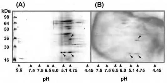
Fig 3 shows that the major allergen at 24 kd had 2
bands with different isoelectric points of 4.75 and 5.1,
whereas the 35-kd allergen had one band with an
iso-electric point of 4.75. Each of the 2 major allergens was
bound to IgE, which was derived from pooled sera of the
study subjects as shown in Fig 3.
N-terminal sequencing
Table II shows the result of amino acid sequencing of the
2 major allergens of molecular weight 24 and 35 kd. The
amino acid sequences of 2 individual bands composing 24
kd were the same for up to 15 amino acid sequences.
FIG 3.Two-dimensional PAGE analysis of CRM extract. A,Coomassie blue staining of 2-dimensional PAGE;
B,IgE immunoblotting of 2-dimensional PAGE with the reactive pooled sera.
TABLE II.Amino acid sequence of the 2 major allergens from CRMs Allergens Sequences 24 kd X-Arg-Leu-His-Asp-Ile-Asn-Gly-Asp-Asp-Val-Arg-Ala-Asp-Gln 35 kd X-Asp-Thr-Pro-Ile-Asp-Phe-X-Phe-Pro-Leu-Phe X, Unidentifiable.
DISCUSSION
Previous studies revealed that CRMs are the source of
the major causative allergen for asthmatic symptoms in
citrus farmers, and IgE-mediated reaction was one of the
major mechanisms because the prevalence of specific IgE
to CRMs was significantly associated with the presence of
asthmatic and rhinitis symptoms.
4,6This is the first study
to identify IgE-binding components and amino acid
sequencing of 2 major allergens within CRM antigens. In
the present study all the patients had high CRM-specific
IgE antibody levels determined by ELISA and ELISA
inhibition tests, confirming the specificity of IgE binding
to CRM antigen. Furthermore, immunoblot analysis
revealed 5 IgE-binding components, and 2 of them may be
considered major allergens because more than 50% of the
subjects had positive binding on these 2 bands. N-terminal
sequence and isoelectric points of the 24- and 35-kd
aller-gens were identified. When the N-terminal sequence of
these 2 allergens was compared with the National Center
for Biological Information database, we could not find any
allergens with a similar N-terminal sequence. These
find-ings indicate that these 2 major allergens at 24 and 35 kd
may be new allergenic materials derived from CRMs.
These findings will be a basis from which to develop
mAbs to major allergens, which might be essential to the
development of diagnostic and therapeutic extracts.
There is agreement in the suggestion that the closer the
taxonomic relationship between the mites, the greater the
level of cross-allergenicity.
10,11CRMs belongs to the order
of mites (Atari), to the suborder of Prostigmata, to the
fam-ily of spider mites (Tetranychidae), and represents its own
species. Bruches et al
2reported that all subjects sensitized
to spider mites were cross-reactive to house dust mites
(HDMs). Our previous study
4revealed that
serum-specif-ic IgE levels to CRMs were higher in the subjects with
positive skin responses to HDMs than to those without
such responses. Most of the asthmatic patients had
con-current positive responses to HDMs, whereas some
patients had an isolated positive response to CRMs with
no response to HDMs. ELISA inhibition tests showed that
minimal inhibitions were noted with additions of HDMs.
However, in subjects with positive skin responses to both
CRMs and HDMs, significant inhibitions were noted with
additions of HDM antigens. Although the 24-kd allergen
of the CRM has the same molecular weight as the group II
antigen of HDMs, amino acid sequencing results in this
study showed that it is species specific and different from
that of HDMs. Further investigations will be needed to
clarify species-specific and common sharing allergens
between CRMs and HDMs.
In conclusion, 5 IgE-binding components and 2 major
allergens (24 and 35 kd) with amino acid sequencing
were identified in CRM antigens.
REFERENCES
1. Reunala T, Bjorksten F, Forstrom L, Kanerva L. IgE-mediated occupa-tional allergy to a spider mite. Clin Exp Allergy 1983;13:383-8. 2. Bruches E, Pelaez A, Morales C, Braso JV, Rochina A, Lopez S, et al.
Occupational allergy due to spider mites: Tetranychus urticae and
Panonychus citri. Clin Exp Allergy 1996;26:1262-7.
3. Ashida T, Ide T, Tabata S, Kunimatsu M, Etoh Y, Yoshikawa T, et al. IgE-mediated allergy to spider mite, Panonychus citri, in occupationally exposed individuals. Arerugi 1995;44:1290-6.
4. Kim YK, Son JW, Kim HY, Park HS, Lee MH, Min KU, et al. Citrus red mite is the most common sensitizing allergen in subjects with asthma and/or aller-gic rhinitis in citrus farmers in Korea. Clin Exp Allergy 1999;29:1102-9. 5. Hong CS, Lee MK, Oh SH. Identification of major allergens from the
house dust mites, Dermatophagoides farinae and Dermatophagoides
pteronyssinus by eletroblotting. Yonsei Med J 1991;32:24-32.
6. Kim YK, Son JW, Kim HY, Park HS, Lee MH, Cho SH, et al. New occu-pational allergen in citrus farmers: citrus red mite (Panonychus citri). Ann Allergy Asthma Immunol 1999;82:223-8.
7. Chai H, Farr RS, Frochlich LA, Mathison DA, Rosenthal RR, Shelter AL, et al. Standardization of bronchial inhalation challenge procedures. J Allergy Clin Immunol 1975;56:534-9.
8. Park HS, Nahm DH. Buckwheat flour hypersensitivity: an occupational asthma in noodle maker. Clin Exp Allergy 1996;26:423-7.
9. Kim HY, Nahm DH, Park HS, Choi DC. Occupational asthma and IgE sensitization to cellulase in a textile industry worker. Ann Allergy Asth-ma Immunol 1999;82:174-8.
10. Platts-Mills TAE, Heymann PW, Chapman MD, Hayden ML, Wilkins SR. Cross-reacting and species-specific determinants on a major allergen from Dermatophagoides pteronyssinus and D. farinae: development of a radioimmunoassay for antigen P1 equivalent in house dust mite extracts. J Allergy Clin Immunol 1986;78:398-407.
11. Arlian LG, Geis DP, Vyszenski-Moher DL, Bernstein IL, Gallagher JS. Cross antigens and allergenic properties of the house-dust mite
Der-matophagoides farinae and the storage mite Tyrophagus putrescentiae. J