COMPARISON OF STRUCTURAL CHANGES ON ENAMEL AFTER Er,Cr:YSGG
AND 37% PHOSPHORIC ACID ETCHING
Rashmi Issar*
1, Shashi Ranjan
1Department of Conservative Dentistry & Endodontics, Patna Dental College & Hospital, Patna, Bihar 2Department of Oral & Maxillofacial Pathology, Dr.B.R.
3Dental Surgeon, Health & Family Welfare, Govt. Of Meghalaya
4Department of Conservative Dentistry & Endodontics, Patna Dental College & Hospital, Patna, Bihar 5,6Dept of Oral and Maxillofacial Pathology,
A R T I C L E I N F O
INTRODUCTION
The existence is governed by evolution as rightly said “the only thing which is constant is change”. The pioneering works of Michael Buonocore (1955), Leon Silverstone (1975), Fusayama (1979), Nakabayashi (1982) has laid the foundation of etching. This concept has evolved alongwith the agents that have been used. Organic agents, polymers, mineral acids and recently lasers have been used to etch the enamel surfaces to achieve predictable bonding. The agent which gained the most popularity was 37% phosphoric acid.
International Journal of Current Advanced Research
ISSN: O: 2319-6475, ISSN: P: 2319-6505,
Available Online at www.journalijcar.org
Volume 8; Issue 11 (B); November 2019
DOI: http://dx.doi.org/10.24327/ijcar.2019
Copyright©2019 Rashmi Issar et al. This is an open access article distributed under the Creative Commons Attribution License, which permits unrestricted use, distribution, and reproduction in any
Article History:
Received 13th August, 2019 Received in revised form 11th September, 2019
Accepted 8th October, 2019
Published online 28th November, 2019
Key words:
Enamel, Er,Cr:YSGG, 37% phosphoric acid, ESEM
*Corresponding author: Rashmi Issar
Department of Conservative Dentistry & Endodontics, Patna Dental College & Hospital, Patna, Bihar
COMPARISON OF STRUCTURAL CHANGES ON ENAMEL AFTER Er,Cr:YSGG
AND 37% PHOSPHORIC ACID ETCHING- AN ESEM ANALYSIS
Shashi Ranjan
2, Deirimika Lakiang
3, Pankaj Singh
4, Masthan
and Aravindha Babu N
6Department of Conservative Dentistry & Endodontics, Patna Dental College & Hospital, Patna, Bihar
Department of Oral & Maxillofacial Pathology, Dr.B.R. Ambedkar Institute of Dental Sciences & Hospital, Patna, Bihar Dental Surgeon, Health & Family Welfare, Govt. Of Meghalaya
Department of Conservative Dentistry & Endodontics, Patna Dental College & Hospital, Patna, Bihar Dept of Oral and Maxillofacial Pathology, Sree Balaji Dental College & Hospital, Chennai, Tamil Nadu
A B S T R A C T
Introduction: Tooth coloured restorations have evolved and so has the concept of bonding. A complete elimination of smear, microporosities and good wetting are the desirable substrate features which is mostly achieved by good enamel etching. Er,Cr:
shown a marked scope in enamel preparation and etching for bonded restoration.
Background: In the present study the conventional 37% phosphoric acid modality of etching is compared by ESEM analysis to Er,Cr:YSGG on enamel surfaces of extracted teeth.
Method: 40 anterior non diseased extracted teeth were collected and the coronal portion vertically sectioned by a diamond disc. The enamel specimens were then reduced to thickness of 1.0±0.5 mm and gauged by bur gauge. The specimens were then divided in GRE1 (control), GrE2 (acid etched) and GrE3 (Er,Cr:YSGG etched).These groups were then assessed for removal of smear layer and the predominant etching pattern exhibited.
Results: Chi-square test revealed that comparison of removal of smear layer in the two groups viz E2 & E3, did not differ significantly (p>.05). However in group E3, absence of smear layer was 88.9% which was significant (2= 5.44, p<0.05) at 5% level & in group E (2= 2.78) the removal of smear layer was significant at 10% level (p<0.10).For the two groups the absence of smear layer was 83.33% and it was significant (z= 2.80, p<0.01).In Type I etching pattern, the proportion in group E2 is higher and significant
(2= 2.78, 0.05<p>0.10) as compared to Group E3. In Type III etching pattern, the proportion in group E3 is higher & significant (2= 4.50, p<0.05) as compared to group E2.
Conclusion: Both 37% phosphoric acid and Er,Cr:YSGG were equally effec
layer removal from enamel specimens with no statistical significance. The predominant etching pattern seen under GrE2 was Type I and for GrE3 was Type III. The surface roughness was higher in laser treated surfaces. The present study advocate
effective alternative to acid etching.
The existence is governed by evolution as rightly said “the only thing which is constant is change”. The pioneering works of Michael Buonocore (1955), Leon Silverstone (1975), Fusayama (1979), Nakabayashi (1982) has laid the foundation cept has evolved alongwith the agents that have been used. Organic agents, polymers, mineral acids and recently lasers have been used to etch the enamel surfaces to achieve predictable bonding. The agent which gained the most
Silverstone et al showed 30
produce retentive enamel surface. With this agent an
bond strength of 15-25 MPa for composite restoration to enamel was achieved.1
Tooth preparation produces gross mechanical roughness but leaves a smear layer of hydroxyapatite crystal and denatured collagen that is approx 1-3µm thick.
removes the whole smear layer & increases the surface area upto 2000 times as compared to the unetched enamel. Acid etching produces uneven dissolution of enamel rods and their sheaths or enamel heads and their tails so that relatively smooth enamel surface becomes pitted and irregular.
International Journal of Current Advanced Research
6505, Impact Factor: 6.614
www.journalijcar.org
2019; Page No.20425-20429
//dx.doi.org/10.24327/ijcar.2019.20429.3990
This is an open access article distributed under the Creative Commons Attribution License, which permits unrestricted use, distribution, and reproduction in any medium, provided the original work is properly cited.
Department of Conservative Dentistry & Endodontics, Patna
COMPARISON OF STRUCTURAL CHANGES ON ENAMEL AFTER Er,Cr:YSGG
AN ESEM ANALYSIS
Masthan K.M.K
5Department of Conservative Dentistry & Endodontics, Patna Dental College & Hospital, Patna, Bihar
titute of Dental Sciences & Hospital, Patna, Bihar
Department of Conservative Dentistry & Endodontics, Patna Dental College & Hospital, Patna, Bihar Sree Balaji Dental College & Hospital, Chennai, Tamil Nadu
Tooth coloured restorations have evolved and so has the concept of bonding. A complete elimination of smear, microporosities and good wetting are the desirable substrate features which is mostly achieved by good enamel etching. Er,Cr: YSGG has
ked scope in enamel preparation and etching for bonded restoration.
In the present study the conventional 37% phosphoric acid modality of ,Cr:YSGG on enamel surfaces of extracted
40 anterior non diseased extracted teeth were collected and the coronal portion vertically sectioned by a diamond disc. The enamel specimens were then reduced to bur gauge. The specimens were then divided in GRE1 (control), GrE2 (acid etched) and GrE3 (Er,Cr:YSGG etched).These groups were then assessed for removal of smear layer and the predominant etching pattern exhibited.
square test revealed that comparison of removal of smear layer in the two groups viz E2 & E3, did not differ significantly (p>.05). However in group E3, absence of = 5.44, p<0.05) at 5% level & in group E2 = 2.78) the removal of smear layer was significant at 10% level (p<0.10).For the two groups the absence of smear layer was 83.33% and it was significant (z= 2.80, p<0.01).In Type I etching pattern, the proportion in group E2 is higher and significant at 10% level 0.05<p>0.10) as compared to Group E3. In Type III etching pattern, the = 4.50, p<0.05) as compared to group E2. Both 37% phosphoric acid and Er,Cr:YSGG were equally effective in smear layer removal from enamel specimens with no statistical significance. The predominant etching pattern seen under GrE2 was Type I and for GrE3 was Type III. The surface roughness was higher in laser treated surfaces. The present study advocates laser as an
showed 30-40% phosphoric acid could produce retentive enamel surface. With this agent an average 25 MPa for composite restoration to
Tooth preparation produces gross mechanical roughness but leaves a smear layer of hydroxyapatite crystal and denatured 3µm thick. 37% phosphoric acid removes the whole smear layer & increases the surface area upto 2000 times as compared to the unetched enamel. Acid etching produces uneven dissolution of enamel rods and their sheaths or enamel heads and their tails so that relatively
surface becomes pitted and irregular.
Research Article
Silverstone et al in 1975 described three patterns of etching on enamel 1) Type I- Prism core material is preferentially removed, leaving the prism peripheries relatively intact, resulting in a honeycomb appearance.2) Type II- The peripheral regions of the prism are dissolved preferentially, leaving the prism cores relatively intact, resulting in a cobblestone appearance.3) Type III- Etching pattern contains areas, which resembles both type I and type II along with some distinct areas where the pattern of etching appears to be unrelated to the enamel prism morphology. Gwinnett mentioned that such losses are not clinically predictable and different etching patterns may occur at adjacent sites in the same tooth.
Studies with polarized light microscope showed that sound enamel etched with phosphoric acid to be affected at 3 distinct levels and may be described in terms of three specific zones (Silverstone 1974). A Superficial etched zone, which is a narrow zone of enamel of about 10µm in depth that is removed by etching. A Qualitative porous zone of about 20µm in depth. It is rendered porous by the acid attack and may be identified qualitatively using polarized light. A Quantitative porous zone of about 20 µm depth.
Although 37% phosphoric acid produced desirable in vivo bonding results and was economical but few drawbacks like demineralization of undesirable sites due to spillage, technique sensitivity due to multiple steps and more chair side time lead to exploring of other alternatives.
In 1989, experimental work by Keller & Hirbst using a pulsed Er:YAG (2,940 nm) laser demonstrated its effectiveness on hard tissue. It became commercially available in UK in 1995, followed by Er,Cr:YSGG (Erbium, Chromium: Ytrium Scandium Gallium Garnet) laser in 1997.The hydrokinetic laser system was introduced by Eversole & Rizoiu. In 1997 with the FDA clearance of Er,Cr:YSGG in US, came the approval for caries removal, cavity preparation and conditioning of the tooth. 2, 3
The proposed mechanisms of ablation were cavitation bubbles, apatite crystal fragmentation and an acceleration of water droplets by laser light called the “hydrokinetic mechanism”. Water absorbs laser light thereby changing its rotational and vibrational states. The increase of energy in the molecules leads to change in length and eigenfrequencies (vibration frequency of a system) of the OH bonds. After a certain lifetime the molecule drops down again to its ground state and releases the absorbed energy. This action is called recombination. As no radiation is emitted this released energy remains in the volume and is converted into a temperature distribution in the water. This increases the volume of the irradiated substrate leading to disruption of crystals in the substrate and thus spallation.
Since enamel is 85% by volume carbonated hydroxyapatite with 12% water (by volume) 2780 nm(wavelength of Er,Cr: YSGG) is highly absorbed generating thermal changes in enamel which may be able to alter its structure chemically and morphologically.4 Etching is through a process of continuous vaporization and microexplosions resulting from vaporization of the water entrapped in the hydroxyapatite matrix. These microexplosions can be explained by- “water molecules that are pressed or pushed into the capillary areas between the enamel prisms absorb the laser energy and expand.3, 5
Usumez & colleagues claimed enamel conditioning at 2W power can be seen to be equivalent in bond strength to acid etching. It is best to use lowest possible energy level just below the ablation threshold, as this reduces the amount of ablation debris or tiny flakes which can be poor surfaces to bond.Lased enamel showed1) Rough surface - the roughness of the lased surface is 150-170µm as compared to 73-94µm of acid etched surface.2) Complete removal of the smear layer 3) Protruding prisms in the enamel without any signs of erosion.1 The present study is an ESEM analysis of the prepared enamel specimens comparing the surface characteristics of acid etched vs laser etched surface.3
MATERIALS AND METHODS
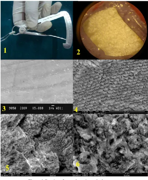
Forty anterior extracted non-diseased human teeth stored in normal saline after cleaning were taken as sample. Vertical sections of enamel surfaces were prepared through labial third of the coronal tooth structure by double sided diamond disk at slow speed. Each specimen was polished by 400 grit SiC paper to produce a smear layer. The thickness of each specimen was 1.0 mm ±0.5mm and was measured by a bur gauge of .1mm sensitivity.(Fig. 1) The enamel specimens were subdivided into GrE1 (4 samples) acting as control, GrE2 (18 samples) acid etched and GrE3 (18 samples) lased.
An acrylic resin platform was made and the specimens were placed in it stabilized by elastomeric impression material during the etching procedure. GrE2 was acid etched using 36% phosphoric acid gel (Conditioner 36, Dentsply). The application time of acid etchant was 15 sec. The specimens were then rinsed with water for 30 sec and dried by oil free compressed air for 15 sec.
Figure 1 Gauging of enamel specimens by bur gauge
Figure 2 Lased enamel specimen showing macroscopic pitting
Figure 3 GrE1 (control group) showing smear layer covered surface
Figure 4 GrE2 (acid etching) showing removal of smear layer and Type I etching pattern at 1200X magnification
Figure 5 GrE3 (laser etching) showing removal of smear layer, ablated, rough, recrystallized surface with evidence of surface cracks at 1200X magnification
GrE3 was subjected to Er,Cr: YSGG laser of 2.78 µm wavelength with the following settings was used-power -3W, air- 70 %, water- 20%. The beam for was aligned perpendicular at 1mm distance and moved in a sweeping fashion over the specimen for 15 sec. The specimens were then dried with an oil free air source for 15 sec.
The specimens were then observed under ESEM at x1200 & ×5000 magnification. Photomicrographs were obtained at both levels of magnification. The impressions of SEM photomicrographs were taken at Bose institute, Kolkata & IISC, Bangalore.
Parameters studied for enamel specimens were 1) removal of smear layer 2) type of etching pattern. The collected data values were subjected to the Chi square analysis.
RESULTS
All the specimens of GrE1 was seen to be covered by smear layer (Fig. 3). GrE2 showed absence of smear layer in fourteen (77.8%) specimens (Fig. 4) where as four specimens showed presence of smear layer. Sixteen (88.9%) specimen of GrE3 specimens showed absence of smear layer (Fig. 5-6) where as two specimen showed presence of smear layer.
Chi-square test reveals that comparison of removal of smear layer in the two groups viz GrE2 & GrE3, did not differ significantly (p>.05). However in GrE3, absence of smear layer is 88.9% which is significant (2= 5.44, p<0.05) at 5% level & in group E2 (2= 2.78) the removal of smear layer is significant at 10% level (p<0.10).For the two groups the absence of smear layer is 83.33% (15 out of 18) and it is significant (z= 2.80, p<0.01). (Graph 1)
GrE2 showed type I etching pattern (Fig. 4) in fourteen (77.8%) specimens, type II & type III etching patterns in two specimens each. GrE3 showed distinguishable type I etching pattern in four specimens & rest fourteen (77.8%) showed type III etching pattern (Fig. 5-6). (Graph 2)
In Type I etching pattern, the proportion in GrE2 is higher and significant at 10% level (2= 2.78,0.05<p>0.10) as compared to GrE3. In Type III etching pattern, the proportion in group E3 is higher & significant (2= 4.50, p<0.05) as compared to group E2
Graph 1 Comparison of absence & presence of smear layer removal between
GrE2 & GrE3
Graph 2 Comparison between GrE2 & GrE3 for type of etching pattern
DISCUSSION
Currently, laser etching is proving to become an alternative to
acid etching of enamel. Er,CR:YSGG laser etching does not involve either vibration or heat; also, the easy handling of the apparatus makes this treatment highly attractive for routine clinical use.6 Surface alterations of the enamel and dentin after Er,Cr:YSGG laser irradiation showed that these surfaces are associated with microirregularities, and there was also the
absence of a smear layer .7
The present study compared the morphological differences of enamel & dentin after surface treatment by 36% phosphoric acid (conventional etching) & Er,Cr:YSGG through SEM analysis. In the present study the specimens of enamel & dentin were polished by 400 grit silicon carbide paper to create a smear layer of around 1-2 µm thickness which is the same as that created by regular grit bur in a clinical situation.8
In the present study phosphoric acid in gel form was used as it is more stable than liquid acids, however there is still a shift of acid on the tooth surface even in the gel form.2
According to T. Dostalova et al at 3 W power settings it is possible to etch the tooth surface without removing the enamel & dentin, the border is well defined & roughness is clearly visible, the same was used in this study.9
D.C. Atrrill et al in his study concluded that the cracks were wider & more prominent were no water coolant was used in Er:YAG. So, laser etching should be accompanied by water if excessive iatrogenic damage to tissues is to be prevented (70% air, 20% water was used in this study).10, 11
Studies by Chousterman et al & Usumez et al have shown that there is no significant difference in the adhesion of composite to laser etched surface for durations ranging from 15sec to 60 sec, so in the present study the minimum etching time of 15 sec was used. 3, 6, 12 The required time for acid etching varies from 15 to 60 seconds. Osorio et al reported total of 60 seconds for each tooth (15 sec etching+ 30 sec rinsing+15 sec drying) is needed with phosphoric acid. The time needed for laser systems is only 15 seconds. From a clinical standpoint, saving chair time also improves adhesion because it reduces the risk of salivary contamination. 6
by SEM interpretation the roughness was more in lased surfaces than in acid etched in this study (Fig. 5-6). According to Nakamura et al higher surface roughness leads to better
bonding.9, 13
Almost all the specimens of enamel showed the absence of smear layer in the present study (Fig. 4-6) & no statistically significant difference was found between acid etched & laser etched surface. Sean Lin et al showed that laser etched enamel & dentin surface did not exhibit a smear layer. Rather, these surfaces were found to be clean with smooth foci that often reported preservation of prism structure.
Four specimens in GrE2 showed presence of smear layer samples alongwith silica gel residue was seen which might be due to methodological error like improper rinsing & drying. According to Perdigão et al. (1994), silica used to thicken the etching gel is not entirely removed by rinsing with water; however according to Dentarois et al the presence of a silica residue did not interfere with bonding of resin to the tooth structure.8
In the present study SEM observation showed that laser irradiation produces recrystallized enamel (Fig. 5-6)which was also reported in studies by M. Hossain et al1, S. Lin et al. 14
On observation of SEM photos of lased enamel in the present study, the lased enamel specimens showed the classic features of laser-treated enamel: grooves, flakes, shelves and sharp edges (Fig. 5-6); all these aspects were more indicative of
microexplosion than of melting .9, 11
In the present study the predominant enamel etching pattern after laser etching seen was Type III (Fig. 5-6) as compared to the Type I in case of acid etching (Fig. 4). Serdar Usumez et al
in their study laser irradiated the enamel surfaces at 2 W and showed type III etching, characterized by a more random etching pattern. There were also regions in which the pattern could not be related to prism morphology.8, 15, 16
However in a study conducted by Torun Ozer et al in 2008, a preferential type I etching pattern was seen. This difference with the present study might be due to different power settings, air water percentages & angulation of the beam used. Further it
was explained by Giovanni Olivi “the morphological
differences of etching pattern appears to be related both to the orientation of the prism with respect to the inclination of the incident laser beam and to the different air/water percentages in the spray & to the energy applied to the tissue by the laser
system.”Also ina study by D.C. Attrill et al they stated that
the etching distribution appears to be selective and broadly follows a series of parallel bands across the surface. The origin of the selective distribution is unclear but may follow incremental developmental lines within the enamel.6, 10, 11
In the present study with acid etching (using phosphoric acid), the enamel surfaces clearly displayed Type I etching pattern i.e. protruding prism sheaths, between which the core of the prism had been eroded away (Fig. 4). This appearance was similar to previous studies by Pashley et al. who reported that this erosive nature of acids on enamel & dentin is not favourable for good adhesion or adaptation of restorative materials with the tooth surfaces.13
In the present study with laser non-erosive type of etching was observed (Fig. 5)According to Pashley the non erosive type of etching favours bonding.9
There was evidence of minor surface cracking on the laser etched enamel due to the high power settings (3W) used in the present study (Fig. 5). According to S.R. Farrar, cracking although is greatly reduced, but not completely eliminated, when a surface water film is included during preparation of the laser etched surfaces. The presence of water appears to act as a surface coolant, reducing the local thermal stresses induced by laser irradiation to a level that may be tolerated by the enamel.
The views of many authors are conflicting in relation to the
effects of cracks on bonding. Kataumi et al, 1998 were the first
to observe cracks in dentin after Er:YAG irradiation.A study
by Bor- Shuinn Lee et al showed that occasional cracks
enhances retention and is ideal for resin penetration. In
opposition to Lee at al, Van Landuyt et al showed that adhesion of composites to laser treated tooth tissue is jeopardized by structural weakening by microcracks.17
CONCLUSION
The effects of Er,Cr: YSGG etching on enamel was removal of smear layer, recrystallized surface, non erosive roughening and Type III etching pattern. Clinical advantages of reduced chair side time and less technique sensitivity makes the application of this laser for etching further encouraging. However further studies for in vitro and in vivo bonding of composite to the laser treated surfaces is needed.
References
1. Perdigao J., Lambrechts P., Meerbek B., Tome A., Vanherle G., Augusto B. Lopes. Morphological field emission-SEM study of the effect of six phosphoric acid etching agents on human dentin. Dent Materials, 1996, 12: pg. 262-272
2. Parker S. Introduction, history of lasers and laser light production.BDJ, 2007 Vol. 202, No 1:21-31
3. Usumez S., Orhan M., Usumez A. . Laser etching of enamel for direct bonding with an Er,Cr:YSGG hydrokinetic laser system. Am J Dentofacial Orthop, 2002;122:649-656
4. Freitas P., Rapozo M., Eduardo C., Featherstone J. . In vitro evaluation of Er,Cr:YSGG laser treated enamel demineralization. Lasers in Med Sci, 2008
5. Mir M., Gutknecht N., Poprawe R., Vanweersch L., Lampert F. Visualizing the procedures in the influence of water on the ablation of dental hard tissue with Er,Cr:YSGG laser pulses. Laser in Med. Sci., 2008 6. Özer T., Basara G., Berk N. Laser etching of enamel for
orthodontic bonding. Am J Dentofacial Orthop, 2008, 134:193-7
7. Secilmis A., Altintas S., Usumez A., Berk G. Evaluation of mineral content of dentin prepared by Er,Cr:YSGG. Laser in Med. Sci., 2007
8. Goes M., Coelho M., Simnoides. Morphological effect of type, concentration & etching time of acid solutions on enamel & dentin surfaces. Braz. Dent. J., 1998, 9(1) 9. Hossain M., Nakumura Y., Yamada Y., Suzuki N.,
Murakami Y., Matsumoto K. Analysis of surface roughness of enamel and dentin after Er,Cr:YSGG laser irradiation. Journal of Clinical Laser Medicine &
Surgery, 2001, Vol. 19, Number 6,pg 297-303
11. Olivi G., Angiero F., Benedicent S. Use of the Er:YAG on human enamel tissues. Influence of the air water spray on the laser tissue interaction: SEM evaluations. Lasers Med Sci Jun, 2009
12. Oliveira M., Zezell D., Apel C. Bond strength of self etching primer to bur cut, E,Cr:YSGG & Er:YAG lased dental surfaces. Phtomedicine & Laser Surgery, 2007, Vol. 25, No. 5
13. Hossain M, Yamada Y., Nakamura Y. A study on surface roughness & microleakage test in cavities prepared by Er:YAG laser irradiation & etched bur cavities. Lasers Med Sci, 2003 18:25-31
14. Lin S., Caputo A., Eversole L., Rizoiu I. Topographical characteristics and shear bond strength of tooth surfaces cut with a laser-powered hydrokinetic system. Journal
of Prosthetic Dentistry, 1999; 82: 451-5
15. Gwinnett A.J., Matsui A., Buonocore M.G. Clinical application of enamel adhesives by D.H. Retief. Operative Dentistry,1969 Supplement. 5, 44-49. 16. Sasaki H.,D.C. Lobo, Moriyama Y., Watanabe S.,
Villaverde A., Tanaka S., Moriyama E., Brugnera A. . Tensile bond strength and SEM analysis of enamel etched with Er:YAG laser and phohsphoric acid: a comparative study In vitro. Brazilian Dental Journal, 2008 Vol. 19, No. 1
17. Lee B., Hsieh T., Lee Y. Bond strengths of orthodontic bracket after acid etched, Er:YAG laser irradiated & combined treatment on enamel surface. The Angle Orthodontist, 2002, Vol. 73, No. 5, 565-570
How to cite this article:
Rashmi Issar et al (2019) 'Comparison of Structural Changes on Enamel After Er,Cr:YSGG And 37% Phosphoric Acid Etching- An ESEM Analysis', International Journal of Current Advanced Research, 08(11), pp.20425-20429.
DOI: http://dx.doi.org/10.24327/ijcar.2019.20429.3990