256
International Journal of Pharmaceutical Sciences and Drug Research
2017; 9(5): 256-262
Review Article
CODEN (USA): IJPSPP
ISSN: 0975-248X
Renal Vascularisation Causing End - Stage Failures and Outcomes: A Pooled
Analysis of Community Based Studies
A. Deevan Paul
*, C. Girish, T. N. Shilpa, Katta Manogna, A. Geetha Susmitha
SVU College of Pharmaceutical Sciences, SV University, Tirupati – 517502, Andhra Pradesh, India
Copyright © 2017 A. Deevan Paul et al. This is an open access article distributed under the terms of the Creative Commons Attribution-NonCommercial-ShareAlike 4.0 International License which allows others to remix, tweak, and build upon the work non-commercially, as long as the author is credited and the new creations are licensed under the identical terms.
ABSTRACT
Although acute renal failure (ARF) is believed to be common in the setting of critical illness and is associated with a high risk of death, little is known about its epidemiology and outcome or how these vary in different regions of the world. Prospective observational study of ICU patients who either were treated with renal replacement therapy (RRT) or fulfilled at least one of the predefined criteria for ARF. The main outcomes are occurrence of ARF, factors contributing to etiologic, illness severity, treatment and need for renal support after hospital discharge and hospital mortality. The marginal effects of acute kidney injury on in-hospital mortality, length of stay (LOS) and costs have not been well described. The acute kidney injury is associated with significantly increased mortality, LOS, and costs across a broad spectrum of conditions. Moreover, outcomes are related directly to the severity of acute kidney injury, whether characterized by nominal or percentage changes in serum creatinine. Individuals should check with a doctor that an exercise program is suited to their age, weight and health. Including abusing alcohol and drugs.
Keywords:Acute renal failure, Replacement therapy, Glomerular filtration rate, ambulatory peritoneal dialysis.
DOI: 10.25004/IJPSDR.2017.090510 Int. J. Pharm. Sci. Drug Res. 2017; 9(5): 256-262
*Corresponding author: Mr. A. Deevan Paul
Address: SVU College of Pharmaceutical Sciences, SV University, Tirupati – 517502, Andhra Pradesh, India E-mail : [email protected]
Relevant conflicts of interest/financial disclosures: The authors declare that the research was conducted in the absence of any commercial or financial relationships that could be construed as a potential conflict of interest.
Received: 11 August, 2017; Revised: 08 September, 2017; Accepted: 12 September, 2017; Published: 15 September, 2017
INTRODUCTION
Dialysis is the development of molecules by dispersion from high focus to low fixation through a semi-porous film. Only those molecules those are small enough to fit through the membrane pores can move through the membrane and reach equilibrium with the entire volume of solution in the system. For instance, if one is dialyzing 1 ml of the test against 200 ml of dialysis
Int. J. Pharm. Sci. Drug Res. September-October, 2017, Vol 9, Issue 5 (256-262)
Fig. 1: Picture representation of healthy kidney and diseased kidney
ACUTE RENAL FAILURE
Acute renal failure represents a rapid decline in renal function sufficient to increase blood levels of nitrogenous wastes and impair fluid and electrolyte balance. It is a common threat to seriously ill persons in intensive care units, with a mortality rate ranging from 42% to 88%. 1 Although treatment methods such as dialysis and renal replacement methods are effective in correcting life-threatening fluid and electrolyte disorders, the mortality rate associated with acute renal failure has not changed substantially since the 1960s. This probably is because acute renal failure is seen more often in older persons than before, and because it frequently is superimposed on other life-threatening conditions, such as trauma, shock, and sepsis. The most common indicator of acute renal failure is azotaemia, an accumulation of nitrogenous wastes (urea nitrogen, uric acid, and creatinine) in the blood. In acute renal failure, the glomerular filtration rate (GFR) is decreased. As a result, excretion of nitrogenous wastes is reduced and fluid and electrolyte balance cannot be maintained. Persons with acute renal failure often are asymptomatic, and the condition is diagnosed by observation of elevations in blood urea nitrogen (BUN) and creatinine. [2]
Pre-renal failure
Prerenal Failure Prerenal failure, the most common form of acute renal failure, is characterized by a marked decrease in renal blood flow. It is reversible if the cause of the decreased renal blood flow can be identified and corrected before kidney damage occurs. Causes of prerenal failure include profound depletion of vascular volume and impaired perfusion caused by heart failure and cardiogenic shock, and decreased vascular filling because of increased vascular capacity. Elderly persons are particularly at risk because of their predisposition to hypovolemia and their high prevalence of renal vascular disorders.
Post- renal failure
Postrenal Failure Postrenal failure results from obstruction of urine outflow from the kidneys. The obstruction can occur in the ureter (i.e., calculi and
strictures), bladder (i.e., tumours or neurogenic bladder), or urethra (i.e., prostatic hypertrophy). Prostatic hyperplasia is the most common underlying problem. Because both ureters must be occluded to produce renal failure, obstruction of the bladder rarely causes acute renal failure unless one of the kidneys already is damaged or a person has only one kidney. The treatment of acute postrenal failure consists of treat-ing the underlying cause of obstruction so that urine flow can be re-established before permanent nephron damage occurs.
Intrinsic Renal failure
Intrinsic Renal Failure Intrinsic or intrarenal renal failure results from conditions that cause damage to structures within the kidney-glomerular, tubular, or interstitial. Injury to the tubules is most common and often is ischemic or toxic in origin. The major causes of intrarenal failure are ischemia associated with prerenal failure, toxic insult to the tubular structures of the nephron, and intratubular obstruction. Acute glomerulonephritis and acute pyelonephritis also are intrarenal causes of acute renal failure.
Acute Tubular Necrosis
Acute tubular necrosis (ATN) is characterized by destruction of tubular epithelial cells with acute suppression of renal function and it is the most common cause of intrinsic renal failure. ATN can be caused by a variety of conditions, including acute tubular damage caused by ischemia, the nephrotoxic effects of drugs, tubular obstruction, and toxins from a massive infection. The tubular injury that occurs in ATN frequently is reversible. The process depends on the recovery of the injured cells, removal of the necrotic cells and intratubular casts, and regeneration of renal cells to restore the normal continuity of the tubular epithelium. However, if the ischemia is severe enough to cause cortical necrosis, irreversible renal failure occurs.
Int. J. Pharm. Sci. Drug Res. September-October, 2017, Vol 9, Issue 5 (256-262)
Afferent arteriolar vasoconstriction, caused in part by tubuloglomular feedback, results in decreased glomerular capillary filtration pressure. Tubular injury and increased intraluminal pressure cause fluid to move from the tubular lumen into the interstitial. [3-4]
CHRONIC RENAL FAILURE
Unlike acute renal failure, chronic renal failure represents progressive and irreversible destruction of kidney structures. Many patients with chronic renal failure progressed to the final stages of the disease and then died. The high mortality rate was associated with limitations in the treatment of renal disease and with the tremendous cost of ongoing treatment. In the United States, there are approximately 400,000 persons with end-stage renal disease who are living today, a product of continued research and advances in treatment renal failure can result from many conditions that cause permanent loss of nephrons, including diabetes, hypertension, glomerulonephritis, and polycystic kidney disease. Typically, the signs and symptoms of renal failure occur gradually and do not become evident until the disease is far advanced. This is because of the amazing compensatory ability of the kidneys. As kidney structures are destroyed, the remaining nephrons undergo structural and functional hypertrophy, each increasing its function as a means of compensating for those that have been lost. It is only when the few remaining nephrons are destroyed that the manifestations of renal failure become evident. [5]
Fig. 3: Number of functioning nephrons in Relation of renal function and nephron mass, each kidney contains 1million tiny nephrons. A proportional relation exists between the number of nephrons affected by disease and the resulting glomerular filtration rate.
Serum albumin is a valuable screening apparatus for perceiving protein energy wasting (PEW) in Dialysis patients. In any case, there are numerous non-wholesome conditions that are significantly more critical determinants of serum egg white’s levels than a patient's healthful state. Nevertheless, we have observed that it is not unusual for caregivers to make a
reflex connection between serum albumin and dietary protein intake and to act on an unfounded belief that protein intake is problematic when serum albumin levels are low or declining. This may be done in the absence of other evidence of PEW or without further nutritional assessment. The recognized limitations in serum albumin as a marker for dietary protein intake deserve further emphasis.
Fig. 4: Manifestations of renal failure
TREATMENT
During the past several decades, an increasing number of persons have required renal replacement therapy with dialysis or transplantation. The growing volume is largely attributable to the improvement in treatment and more liberal policies regarding who is treated. Medical Management Chronic renal failure can be treated by conservative management of renal insufficiency and by renal replacement therapy with dialysis or transplantation. Conservative treatment consists of measures to prevent or retard deterioration in remaining renal function and to assist the body in compensating for the existing impairment. Interventions that have been shown to significantly retard the progression of chronic renal insufficiency include dietary protein restriction and blood pressure normalization. Various interventions are used to compensate for reduced renal function and correct the resulting anaemia, hypocalcaemia and acidosis. [6-8] Haemodialysis
Int. J. Pharm. Sci. Drug Res. September-October, 2017, Vol 9, Issue 5 (256-262)
To be more specific, a surgical procedure will be done to connect an artery to a vein to create a site through which you will receive your dialysis treatments. An evaluation of your veins and how soon you need to start dialyzing may assist the doctors in determining the type of access placed. Sometimes a doctor will use “vein mapping” to determine which type of access is best. Vein mapping uses a painless sound wave (ultrasound) to look at your veins. [9-11]
Some type of access will be necessary for you to receive your dialysis treatments.
There are 3 types of accesses.
Fistula: One of your veins is reconnected to an artery,
allowing greater blood flow through the vein. Because it is your own vein it often last longer and may have fewer problems than the other two types.
Graft: An artificial tube is inserted just under the skin
and is connected at one end to an artery and the other end to a vein. Sometimes this access requires more maintenance and does include an increased risk of clotting.
Catheter: Sometimes used for temporary access, this is
a long, two-sided tube inserted through the skin and into a vein.
Fig. 5: Drafting representation of AV Fistula, AV Graft and Catheter
The basic principles of haemodialysis have remained unchanged throughout the years, although new technology has improved the efficiency and speed of dialysis. A haemodialysis system, or artificial kidney, consists of three parts
Blood compartment Dialysis fluid compartment Cellophane membrane
These cellophane membranes are semipermeable, permitting all molecules except blood cells and plasma proteins to move freely in both directions from the blood into the dialyzing solution and from the dialyzing solution into the blood. The direction of flow is determined by the concentration of the substances contained in the two solutions. The waste products and excess electrolytes in the blood normally diffuse into the dialyzing solution. If there is a need to replace or add substances, such as bicarbonate to the blood these can be added to the dialyzing solution.
During dialysis, blood moves form an artery through the tubing and blood chamber in the dialysis machine and then back into the body through a vein. Access to the vascular system is accomplished through an external arteriovenous shunt (ie., tubing implanted into
an artery and a vein) or more commonly through an internal arteriovenous fistula (i.e., anastomosis of a vein to an artery, usually in the forearm). Heparin is used to prevent clotting during the dialysis treatment; it can be administered continuously or intermittently.
Fig. 6: Schematic diagram of a haemodialysis system. The blood compartment and dialysis solution compartment are separated by a cellophane membrane. This membrane is porous enough to allow all the constituents, except the plasma proteins and blood cells, to diffuse between the two compartments.
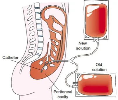
Fig. 7: Peritoneal dialysis. A semipermeable membrane, richly supplied blood vessels, lines the peritoneal cavity. With dialysate dwelling in the peritoneal cavity, waste products diffuse form the network of blood cells into the dialysate.
Peritoneal Dialysis
Int. J. Pharm. Sci. Drug Res. September-October, 2017, Vol 9, Issue 5 (256-262)
out of the peritoneal cavity by gravity into a sterile bag. Glucose in the dialysis solution accounts for water removal. Commercial dialysis solution is available in 1.5%, 2.5%, and 4.25% dextrose concentrations. Solutions with higher dextrose levels increase osmosis, causing more fluid to be removed. The most common method is continuous ambulatory peritoneal dialysis (CAPD), a self-care procedure in which the person manages the dialysis procedure and the type of solution (i.e., dextrose concentration) used at home. [12-14]
Kidney transplantation
A kidney transplant is not a cure, it is a treatment. A kidney transplant is when a kidney is removed from one person (called a donor) and surgically placed into another person (called a recipient). Many people prefer a transplant because of the benefits it provides. A successful kidney transplant may prevent you from ever needing dialysis. If you have started dialysis, a successful transplant should allow you to stop the dialysis treatments. Your energy level should improve as your new kidney will promote the production of red blood cells. You will have fewer restrictions with your diet and with your fluid intake. You will hopefully be able to return to a more normal lifestyle with increased activity and independence.
Your physician or dialysis unit can refer you to a transplant centre for evaluation. This evaluation process includes meeting with several members of the transplant team. You will meet with a transplant nurse, social worker, dietician, financial specialist, transplant nephrologists (kidney doctor) and a surgeon. Success rates are slightly better and the surgery is scheduled at a convenient time. But not everyone who needs a kidney has a live donor available. Unfortunately, there are not enough deceased donors to meet the huge demand and there is a waiting time for these transplants. Your transplant team will explain to you how kidneys are distributed and the average waiting time for a kidney. Kidney transplant surgery is approximately a 3-4-hour operation. Your own kidneys (native kidney) are not usually removed. Your new kidney is placed in your lower abdomen. There are some circumstances that may require removal of native kidneys. If you fall into one of these situations, your transplant team will discuss this with you. After surgery, you will usually be in an intensive care area overnight and most likely be in the hospital for approximately 5-7 days. Although, this is a major operation, most patients begin to feel better almost immediately.
The disadvantages include multiple medicines, the follow-up and increased risks of infections and a slight increased risk of cancer. As with any transplant there is always a risk the body would reject the new kidney at any point. The transplant teams will discuss the risks and benefits in greater detail with you. The success rates for kidney transplantation are very good. [15]
SOCIAL ADJUSTMENT AND AWARENESS TO THE RENAL PATIENT
It is very common to experience a flood of emotions when dealing with an incurable disease such as kidney failure. Everyone experiences different emotions at different times and adjust at varying rates. It is important to remember that you have a support system at your dialysis unit available to you. Expect to feel emotions such as denial, guilt, fear, anxiety and acceptance at different times. Remember that a chronic illness also affects family members and those that love you. They can be your biggest supporter and provide you the encouragement you need for all that you will be balancing with your emotions, treatment schedule, diet and medications. We understand that there are many questions running through your mind about how this process works and how it may affect your life. Changes in your lifestyle will be required of you as a kidney patient – this varies somewhat by the treatment option you select. [16]
AWARENES TO THE RENAL PATIENT
The high morbidity and mortality rates associated with acute renal failure, attention should be focused on prevention and early diagnosis. This includes assessment measures to identify persons at risk for the development of acute renal failure, including those with pre-existing renal insufficiency and diabetes. Elderly persons are susceptible to all forms of acute renal failure because of the effects of aging on renal reserve. Careful observation of urine output is essential for persons at risk for the development of acute renal failure. Urine output and urine osmolality or specific gravity should be carefully monitored. One of the earliest manifestations of tubular damage is the inability to concentrate the urine. Further diagnostic information that can be obtained from the urinalysis includes evidence of proteinuria, haemoglobinuria, and casts or crystals in the urine. Blood tests for creatinine provide information regarding the ability to remove nitrogenous wastes from the blood. A major concern in the treatment of acute renal failure is identifying and correcting the cause (e.g., improving renal perfusion, discontinuing nephrotoxic drugs). Fluids are carefully regulated to maintain normal fluid volume and electrolyte concentrations. Adequate caloric intake is needed to prevent the breakdown of body proteins, which increases the need for elimination of nitrogenous wastes. Parenteral hyperalimentation may be used for this purpose. Because secondary infections are a major cause of death in persons with acute renal failure, constant effort is needed to prevent and treat such infections. Dialysis or continuous renal replacement therapy (CRRT) may be indicated when nitrogenous wastes and the water and electrolyte balance cannot be kept under control by other means. [17-19]
EFFECTS OF AGE ON THE OUTCOME OF
PERITONEAL DIALYSIS – ASSOCIATED
Int. J. Pharm. Sci. Drug Res. September-October, 2017, Vol 9, Issue 5 (256-262)
Peritoneal dialysis (PD) has been reported as a practical renal replacement therapy in elderly patients with end-stage renal disease (ESRD). The most related studies have evaluated the overall outcome of geriatric ESRD in patients who have received PD therapy.
This review undergoes the 180 cases (97 women and 83 men; mean age 54.0 + 13.6 years). Among them, 39 patients older than 65 years were assigned to a geriatric group and the remaining 141 were assigned to a nongeriatric group. We collected each patients data on general characteristics (age, sex and co-morbities); vital signs in the Emergency departments ED ( temperature, heartrate, respiratory rate and systolic arterial pressure); laboratory tests (white blood cells counts of effluent and blood, serum glucose, sodium, potassium, C-reactive protein and alanine aminotransferase) and hospital courses and outcomes (hospital days, requirement for intensive care and removal of the peritoneal catheter, rate of recurrent/relapsing/repeat/ refractory peritonitis, shift to haemodialysis therapy and mortality). [20-22] These variables were compared
between the geriatric and nongeriatric groups. The observation with no statically significant differences in the general characteristics, vital signs in the ED and laboratory tests between the 2 groups, indicating that the groups had similar initial disease severities and physiological derangements. All the patients received intraperitoneal antibiotics therapy and the choice of antibiotics was based on the results of the effluent culture were negative. Decisions to remove the peritoneal catheter were made after considering the following reasons; Peritonitis refractory to adequate antibiotic treatment. Decisions to shift from PD to haemodialysis were made after agreement between the patients and nephrologists.
Regarding the hospital courses and outcomes, the geriatric group showed a slightly longer hospital stay (10.1 ± 8.9 vs 9.1 ± 8.9days, P=0.538), a higher likelihood of requiring intensive care and removal of the peritoneal vathetar (8% vs 6%, P= 0.772 and 26% vs 21%, P=0.562, respectively) and higher mortality (8% vs 4%, P= 0.383). However, these variables did not differ significantly between the 2 groups. The presented data revealed similar outcomes of PD peritonitis in a single hospitalization in the geriatric and nongeriatric groups. The mortality rate in patients with geriatric ESRD was higher than that in patients without nongeriatric ESRD. However, death in most cases was caused by cardiovascular disease. PD peritonitis may play a limited role in the overall outcome in patients with geriatric ESRD. Elderly patients typically have multiple co-morbities, which may result in deteriorated consequences. [23-24]
Chronic kidney disease, also known as chronic renal failure is much across the board than individuals acknowledge, it frequently goes undetected and undiscovered until the point that the sickness is all around cutting edge. Treatment is aimed at stopping or
slowing down the progression of the disease – this is usually done by controlling its underlying cause. It is important that people who are at high risk of developing kidney disease have their kidney functions regularly checked. Early detection can significantly help prevent serious kidney damage. A kidney disappointment propels and the organ work is extremely weakened, unsafe levels of waste and liquid can quickly develop in the body. Treatment is gone for halting or backing off the movement of the illness – this is normally done by controlling its hidden reason. It is essential that individuals who are at high danger of creating kidney ailment have their kidney capacities consistently checked. Early location can fundamentally help anticipate genuine kidney harm. The kidneys of patients with end-organize kidney malady can't stay aware of the waste and liquid disposal process without anyone else the patient will require dialysis or a kidney transplant to survive. A solid eating routine, including a lot of foods grown from the ground, entire grains and lean meats or fish will enable keep to circulatory strain down. Customary Physical exercise is perfect for keeping up sound circulatory strain levels, it additionally helps control ceaseless conditions, for example, diabetes and coronary illness. People should check with a specialist that an activity program is suited to their age, weight and wellbeing, counting mishandling liquor and medications. Keep away from long haul introduction to substantial metals, for example, lead. Maintain a strategic distance from long haul presentation to fills, solvents and other lethal chemicals.
REFERENCES
1. Guyton A, Hall JE. Textbook ofmedical physiology. 10th edition, Philadelphia: W. B. Saunders, 2000, pp. 369–371, 373– 378.
2. Cotran RS, Kumar V, Collins T. Robbinspathologicbasisof disease.6th ed., Philadelphia: W. B. Saunders, 1999, pp. 932– 933, 969–971, 1229.
3. Bailie GR. Acute renal failure. In Young LY, Koda-Kimble MA. (Eds.), applied therapeutics: The clinical use of drugs (6th edition). Vancouver, WA: Applied Therapeutics, 1996, pp. 29-6–29-17.
4. Hruska KA, Teitelbaum SL. Renal osteodystrophy. New England Journal of Medicine. 1995 Jul 20;333(3):166-75. 5. Couttenye MM, D'haese PC, Verschoren WJ, Behets GJ,
Schrooten I, De Broe ME. Low bone turnover in patients with renal failure. Kidney International. 1999 Dec 31;56:S70-6. 6. Brenner BM, Lazarus JM. Chronic renal failure. In Wilson JD,
Braunwald E, Isselbacher KJ, et al. (Eds.), Harrison’s principlesofinternalmedicine.12th ed., New York: McGraw-Hill, pp. 1150–1156.
7. Drüeke TB. Control of secondary hyperparathyroidism by vitamin D derivatives. American journal of kidney diseases. 2001 Jan 31;37(1):S58-61.
8. Tong EM, Nissenson AR. Erythropoietin and anemia. InSeminars in nephrology 2001 Mar 1 (Vol. 21, No. 2, pp. 190-203). Elsevier.
9. Besarab A, Levin A. Defining a renal anemia management period. American journal of kidney diseases. 2000 Dec 31;36(6):S13-23.
Int. J. Pharm. Sci. Drug Res. September-October, 2017, Vol 9, Issue 5 (256-262)
11. Preston RA, Singer I, Epstein M. Renal parenchymal hypertension: current concepts of pathogenesis and management. Archives of internal medicine. 1996 Mar 25;156(6):602-11.
12. Al-Ahmad A, Sarnak MJ, Salem DN, Konstam MA. Cause and management of heart failure in patients with chronic renal disease. InSeminars in nephrology 2001 Jan 1 (Vol. 21, No. 1, pp. 3-12). Elsevier.
13. Gunukula SR, Spodick DH. Pericardial disease in renal patients. InSeminars in nephrology 2001 Jan 1 (Vol. 21, No. 1, pp. 52-56). Elsevier.
14. Rickus MA. Sexual dysfunction in the female ESRD patient. American Nephrology Nurses’ Association Journal. 1987; 14: 185–186.
15. Agodoa LY, Eggers PW. Renal replacement therapy in the United States: data from the United States renal data system. American journal of kidney diseases. 1995 Jan 1;25(1):119-33. 16. Daelemans RA, D'Haese PC, De Broe ME. Dialysis. In
Seminars in nephrology 2001 Mar 1 (Vol. 21, No. 2, pp. 204-212). Elsevier.
17. Ramanathan V, Goral S, Helderman JH. (2001). Renal transplantation. Seminars in Nephrology 2001 Mar 1 (Vol. 21, No. 2, pp. 213-219). Elsevier.
18. Hanna JD, Krieg Jr RJ, Scheinman JI, Chan JC. Effects of uremia on growth in children. InSeminars in nephrology 1996 May (Vol. 16, No. 3, pp. 230-241).
19. Abitbol C, Chan JC, Trachtman H, Strauss J, Greifer I. Growth in children with moderate renal insufficiency: measurement, evaluation, and treatment. The Journal of pediatrics. 1996 Aug;129(2):s3-8.
20. Haffner D, Schaefer F, Nissel R, Wühl E, Tönshoff B, Mehls O. Effect of growth hormone treatment on the adult height of children with chronic renal failure. New England Journal of Medicine. 2000 Sep 28;343(13):923-30.
21. Hiramatsu M, Japanese Society for Elderly Patients on Peritoneal Dialysis. How to improve survival in geriatric peritoneal dialysis patients. Peritoneal Dialysis International. 2007 Jun 1;27(Supplement 2):S185-9.
22. Lin PC, Tsai CC, Hsu CC, Chen KT. Aging and comorbidity augment disease severity and requirements for treatment resources in older adults with lower extremity skin and soft tissue infection. Journal of the American Geriatrics Society. 2016 Jul 1;64(7):1515-6.
23. Lin PC, Liao YY, Hsu CC, Vong SC, Chen KT. Comorbidities and complications influence the diagnosis and management of geriatric supraglottitis. The American journal of emergency medicine. 2014 Nov 30;32(11):1334-8.
24. Castrale C, Evans D, Verger C, Fabre E, Aguilera D, Ryckelynck JP, Lobbedez T. Peritoneal dialysis in elderly patients: report from the French Peritoneal Dialysis Registry (RDPLF). Nephrology Dialysis Transplantation. 2009 Aug 7;25(1):255-62.
HOW TO CITE THIS ARTICLE: Deevan Paul A, Girish C, Shilpa TN, Manogna K, Geetha Susmitha A. Renal Vascularisation Causing End - Stage Failures and Outcomes: A Pooled Analysis of Community Based Studies.