R E S E A R C H A R T I C L E
Open Access
Organ-specific and tumor-size-dependent
responses to sunitinib in clear cell renal cell
carcinoma
Norihiko Tsuchiya
1, Takeshi Yuasa
2, Shinya Maita
1, Shintaro Narita
1, Takamitsu Inoue
1, Kazuyuki Numakura
1,
Mitsuru Saito
1, Shigeru Satoh
1, Junji Yonese
2and Tomonori Habuchi
1*Abstract
Background:Tyrosine kinase inhibitors (TKIs) have been used as standard therapy for patients with advanced renal cell carcinoma (RCC). However, information on factors predicting response to treatment with TKIs is lacking. This study aimed to assess the association between initial tumor size, involved organs, pre-treatment C-reactive protein (CRP) levels, and reduction in tumor size in patients with clear cell RCC (CCRCC) treated with sunitinib.
Methods:Patients with advanced CCRCC with target lesions with a maximum diameter≥10 mm treated with sunitinib were evaluated. The tumor diameter representing the best overall response was designated as the post-treatment tumor diameter.
Results:A total of 179 lesions in 38 patients were analyzed. Organ-specific analysis demonstrated that
pre-treatment diameter of lung metastatic lesions had a moderate inverse association with percent reduction in post-treatment tumor diameter (R = 0.341). Lung lesions showed significantly greater percent reductions in diameter than liver and kidney lesions (P= 0.007 and 0.002, respectively). Furthermore, based on a CRP cut-off level of 2.0 mg/dl, mean tumor size reduction was significantly greater in patients with low CRP levels than in patients with high CRP levels in lesions with diameters < 20 mm (P= 0.002). CRP level had no effect on mean size reduction in lesions with a diameter≥20 mm.
Conclusions:Patients with CCRCC with smaller lung metastatic lesions and lower CRP levels may achieve greater percent reductions in tumor size with sunitinib therapy than patients with extra-pulmonary lesions, large lung lesions, and/or higher CRP levels.
Keywords:Advanced renal cell carcinoma, Sunitinib, Tumor size, Tumor response, C-reactive protein
Background
In the era of cytokine therapy, tumor response to treat-ment in advanced or metastatic renal cell carcinoma (RCC) has been reported to vary according to the organs involved [1,2]. Longer overall survival and a higher re-sponse rate to therapy with interferon-α or a combin-ation of interleukin-2 and interferon-αwere observed in patients with only lung metastasis, compared with those with extra-pulmonary metastasis [1,2]. Complete remis-sion (CR) after treatment with tyrosine kinase inhibitors
(TKIs), which mainly target vascular endothelial growth factor receptors, remains a rare event, but most patients who do achieve CR have either lung metastasis alone, or only lymph node involvement [3,4]. However, most can-cer clinical trials evaluate tumor response using the re-sponse evaluation criteria in solid tumors (RECIST), in which the longest diameters of target lesions in multiple organs are summed. Tumor response in individual meta-static lesions in specific organs has not been delineated.
A reduction in tumor size >10%, calculated as the sum of the longest diameter of the target lesions, was signifi-cantly associated with both time to treatment failure and overall survival, suggesting that size reduction of target lesions may predict the outcome of treatment with TKIs * Correspondence:thabuchi@doc.med.akita-u.ac.jp
1
Department of Urology, Akita University Graduate School of Medicine, Akita, Japan
Full list of author information is available at the end of the article
[5]. In addition, Yuasa et al. recently demonstrated that a smaller initial tumor size predicted a good response to TKIs, and that the maximum response was achieved in lung lesions [6]. TKIs have shown significant clinical benefit in advanced clear cell RCC (CCRCC) in large randomized trials [7-9]. However, the reported objective responses vary according to the different types of TKIs, and a recent phase II trial failed to demonstrate any clin-ical efficacy of sunitinib in non-CCRCC [10]. Tumor size reduction may thus be affected by many factors, includ-ing initial tumor size, involved organs, tumor histology, tumor aggressiveness, or type of TKI used. In this study, we evaluated the association between initial tumor size of individual lesions in specific organs and reduction in tumor size in patients with CCRCC treated with sunitinib.
Methods
Patients and tumor measurement
A total of 38 patients with advanced CCRCC, who re-ceived at least two cycles of sunitinib at Akita University Hospital and at the Cancer Institute Hospital of the Japanese Foundation for Cancer Research, were enrolled in this institutional, review-board-approved, retrospect-ive study. Pathological diagnosis was made by radical nephrectomy in 30 patients and by percutaneous biopsy in eight patients who were not indicated for surgical treatment, because of a significantly higher total volume of metastatic lesions compared with the primary lesion. The initial dose of sunitinib was 50 mg/day, which was reduced to 37.5 mg/day based on the patient’s physique, age, and performance status. Sunitinib was initiated on a 28 days on/14 days off schedule, and a dose reduction to 25 mg/day or complete cessation was considered in the event of grade 3 or higher toxicity, according to the Common Terminology Criteria for Adverse Events (CTC-AE). All lesions were evaluated using a multi-detector computed tomography scanner, and lesions
≥10 mm in diameter were considered target lesions. The maximum diameter of each target lesion was mea-sured before treatment with sunitinib (pre-treatment tumor diameter) and every 2–3 months thereafter. The tumor diameter at the point when best overall response was achieved, based on the RECIST version 1.0, was adopted as the post-treatment tumor diameter. In this study, the most common metastatic organs, including lung, liver, and lymph nodes, as well as the kidney, were subjected to analysis.
Statistical analysis
The association between pre-treatment tumor diameter and percent change between pre- and post-treatment tumor diameters for each lesion was assessed by Pearson’s correlation coefficient. The Kruskal Wallis test
was used to compare differences in percent change in tumor diameter between the four different organs. The Mann–Whitney U test was used to compare differences between two groups. A receiver-operator curve (ROC) was constructed to find the pre-treatment tumor diameter predicting tumor response to sunitinib treatment. A value ofP< 0.05 was considered statistically significant.
Results
Patients and target lesions
The patients included 30 men and eight women with a median age of 62 years (range 27–81 years). The pa-tients’characteristics are listed in Table 1. The best re-sponse to sunitinib treatment was CR in one patient (3%), partial response (PR) in 11 (29%), stable disease (SD) in 23 (61%), and progressive disease (PD) in three (8%). The objective response rate was 32% and the cli-nical benefit rate (CR + PR + SD for at least 3 months) was 92%. A total of 179 lesions ranging from 10 to 106 mm were measured and analyzed in 38 patients. These lesions were localized as follows: 124 in the lung,
Table 1 Patients’characteristics
Characteristic No. of patients (%)
Sex
Male 30 (78.9)
Female 8 (21.1)
Age, y
Median [range] 62 [27–81]
ECOG performance status
0 25 (65.8)
1 7 (18.4)
> 1 6 (15.8)
MSKCC risk category
Favorable 8 (21.1)
Intermediate 20 (52.6)
Poor 10 (26.3)
Target organs
Lung 31 (81.6)
Liver 6 (15.8)
Lymph node 11 (28.9)
Kidney 15 (39.5)
Nephrectomy
Yes 30 (78.9)
No (biopsy) 8 (21.1)
Prior treatments
None 27 (71.1)
Cytokines alone 4 (10.5)
12 in the liver, 24 in the lymph nodes, and 19 in the kid-ney. Of the 15 patients with kidney tumors, seven who underwent nephrectomy had target lesions in the contra-lateral kidney, including two patients with multiple lesions. The remaining eight patients had primary kidney tumors that were diagnosed by percutaneous needle biopsy.
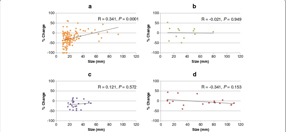
Associations between pre-treatment tumor diameter and percent change in target lesion size in different organs
The associations between pre-treatment tumor diameter and percent change in size of each target lesion in each of four organs were analyzed separately. Organ-specific analysis demonstrated that pre-treatment diameter of lung metastatic lesions had a moderately positive association with percent change in post-treatment tumor diameter (R = 0.341, Figure 1). There were fewer target lesions in the other three organs than in the lung, and there was no association between liver, lymph node, or kidney lesion size and percent change in post-treatment tumor diameter (Figure 1). The percent changes in target lesion size dif-fered significantly between the four individual organs (P= 0.0007 by the Kruskal Wallis test). The mean (± SD) per-cent changes in target lesion size in the lung, liver, lymph nodes, and kidney were −27.1 ± 33.5, 0.5 ± 29.4, −16.7 ± 19.6, and −2.7 ± 21.7, respectively. Target lesions in the lung showed the greatest change in size, which was signifi-cantly greater than in the liver and kidney (P= 0.007 and 0.002, respectively). There was no difference in the per-cent change in target lesion size between the lung and lymph nodes (P= 0.114) (Figure 2).
Lesions in each organ were divided into two groups according to the median pre-treatment diameter, and percent changes in tumor diameter were compared be-tween the two groups. Lung lesions with a pre-treatment diameter less than the median value of 17.3 mm showed a significant percent reduction in diameter compared with tumors≥17.3 mm (P= 0.002). There were no dif-ferences in the percent change in relation to size above or below the median value in other organs (Figure 2).
Cut-off value for pre-treatment tumor diameter predicting response to sunitinib in lung metastasis
ROC curves were drawn to determine cut-off values pre-dicting 30% and 50% reductions in the diameter of lung metastatic lesions (Figure 3). The cut-off predicting a 30% reduction in diameter was 16.5 mm, with a sensitiv-ity of 69.6%, specificsensitiv-ity of 58.2%, and an area under the curve (AUC) of 0.662. The cut-off predicting a 50% re-duction in diameter was also 16.5 mm, with a sensitivity of 67.0%, specificity of 77.8%, and an AUC of 0.752. Using this cut-off value, the percent change in lesion size for lesions < 16.5 mm was −41.7 ± 35.5%, while that for lesions≥16.5 mm was−16.2 ± 26.9% (P= 0.0005).
Influence of pre-treatment CRP value, cytoreductive nephrectomy, and treatment line on percent change in target lesion size in the lung
Metastatic lung lesions were categorized into two groups based on pre-treatment C-reactive protein (CRP) levels. The mean diameter and percent change in lesion size
-100 -50 0 50 100
0 20 40 60 80 100 120
% Change
Size (mm)
a
-100 -50 0 50 100
0 20 40 60 80 100 120
% Change
Size (mm)
b
-100 -50 0 50 100
0 20 40 60 80 100 120
% Change
Size (mm)
c
-100 -50 0 50 100
0 20 40 60 80 100 120
% Change
Size (mm)
d
R = 0.341, P= 0.0001 R = -0.021, P= 0.949
R = -0.341, P= 0.153 R = 0.121, P= 0.572
in patients with CRP < 2.0 mg/ml were 19.0 ± 9.3 mm and −38.3 ± 30.5%, respectively, while those in patients with CRP≥2.0 mg/ml were 28.3 ± 20.6 mm and −9.6 ± 33.9%, respectively. The lesions were further divided into three subgroups according to pre-treatment diameter < 20 mm,≥ 20 to < 40 mm, and≥40 mm. Percent changes were compared between lesions in patients with CRP < 2.0 mg/dl (low CRP) and those with CRP≥2.0 mg/dl (high
CRP) in each subgroup. For lesions < 20 mm, patients with low CRP had significantly greater reductions in tumor diameter than patients with high CRP (−43.8 ± 33.4% vs.−15.3 ± 37.7%,P= 0.0019). In patients with lesions≥20 to < 40 mm, there was a tendency towards a greater reduc-tion in patients with low CRP compared with high CRP, though the difference was not significant (−29.0 ± 17.3% vs. −12.4 ± 30.2%, P= 0.054), and similarly there was no significant association between tumor reduction and CRP level in patients with lesions≥40 mm (−13.3 ± 20.6 vs. 6.8 ± 17.4, P= 0.151) (Figure 4).
Percent changes in size were compared between lung lesions in patients in each diameter subgroup who did and did not undergo cytoreductive nephrectomy. There were no lung lesions≥40 mm in patients who did not undergo cytoreductive nephrectomy. There was no sig-nificant difference between mean percent change in lesion size in patients with and without cytoreductive nephrec-tomy (−36.1 ± 32.6 vs.−28.2 ± 41.9,P= 0.421 and−20.4 ± 25.1 vs. −32.0 ± 21.9, P= 0.307 in lesions < 20 mm and
≥20 mm, respectively).
Similarly, there was no significant difference in percent change in lesion size between lung lesions in patients treated as first-line therapy and those treated as second-line or later therapy in any diameter subgroup (−38.1 ± 37.7 vs. −32.1 ± 33.9, P= 0.280; −23.5 ± 21.6 vs. −17.5 ± 23.3, P= 0.803; and 3.5 ± 2.6 vs. 1.9 ± 22.9, P> 0.9 in lesions < 20 mm, ≥ 20 to < 40 mm, and≥40 mm, respectively).
Discussion
A previous study on metastatic RCC by Yuasa et al. de-monstrated that: 1) smaller initial tumor size predicted a -80
-60 -40 -20 0 20
%Change
62 62 12 12
9 10 6
6
Lung Liver Lymph node Kidney
Median (mm) 17.3 30.0 25.8 63.3
P = 0.002 P = 0.245 P = 0.773 P = 0.668 P = 0.007
P = 0.114 P = 0.002
Total < Median > Median
124 12 24
19
Figure 2Percent change in target lesion size in different organs.The reduction in lesion size was significantly greater in lung lesions compared with liver and kidney lesions. Lung lesions with an initial diameter < 17.3 mm (median) showed a significant percent reduction in size compared with lesions≥17.3 mm. No significant differences in relation to initial lesion size were observed in the liver, lymph nodes, and kidney.
1.0
0.8
0.6
0.4
0.2
0.0
0.0 0.2 0.4 0.6 0.8 1.0
Cut-off of %reduction 50% 30%
Sensitivity
1 - Specificity 16.5 mm
16.5 mm
good response to TKIs; 2) the greatest response was achieved in patients with lung lesions; and 3) there was no difference in tumor response between patients treated with sorafenib and sunitinib [6]. However, these results raised several specific questions. First, tumor histology and progression risk may affect the response to TKIs. TKIs are associated with a good response in patients with CCRCC, but are less effective against non-CCRCC [10]. Similarly, patients with favorable risk factors have a greater chance of a good tumor response than those with poorer risk factors. Second, there is the possibility of bias in terms of the types of TKI selected; given that sunitinib showed a higher response rate than sorafenib [7,8], pa-tients with larger or more rapidly-growing tumors may be allocated sunitinib rather than sorafenib in clinical prac-tice. Third, efficacy based on initial tumor size may dif-fer between difdif-ferent organs; although the previous study compared mean lesion-size reductions between different organs, they did not compare the effect of initial tumor size in individual organs. It is therefore unclear if the asso-ciation between initial lesion size and tumor response was observed in each organ, or if the association could be at-tributed to the fact that most of the small lesions were lung metastases, which showed a good response to TKIs. The current study only included CCRCC patients treated with sunitinib. We found that lung lesions showed the greatest response to sunitinib, and detected a modest cor-relation between initial tumor diameter and reduction in lesion size, while even small lesions in other organs failed to respond. However, the number of extra-pulmonary tu-mors assessed was too small to determine statistical sig-nificance, and further studies with larger numbers of tumors are needed to obtain conclusive results.
Only lesions with an initial diameter < 20 mm achieved a CR in this study, indicating that a lung-tumor reduc-tion of > 50% might be limited to smaller lesions. The cut-off value of 16.5 mm for a > 50% reduction in diam-eter was calculated using ROC analysis, with a sensitivity of 67.0% and a specificity of 77.8%. Some physicians may prefer conservative therapies without TKIs, or a watchful waiting strategy, in CCRCC patients with only small lung metastatic lesions [11]. Furthermore, cytokine ther-apies are still employed in CCRCC patients, especially in Japan, because of their low toxicity and ability to achieve long-term stable disease [12]. However, the present re-sults suggest that smaller lung lesions are associated with a greater chance of response to TKIs, and it is therefore important not to miss the opportunity for early initiation of TKI treatment in patients with PD during watchful waiting periods or cytokine therapy.
Several studies have investigated the response of pri-mary kidney lesions to TKIs [13-15]. Kroon et al. re-ported that smaller primary lesions were more responsive to treatment, and that tumors of 5–7 cm may benefit from neoadjuvant treatment followed by nephron-sparing sur-gery. In contrast, our results showed that the response of kidney lesions to sunitinib was independent of initial tumor size, and many smaller lesions exhibited no res-ponse. A possible explanation for this difference may be the selection of patients; most of the kidney lesions were investigated in the neoadjuvant setting in Kroon et al.’s study, while all the patients with kidney lesions in the current study had an extensive metastatic tu-mor burden. The different patient backgrounds may have led to different responses to TKIs, particularly in small kidney lesions.
-80 -60 -40 -20 0 20
54 23 24 11 3 9
< 20 20 - 40 > 40
%Change
P = 0.0019 P = 0.054 P = 0.151
Tumor size (mm)
CRP < 2.0 CRP ≥ 2.0 Total
77 35 12
CRP is an acute phase protein produced by the liver in response to various conditions, such as inflammation, in-fection, and malignancy [16]. In the cytokine era, elevated serum CRP level has been suggested as a biomarker for predicting poor survival in RCC patients [17-19]. Yasuda et al. recently demonstrated that CRP was a significant predictive marker for prognosis in metastatic RCC pa-tients treated with TKIs [20]. In the current study, the size reduction of lung lesions in patients with high serum CRP levels was lower than that in patients with low CRP levels, irrespective of the initial size. This lower response to suni-tinib in patients with higher serum CRP levels may be at-tributed to an aggressive disease status, reflected by higher CRP levels, the acquisition of resistance to therapeutic agents through an increase in inflammatory mediators in the cancer-cell microenvironment, or compromised drug metabolism induced by such mediators associated with CRP [21].
Tumor response to treatment is currently assessed by imaging based on RECIST criteria [22]. However, al-though marked central necrosis is often detected in le-sions with a small size reduction after treatment with TKIs, RECIST only considers one-dimensional lesional size changes, suggesting that it may substantially un-derestimate the actual tumor response. Several studies recently reported novel criteria, which may improve response assessment by evaluating changes in tumor attenuation and morphology on contrast-enhanced com-puted tomography scans in addition to size changes [5,22-26]. The results of this study therefore need to be interpreted carefully, because lesions in different organs may exhibit distinct response patterns in imaging. More-over, the current study did not demonstrate an association between tumor response and patient survival, and it is possible that percent change in tumor size might not cor-relate directly with survival. Further studies are needed to determine the influence of organ-specific response pat-terns to TKI treatment on survival.
Conclusions
The results suggest that tumor-size reduction depends on initial tumor size and the organs involved, as well as systemic reaction to the lung tumor, as indicated by CRP levels. CCRCC patients with lung metastatic lesions < 20 mm in diameter and lower CRP levels may achieve greater reductions in tumor size with sunitinib therapy than those with extra-pulmonary lesions, lung lesions≥ 20 mm in diameter, and/or higher CRP levels.
Abbreviations
RCC:Renal cell carcinoma; TKI: Tyrosine kinase inhibitor; RECIST: Response evaluation criteria in solid tumors; CCRCC: Clear cell RCC; CTC-AE: Common Terminology criteria for Adverse Events; ROC: Receiver-operator curve; AUC: Area under the curve; CRP: C-reactive protein.
Competing interests
Norihiko Tsuchiya and Tomonori Habuchi received honoraria from Pfizer Japan Inc.
Authors’contributions
NT, TY, JY, and TH were involved in the conception and design of the study. TY, KN, MS, and SM were involved in the provision of patients’clinical data. NT and TH drafted the manuscript. SN, TI, SS supported the manuscript writing. All authors have read and approved the final manuscript.
Acknowledgements
There were no external sources of funding.
Author details
1Department of Urology, Akita University Graduate School of Medicine, Akita, Japan.2Department of Urology, Cancer Institute Hospital, Japanese Foundation for Cancer Research, Tokyo, Japan.
Received: 20 July 2013 Accepted: 28 February 2014 Published: 11 March 2014
References
1. Akaza H, Kawai K, Tsukamoto T, Fujioka T, Tomita Y, Kitamura T, Ozono S, Miki T, Naito S, Zembutsu H:Successful outcomes using combination therapy of interleukin-2 and interferon-alpha for renal cell carcinoma patients with lung metastasis.Jpn J Clin Oncol2010,40(7):684–689. 2. Flanigan RC, Salmon SE, Blumenstein BA, Bearman SI, Roy V, McGrath PC,
Caton JR Jr, Munshi N, Crawford ED:Nephrectomy followed by interferon alfa-2b compared with interferon alfa-2b alone for metastatic renal-cell cancer.N Engl J Med2001,345(23):1655–1659.
3. Albiges L, Oudard S, Negrier S, Caty A, Gravis G, Joly F, Duclos B, Geoffrois L, Rolland F, Guillot A, Laguerre B, Legouffe E, Kohser F, Dietrich PY, Theodore CA, Escudier B:Complete remission with tyrosine kinase inhibitors in renal cell carcinoma.J Clin Oncol2012,30(5):482–487.
4. Staehler M, Haseke N, Zilinberg E, Stadler T, Karl A, Siebels M, Durr HR, Siegert S, Jauch KW, Bruns CJ, Stief CG:Complete remission achieved with angiogenic therapy in metastatic renal cell carcinoma including surgical intervention.Urol Oncol2010,28(2):139–144.
5. Krajewski KM, Guo M, Van den Abbeele AD, Yap J, Ramaiya N, Jagannathan J, Heng DY, Atkins MB, McDermott DF, Schutz FA, Pedrosa I, Choueiri TK: Comparison of four early posttherapy imaging changes (EPTIC; RECIST 1.0, tumor shrinkage, computed tomography tumor density, Choi criteria) in assessing outcome to vascular endothelial growth factor-targeted therapy in patients with advanced renal cell carcinoma.Eur Urol 2011,59(5):856–862.
6. Yuasa T, Urakami S, Yamamoto S, Yonese J, Nakano K, Kodaira M, Takahashi S, Hatake K, Inamura K, Ishikwa Y, Fukui I:Tumor size is a potential predictor of response to tyrosine kinase inhibitors in renal cell cancer. Urology2011,77(4):831–835.
7. Motzer RJ, Hutson TE, Tomczak P, Michaelson MD, Bukowski RM, Rixe O, Oudard S, Negrier S, Szczylik C, Kim ST, Chen I, Bycott PW, Baum CM, Figlin RA:Sunitinib versus interferon alfa in metastatic renal-cell carcinoma. N Engl J Med2007,356(2):115–124.
8. Escudier B, Eisen T, Stadler WM, Szczylik C, Oudard S, Siebels M, Negrier S, Chevreau C, Solska E, Desai AA, Rolland F, Demkow T, Hutson TE, Gore M, Freeman S, Schwartz B, Shan M, Simantov R, Bukowski RM:Sorafenib in advanced clear-cell renal-cell carcinoma.N Engl J Med2007, 356(2):125–134.
9. Rini BI, Escudier B, Tomczak P, Kaprin A, Szczylik C, Hutson TE, Michaelson MD, Gorbunova VA, Gore ME, Rusakov IG, Negrier S, Ou YC, Castellano D, Lim HY, Uemura H, Tarazi J, Cella D, Chen C, Roosbrook B, Kim S, Motzer RJ: Comparative effectiveness of axitinib versus sorafenib in advanced renal cell carcinoma (AXIS): a randomised phase 3 trial.Lancet2011, 378(9807):1931–1939.
10. Tannir NM, Plimack E, Ng C, Tamboli P, Bekele NB, Xiao L, Smith L, Lim Z, Pagliaro L, Araujo J, Aparicio A, Matin S, Wood CG, Jonasch E:A phase 2 trial of sunitinib in patients with advanced Non-clear cell renal cell carcinoma.Eur Urol2012,62(6):1013–1019.
12. Fujioka T, Obara W:Evidence-based clinical practice guideline for renal cell carcinoma: the Japanese Urological Association 2011 update. Int J Urol2012,19(6):496–503.
13. van der Veldt AA, Meijerink MR, van den Eertwegh AJ, Bex A, de Gast G, Haanen JB, Boven E:Sunitinib for treatment of advanced renal cell cancer: primary tumor response.Clin Cancer Res2008,14(8):2431–2436. 14. Kroon BK, de Bruijn R, Prevoo W, Horenblas S, Powles T, Bex A:Probability
of downsizing primary tumors of renal cell carcinoma by targeted therapies is related to size at presentation.Urology2012,81(1):111–115. 15. Abel EJ, Culp SH, Tannir NM, Tamboli P, Matin SF, Wood CG:Early primary
tumor size reduction is an independent predictor of improved overall survival in metastatic renal cell carcinoma patients treated with sunitinib.Eur Urol2011,60(6):1273–1279.
16. Gabay C, Kushner I:Acute-phase proteins and other systemic responses to inflammation.N Engl J Med1999,340(6):448–454.
17. Bromwich E, McMillan DC, Lamb GW, Vasey PA, Aitchison M:The systemic inflammatory response, performance status and survival in patients undergoing alpha-interferon treatment for advanced renal cancer. Br J Cancer2004,91(7):1236–1238.
18. Casamassima A, Picciariello M, Quaranta M, Berardino R, Ranieri C, Paradiso A, Lorusso V, Guida M:C-reactive protein: a biomarker of survival in patients with metastatic renal cell carcinoma treated with subcutaneous interleukin-2 based immunotherapy.J Urol2005,173(1):52–55.
19. Ramsey S, Lamb GW, Aitchison M, Graham J, McMillan DC:Evaluation of an inflammation-based prognostic score in patients with metastatic renal cancer.Cancer2007,109(2):205–212.
20. Yasuda Y, Saito K, Yuasa T, Kitsukawa S, Urakami S, Yamamoto S, Yonese J, Takahashi S, Fukui I:Prognostic impact of pretreatment C-reactive protein for patients with metastatic renal cell carcinoma treated with tyrosine kinase inhibitors.Int J Clin Oncol2012,18(5):884–889.
21. Slaviero KA, Clarke SJ, Rivory LP:Inflammatory response: an unrecognised source of variability in the pharmacokinetics and pharmacodynamics of cancer chemotherapy.Lancet Oncol2003,4(4):224–232.
22. Eisenhauer EA, Therasse P, Bogaerts J, Schwartz LH, Sargent D, Ford R, Dancey J, Arbuck S, Gwyther S, Mooney M, Rubinstein L, Shankar L, Dodd L, Kaplan R, Lacombe D, Verweij J:New response evaluation criteria in solid tumours: revised RECIST guideline (version 1.1).Eur J Cancer2009, 45(2):228–247.
23. Han KS, Jung DC, Choi HJ, Jeong MS, Cho KS, Joung JY, Seo HK, Lee KH, Chung J:Pretreatment assessment of tumor enhancement on contrast-enhanced computed tomography as a potential predictor of treatment outcome in metastatic renal cell carcinoma patients receiving antiangiogenic therapy.Cancer2010,116(10):2332–2342. 24. Nathan PD, Vinayan A, Stott D, Juttla J, Goh V:CT response assessment
combining reduction in both size and arterial phase density correlates with time to progression in metastatic renal cancer patients treated with targeted therapies.Cancer Biol Ther2010,9(1):15–19.
25. Hittinger M, Staehler M, Schramm N, Ubleis C, Becker C, Reiser M, Berger F: Course of size and density of metastatic renal cell carcinoma lesions in the early follow-up of molecular targeted therapy.Urol Oncol2012, 30(5):695–703.
26. Choi H, Charnsangavej C, Faria SC, Macapinlac HA, Burgess MA, Patel SR, Chen LL, Podoloff DA, Benjamin RS:Correlation of computed tomography and positron emission tomography in patients with metastatic gastrointestinal stromal tumor treated at a single institution with imatinib mesylate: proposal of new computed tomography response criteria.J Clin Oncol2007,25(13):1753–1759.
doi:10.1186/1471-2490-14-26
Cite this article as:Tsuchiyaet al.:Organ-specific and tumor-size-dependent responses to sunitinib in clear cell renal cell carcinoma.BMC Urology201414:26.
Submit your next manuscript to BioMed Central and take full advantage of:
• Convenient online submission
• Thorough peer review
• No space constraints or color figure charges
• Immediate publication on acceptance
• Inclusion in PubMed, CAS, Scopus and Google Scholar
• Research which is freely available for redistribution