C A S E R E P O R T
Open Access
Primary Ewing
’
s sarcoma/primitive
neuroectodermal tumor of the ileum: case
report of a 16-year-old Chinese female and
literature review
Teng Li
1, Fang Zhang
1, Yarui Cao
1, Shoubin Ning
2, Yongmin Bi
3, Weicheng Xue
4and Li Ren
1*Abstract
Background:
Ewing
’
s sarcoma (ES) and primitive neuroectodermal tumors (PNET) are closely related tumors.
Although soft tissue ES/PNET are common in clinical practice, they are rare in the small intestine. Because of the
absence of characteristic clinical symptoms, they are easily misdiagnosed as other benign or malignant diseases.
Case presentation:
Here, we present the case of a 16-year-old female who complained of anemia and interval
hematochezia. Her serum test results showed only a slight elevation of CA-125 and a low level of hemoglobin.
Computer tomography and magnetic resonance imaging revealed a cystic and solid mass in the lower abdominal
quadrant and pelvic region, which prompted suspicion of a malignant gastrointestinal stromal tumor of the small
intestine. After resection, the tumor
’
s histology and immunohistochemistry (positive for CD99, vimentin and synaptophysin)
results suggested ES/PNET. Fluorescent
in situ
hybridization tests proved the breakpoint rearrangement of the
EWSR1
gene
in chr 22.Ultrastructural analysis revealed neurosecretory and glycogen granules in the tumor cell cytoplasm.
Conclusions:
Together, these data supported the diagnosis of a rare case of localized ES/PNET in the small intestine
without adjuvant chemo- or radiotherapy. To our knowledge, this is the first report from China of a primary small
bowel ES/PNET in the English-language literature. In addition, on the basis of findings from previous publications
and the current case, the optimal treatment for localized gastrointestinal ES/PNET is discussed.
Keywords:
Ewing
’
s sarcoma, Primitive neuroectodermal tumor, Extraosseous, Small intestine, FISH, EWS gene
Background
Ewing
’
s sarcoma (ES)/primitive neuroectodermal tumor
(PNET) is a small round cell tumor with simple
sarcoma-specific genetic alterations resulting in
TET/FET
family
member and
ETS
family member fusion proteins [1].
Pathologists no longer categorize ES and PNET as
differ-ent tumors because their genetic abnormalities overlap.
Instead, they are termed the Ewing
’
s sarcoma family of
tumors [2, 3], together with the Askin tumor. ES/PNET
are most commonly seen in patients younger than 20 years
of age and are derived mainly from bone [4]. The tumor
has been discovered in most organs, including the pancreas,
liver, adrenal gland, esophagus, and uterus [5
–
11]. However,
ES/PNET is extremely rare in the small bowel. Although it
has been reported previously in this location [12
–
17], none
of these reports came from China. Here, we present the
first reported case in China of primary ES/PNET in the
ileum with EWS rearrangement.
Case presentation
Clinical history
A 16-year-old Chinese girl presented complaining of
anemia and interval hematochezia. Her hemoglobin was
54 g/L on admission. Capsule endoscopy and
double-balloon enteroscopy showed mucosal hyperemia, edema
and mass protrusion on the ileal wall. Computed
tomog-raphy (CT) scans and three-dimensional reconstruction
revealed a 10.0 × 7.3 × 5.3 cm irregular mass that had
* Correspondence:[email protected]
1Department of Pathology, The General Hospital of Air force, PLA, Fucheng Road 30th, Beijing, China
Full list of author information is available at the end of the article
developed from the ileal wall in the right lower quadrant
(Fig. 1a-h). The lesion showed intense but
inhomogen-eous enhancement following contrast administration
(Fig. 1e-h), particularly in the arterial phase. There was a
small amount of effusion in the pelvic cavity. Pelvic
mag-netic resonance imaging (MRI) indicated a right ovarian
cyst in addition to the above mass. Both CT and MRI
prompted suspicion of malignant GIST of the small
bowel. Her serum CA-125 was slightly increased (50.5
U/mL, standard 0
–
36 U/mL), but the other markers
were within normal limits. The tumor and a loop of
small intestine were resected through a right ventral
midline incision. The patient recovered uneventfully.
Postoperative bone scintigraphy proved that there was
no lesion in the skeletal system (Fig. 1 d). Her chest CT
scan and cerebral MRI were also unremarkable. Thus,
the patient was classified as T2aN0M0 according to the
8th edition of the AJCC Cancer Staging Manual.
Gross features
On laparotomy, a large cystic and solid mass 10.5 cm in
diameter was found arising from the ileal wall. The cut
tumor surface showed large central hemorrhagic and
necrotic changes and pseudocystic degeneration. The
tumor tissue was mostly light gray and solid, with some
softer and more friable reddish congested areas (Fig. 2).
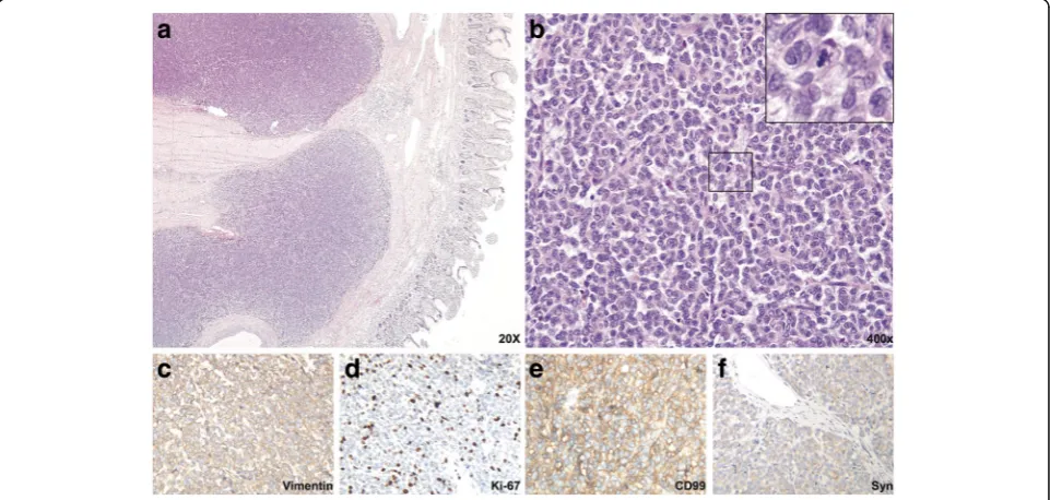
Histological features
Cross sections revealed solid nests of small round tumor
cells arising from the muscular layer and infiltrating all
layers of the ileum wall. Cystic and hemorrhagic changes
were seen on part of the sections, as were sharply
de-marcated borders that were frequently covered by
intact serosa. No vascular tumor embolus or
perineur-onal invasion were observed. The serosal layer and the
surgical margins of the specimen were free of disease.
Under high-power view, tumor cells were round or
elliptical, possessing scant eosinophilic cytoplasm and
abortive pseudorosette formation. The tumor cell
nu-clei were round, with exquisite chromatin, ambiguous
nucleoli, and 9/10 high-power-field pathological mitoses
(Fig. 3a-c). Tumor cells showed positive immunoreactivity
for Vimentin and CD99 (Fig. 3 d, j) and moderate staining
for Cam5.2, Syn and PR (Fig. 3 f, g, k). Results were
nega-tive for CKpan, LCA, S-100, HMB45, Melan-A, CD31,
CD34, NSE, P53, CD56, CgA, SMA, Desmin, CD117,
Dog-1, ER, Bcl-2, and alpha-inhibin.
Fig. 1Abdominal and pelvic CT scan, 3D reconstruction and ECT demonstrating the tumor originating in the ileum of the patient. CT scan was
performed immediately after enteroscopy. Thus, the patient’s intestine was dilated.aCoronal scan arterial phase reveals that the tumor was derived from ileal wall.b3D reconstruction with volume rendering technique illustrates the supporting vasculature.c3D reconstruction with maximum intensity projection demonstrates major vascular support of the tumor.dPostoperative bone scintigraphy proved that there was no lesion in her skeletal system.ePlain scan revealed pelvic a 10.0 × 7.6 × 5.3 cm mass with areas of necrosis. Most of the mass was had clear boundaries with surrounding tissues, although part of it obliterated the lumen of the terminal ileum. Workup for metastasis was negative.f-h
FISH
Dual color break-apart probe FISH examination showed
that 90% of the cells (100 counted cells per slide)
exhib-ited 1 yellow and 1 red signal (1F1R) and that 6% of the
cells exhibited 1 yellow, 1 red and 1 green (break-apart)
signal (1F1G1R). However, only 4% cell had two yellow
signals, which proved a break of the
EWSR1
locus (2 F)
(Fig. 4a-c).
EM
Transmission electron microscopy revealed dense
clus-ters of tumor cells, inclus-terspersed with a few inclus-terstitial
cells (Fig. 5 a). The tumor cells were small and
irregu-lar, with scant cytoplasm and organelles, and significant
nuclear atypia (Fig. 5 a). Some cells had small nucleoli
(Fig. 5 a). Occasionally, gap junctions between the cells
were observed (Fig. 5 b), but neuroendocrine granules
Fig. 2Gross features of the tumor with the resected ileum
Fig. 3Histological and immunohistochemical features of the intestinal tumor.aLow-power view with HE staining indicates sheets of tumor cells
in the cytoplasm were rarely seen (Fig. 5 c). Most cells
had glycogen particles attached to the endoplasmic
reticulum (Fig. 5 d).
Treatments and outcome
The patient underwent an exploratory laparotomy, and
tumor resection was performed along with 60 cm of ileum.
The patient refused chemotherapy and/or radiotherapy as
adjuvant treatments. She is currently alive (10 months after
the surgery) without any signs of recurrence.
Discussion
ES/PNET belongs to a family of tumors that harbor the
EWSR1
-
ETS
fusion protein, according to recent studies
Fig. 4Dual color (red/green) break-apart probe FISH test of the tumor.aNormal karyotype cells have two yellow (red/greenmerged) signals (arrowheads).
bMost tumor cells (90%) had one yellow (red/greenmerged) signal and one red signal (arrows). C. Consecutive sections were HE-stained
Fig. 5Ultrastructure analysis of the tumor.aAt lower magnification, EM shows the general tumor ultrastructure.bCell-cell gap junctions (blue
Table
1
Review
of
reported
cases
of
gastrointestinal
ES/PNET
(Con
tinued)
Small
bowe
l
42
M
+
+
+
+
+
+
+
+
-Sx
+
Cx
Die
d
11
mont
hs
afte
r
dia
gnosi
s
Mi
lione
M
et
al.
Small
bowe
l
45
M
+
+
+
+
+
+
+
+
-Sx
+
Cx
Die
d
13
mont
hs
afte
r
dia
gnosi
s
Mi
lione
M
et
al.
Small
bowe
l
15
F
+
+
+
+
+
+
+
+
-Sx
+
Cx
+
Rx
28
mont
hs
D
FS
Mi
lione
M
et
al.
Small
bowe
l
57
M
+
+
+
+
+
+
+
+
-Lost
Lost
Mi
lione
M
et
al.
Small
bowe
l
28
F
+
+
+
+
-+
+
Liver
Sx
+
Cx
204
month
s
DFS
Mi
lione
M
et
al.
F
Female,
M
Male,
ND
Not
done,
Sx
Surgery,
Cx
Chemotherapy,
ImCx
Immuno
chemotherapy,
StemCx
Stem
cell
based
chemotherapy,
Rx
Radiotherapy,
DFS
Disease
free
[18]. It is the second most common pediatric sarcoma of
bone. It most commonly arises from bone but can develop
in extraskeletal sites [19]. The
EWSR1
gene, together with
several other genes, forms the TET family [20]. Their motif
of RNA binding activity enables the
EWSR1
-
ETS
fusion
protein to regulate target genes as transcription factors
[21, 22]. Previous research provided evidence that
mes-enchymal stem cells may be candidate cells from which
ES/PNET originate and that
EWSR1
–
FLI1
may be the
sole initiating factor in the pathogenesis of these tumors
[20, 23]. Such expression results in cell transformation,
with the subsequent emergence of tumors bearing the
morphological and gene expression hallmarks of Ewing
’
s
sarcoma [24].
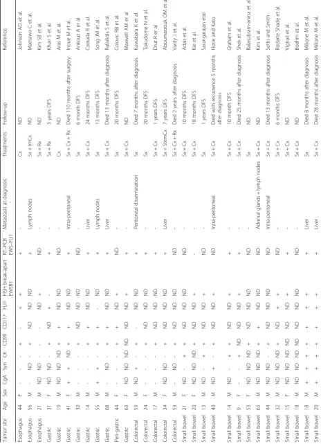
Gastroenterological ES/PNET is extremely rare. Here,
we have summarized all previous publications of
gastro-intestinal ES/PNET in Table 1 [7, 12, 13, 17, 25
–
49].
Among the 36 cases, 3 cases were derived from the
esophagus, 9 from the stomach, 5 were of colorectal
ori-gin, and 19 arose from the small intestine. The patient
gender ratio (female/male) was 22/14, and the ages
ranged from 9 to 68 years. Thirty-one of 32 cases were
positive for CD99 immunoreactivity. Fluorescent in situ
hybridization or real-time PCR tests confirmed that
most cases had the
EWSR1
-
ETS
fusion protein.
Intri-guingly, however, only 4 non-metastatic gastrointestinal
ES/PNET cases were treated only by resection of the
tumor. Follow-up of these cases suggested that the
pa-tients were relatively younger and had up to 20 months
of disease-free survival. In the current case, the young
patient also refused to take adjuvant chemo- or
radiother-apy. To our delight, after the 10-month follow-up
examin-ation, the patient is currently alive and well, without any
sign of recurrence.
To date, the 5-year survival rate of localized ES/PNET
is relatively high (65%-75%). However, the outcome for
metastatic patients is usually poor (<30%), despite the use
of chemo- and/or radiotherapy [50]. Several studies have
indicated that localized extraskeletal ES/PNET has a more
favorable outcome than skeletal tumors [51, 52]. The
opti-mal management for localized ES/PNET is still debated.
The National Comprehensive Cancer Network guidelines
recommend that any ES/PNET should be treated with
local treatment (surgery and/or radiotherapy) plus
chemo-therapy [53]. Nevertheless, consistent with our findings in
Table 1, others have suggested that complete surgery, if
feasible, may be a better option for local disease
consider-ing the late side effects of high-dose radiotherapy
espe-cially for children [52, 54]. Because small bowel ES/PNET
is extremely rare and difficult to cure, our case will
con-tribute to the understanding of the prognosis and
deter-mination of optimal management.
In the current case, the 16-year-old female patient was
initially misdiagnosed with malignant GIST because of the
clinical symptoms and imaging results. To differentiate
among ES/PNET, malignant GIST, clear-cell sarcoma,
and synovial sarcoma, immunohistochemistry,
ultra-structure analysis and FISH tests were performed.
Malignant GIST usually expresses CD117, Dog-1 and
CD34, which were all negative in this case. Although
both synovial sarcoma and ES/PNET could have genetic
rearrangements, the regions of these translocations are
quite different. In ES/PNET, Chr22
EWS-FLI
or
EWS-FEV
translocations are commonly reported [16]. However, in
synovial sarcoma,
SYT-SSX
translocation is frequently
observed [55]. Clear-cell sarcoma could be ruled out by
negative immunohistochemistry for HMB45, S-100 and
Melan A. A previous study also indicated the necessity
of distinguishing from an intraabdominal desmoplastic
small round cell tumor (IDSRCT) by histological and
immunohistochemical characteristics when ES/PNET
occurs in the abdominal cavity [13].
Previous demographic research has suggested that
Ewing
’
s sarcoma is far less frequent in China than in
the United States Caucasian population [56]. However,
whether this finding is related to genetic background
differences remains to be studied. Two recent
publica-tions noted a difference in Ewing
’
s sarcoma occurrence
between Caucasian and Hispanic populations [57, 58].
However, they did not include a reason to explain this
differences.
Conclusions
In conclusion, we have described for the first time a rare
case of localized ES/PNET occurring in the small intestine
in the Chinese population, as confirmed by ultrastructure
and genetic analyses. This case, together with previous
reports, has expanded the spectrum of tumors in the small
intestine.
Abbreviations
Bcl-2:B cell lymphoma 2; CA-125: Cancer antigen- 125; CD: Cluster of differentiation; CgA: Glycoprotein hormone alpha chain; CKpan: Pan-cytokeratin; CT: Computed tomography; ER: Estrogen hormone receptor; ES: Ewing’s sarcoma; GIST: Gastrointestinal stromal tumor; LCA: Lymphocyte common antigen; MRI: Magnetic resonance imaging; NSE: Neuron-specific enolase; PNET: Primitive neuroectodermal tumor; SMA: Alpha smooth muscle actin
Acknowledgments
The authors thank Dr. Na Jia from the Department of Pathology, Beijing Cancer Hospital, for his expert technical assistance performing FISH analyses.
Funding
This study was supported by a grant-in-aid for Scientific Research from the General Hospital of the Air Force, PLA to Ren Li and from the Junior Scientists Fund (16QNP025) to Li Teng.
Availability of data and materials
Authors’contributions
T Li performed histopathological evaluations and drafted the manuscript. L Ren conceived the study and participated in the design and preparation of the manuscript. F Zhang collected, evaluated and interpreted the clinical and surgical data. YR Cao evaluated the FISH and electron microscopic findings. SB Ning performed the gastroenterological examination and provided related data. YM Bi examined the patient with CT, MRI, and bone scintigraphy and provided related data. WC Xue discussed the pathological diagnosis and supported the FISH test. All authors read and approved the final manuscript.
Competing interests
The authors declare that they have no competing interests.
Consent for publication
Written informed consent was obtained from the patient for the publication of this case report and any accompanying images.
Ethical approval and consent to participate
The ethical approval and documentation for the participation and publication of this case report were waived with approval of the Institutional Review Board at the General Hospital of the Air Force, PLA.
Publisher
’
s Note
Springer Nature remains neutral with regard to jurisdictional claims in published maps and institutional affiliations.
Author details
1
Department of Pathology, The General Hospital of Air force, PLA, Fucheng Road 30th, Beijing, China.2Department of Gastroenterology, The General Hospital of Air force, PLA, Fucheng Road 30th, Beijing, China.3Department of Radio and Imaging, The General Hospital of Air force, PLA, Fucheng Road 30th, Beijing, China.4Department of Pathology, Beijing Cancer Hospital, Fucheng Road 52nd, Beijing, China.
Received: 28 November 2016 Accepted: 17 April 2017
References
1. Kim SK, Park YK. Ewing sarcoma: a chronicle of molecular pathogenesis. Hum Pathol. 2016;55:91–100.
2. Dehner LP. Primitive neuroectodermal tumor and Ewing’s sarcoma. Am J Surg Pathol. 1993;17(1):1–13.
3. Dehner LP. The evolution of the diagnosis and understanding of primitive and embryonic neoplasms in children: living through an epoch. Mod Pathol. 1998;11(7):669–85.
4. Collini P, Mezzelani A, Modena P, Dagrada P, Tamborini E, Luksch R, et al. Evidence of neural differentiation in a case of post-therapy primitive neuroectodermal tumor/Ewing sarcoma of bone. Am J Surg Pathol. 2003; 27(8):1161–6.
5. Jimenez RE, Folpe AL, Lapham RL, Ro JY, O’Shea PA, Weiss SW, et al. Primary Ewing’s sarcoma/primitive neuroectodermal tumor of the kidney: a clinicopathologic and immunohistochemical analysis of 11 cases. Am J Surg Pathol. 2002;26(3):320–7.
6. Harimaya K, Oda Y, Matsuda S, Tanaka K, Chuman H, Iwamoto Y. Primitive neuroectodermal tumor and extraskeletal Ewing sarcoma arising primarily around the spinal column: report of four cases and a review of the literature. Spine (Phila Pa 1976). 2003;28(19):E408–12.
7. Johnson AD, Pambuccian SE, Andrade RS, Dolan MM, Aslan DL. Ewing sarcoma and primitive neuroectodermal tumor of the esophagus: report of a case and review of literature. Int J Surg Pathol. 2010;18(5):388–93. 8. Cambruzzi E, Guerra EE, Hilgert HC, Schmitz HJ, Silva VL, Milani DM, et al.
Primitive neuroectodermal tumor of the liver: a case report. Case Rep Med. 2011;2011:748194.
9. Ren YL, Tang XY, Li T. Ewing sarcoma-primitive neuroectodermal tumor of the uterus: a clinicopathologic, immunohistochemical and ultrastructural study of one case. Arch Gynecol Obstet. 2011;283(5):1139–43.
10. Bose P, Murugan P, Gillies E, Holter JL. Extraosseous Ewing’s sarcoma of the pancreas. Int J Clin Oncol. 2012;17(4):399–406.
11. Abi-Raad R, Manetti GJ, Colberg JW, Hornick JL, Shah JG, Prasad ML. Ewing sarcoma/primitive neuroectodermal tumor arising in the adrenal gland. Pathol Int. 2013;63(5):283–6.
12. Adair A, Harris SA, Coppen MJ, Hurley PR. Extraskeletal Ewings sarcoma of the small bowel: case report and literature review. J R Coll Surg Edinb. 2001; 46(6):372–4.
13. Shek TW, Chan GC, Khong PL, Chung LP, Cheung AN. Ewing sarcoma of the small intestine. J Pediatr Hematol Oncol. 2001;23(8):530–2.
14. Batziou C, Stathopoulos GP, Petraki K, Papadimitriou C, Rigatos SK, Kondopodis E, et al. Primitive neurectodermal tumors: a case of extraosseous Ewing’s sarcoma of the small intestine and review of the literature. J BUON. 2006;11(4):519–22. 15. Prasertvit S, Stoikes N. A rare case of ewing’s sarcoma of the small intestine.
Am Surg. 2013;79(2):E78–9.
16. Milione M, Gasparini P, Sozzi G, Mazzaferro V, Ferrari A, Casali PG, et al. Ewing sarcoma of the small bowel: a study of seven cases, including one with the uncommonly reported EWSR1-FEV translocation. Histopathology. 2014;64(7):1014–26.
17. Vignali M, Zacche MM, Messori P, Natale A, Busacca M. Ewing’s sarcoma of the small intestine misdiagnosed as a voluminous pedunculated uterine leiomyoma. Eur J Obstet Gynecol Reprod Biol. 2012;162(2):234–5. 18. Thompson LD. Ewing sarcoma and primitive neuroectodermal tumor. Ear
Nose Throat J. 2007;86(2):79–80.
19. Sandberg AA, Bridge JA. Updates on cytogenetics and molecular genetics of bone and soft tissue tumors: Ewing sarcoma and peripheral primitive neuroectodermal tumors. Cancer Genet Cytogenet. 2000;123(1):1–26. 20. Riggi N, Cironi L, Suva ML, Stamenkovic I. Sarcomas: genetics, signalling, and
cellular origins. Part 1: The fellowship of TET. J Pathol. 2007;213(1):4–20. 21. Crozat A, Aman P, Mandahl N, Ron D. Fusion of CHOP to a novel RNA-binding
protein in human myxoid liposarcoma. Nature. 1993;363(6430):640–4. 22. Ohno T, Ouchida M, Lee L, Gatalica Z, Rao VN, Reddy ES. The EWS gene,
involved in Ewing family of tumors, malignant melanoma of soft parts and desmoplastic small round cell tumors, codes for an RNA binding protein with novel regulatory domains. Oncogene. 1994;9(10):3087–97. 23. Riggi N, Suva ML, Suva D, Cironi L, Provero P, Tercier S, et al. EWS-FLI-1
expression triggers a Ewing’s sarcoma initiation program in primary human mesenchymal stem cells. Cancer Res. 2008;68(7):2176–85.
24. Castillero-Trejo Y, Eliazer S, Xiang L, Richardson JA, Ilaria Jr RL. Expression of the EWS/FLI-1 oncogene in murine primary bone-derived cells Results in EWS/FLI-1-dependent, ewing sarcoma-like tumors. Cancer Res. 2005;65(19): 8698–705.
25. Horie Y, Kato M. Peripheral primitive neuroectodermal tumor of the small bowel mesentery: a case showing perforation at onset. Pathol Int. 2000; 50(5):398–403.
26. Sarangarajan R, Hill DA, Humphrey PA, Hitchcock MG, Dehner LP, Pfeifer JD. Primitive neuroectodermal tumors of the biliary and gastrointestinal tracts: clinicopathologic and molecular diagnostic study of two cases. Pediatr Dev Pathol. 2001;4(2):185–91.
27. Balasubramanian B, Dinakarababu E, Molyneux AJ. Primary primitive neuroectodermal tumour of the small bowel mesentery: case report. Eur J Surg Oncol. 2002;28(2):197–8.
28. Graham DK, Stork LC, Wei Q, Ingram JD, Karrer FM, Mierau GW, et al. Molecular genetic analysis of a small bowel primitive neuroectodermal tumor. Pediatr Dev Pathol. 2002;5(1):86–90.
29. Maesawa C, Iijima S, Sato N, Yoshinori N, Suzuki M, Tarusawa M, et al. Esophageal extraskeletal Ewing’s sarcoma. Hum Pathol. 2002;33(1):130–2. 30. Tokudome N, Tanaka K, Kai MH, Sueyoshi K, Matsukita S, Setoguchi T.
Primitive neuroectodermal tumor of the transverse colonic mesentery defined by the presence of EWS-FLI1 chimeric mRNA in a Japanese woman. J Gastroenterol. 2002;37(7):543–9.
31. Drut R, Drut M, Muller C, Marron A. Rectal primitive neuroectodermal tumor. Pediatr Pathol Mol Med. 2003;22(5):391–8.
32. Czekalla R, Fuchs M, Stolzle A, Nerlich A, Poremba C, Schaefer KL, et al. Peripheral primitive neuroectodermal tumor of the stomach in a 14-year-old boy: a case report. Eur J Gastroenterol Hepatol. 2004;16(12):1391–400. 33. Soulard R, Claude V, Camparo P, Dufau JP, Saint-Blancard P, Gros P. Primitive
neuroectodermal tumor of the stomach. Arch Pathol Lab Med. 2005;129(1):107–10. 34. Vardy J, Joshua AM, Clarke SJ, Yarrow PM, Lin BP. Small blue cell tumors of the rectum. Case 1. Ewing’s sarcoma of the rectum. J Clin Oncol. 2005;23(4):910–2. 35. Kuwabara K, Ishida H, Shirakawa K, Yokoyama M, Nakada H, Hayashi Y, et al. Primitive neuroectodermal tumor arising in the colon: report of a case. Surg Today. 2006;36(2):193–7.
37. Sethi B, Smith GT. Primary primitive neuroectodermal tumour arising in the small bowel. Histopathology. 2007;50(5):665–6.
38. Colovic RB, Grubor NM, Micev MT, Matic SV, Atkinson HD, Latincic SM. Perigastric extraskeletal Ewing’s sarcoma: a case report. World J Gastroenterol. 2009;15(2):245–7.
39. Rafailidis S, Ballas K, Psarras K, Pavlidis T, Symeonidis N, Marakis G, et al. Primary Ewing sarcoma of the stomach–a newly described entity. Eur Surg Res. 2009;42(1):17–20.
40. Ankouz A, Elbouhadouti H, Lamrani J, Bouassria A, Louchi A, Taleb KA. [Peripheral primitive neuroectodermal tumor with gastric primary location: about a new case]. Pan Afr Med J. 2010;6:15.
41. Inoue M, Wakai T, Korita PV, Sakata J, Kurosaki R, Ogose A, et al. Gastric Ewing sarcoma/primitive neuroectodermal tumor: A case report. Oncol Lett. 2011;2(2):207–10.
42. Rodarte-Shade M, Palomo-Hoil R, Vazquez J, Ancer A, Vilches N, Flores-Gutierrez JP, et al. Primitive Neuroectodermal Tumor (PNET) of the Small Bowel in a Young Adult with Lower Gastrointestinal Bleeding. J Gastrointest Cancer. 2012;43 Suppl 1:S243–5.
43. Aras M, Dede F, Dane F, Aktas B, Turoglu HT. FDG PET/CT appearance of portal vein tumor thrombus in the gastric primitive neuroectodermal tumor: uncommon primary tumor site with rare finding. Clin Nucl Med. 2013;38(1):47–9. 44. Insabato L, Guadagno E, Natella V, Somma A, Bihl M, Pizzolorusso A, et al.
An unusual association of malignant gastrointestinal neuroectodermal tumor (clear cell sarcoma-like) and Ewing sarcoma. Pathol Res Pract. 2015; 211(9):688–92.
45. Kim SB, Lee SH, Gu MJ. Esophageal subepithelial lesion diagnosed as malignant gastrointestinal neuroectodermal tumor. World J Gastroenterol. 2015;21(18):5739–43.
46. Boland JM, Folpe AL. Oncocytic variant of malignant gastrointestinal neuroectodermal tumor: a potential diagnostic pitfall. Hum Pathol. 2016;57:13–6. 47. Khuri S, Gilshtein H, Sayidaa S, Bishara B, Kluger Y. Primary Ewing Sarcoma/ Primitive Neuroectodermal Tumor of the Stomach. Case Rep Oncol. 2016; 9(3):666–71.
48. Maxwell AW, Wood S, Dupuy DE. Primary extraskeletal Ewing sarcoma of the stomach: a rare disease in an uncommon location. Clin Imaging. 2016; 40(5):843–5.
49. Song MJ, An S, Lee SS, Kim BS, Kim J. Primitive Neuroectodermal Tumor of the Stomach: A Case Report. Int J Surg Pathol. 2016;24(6):543–7. 50. Gaspar N, Hawkins DS, Dirksen U, Lewis IJ, Ferrari S, Le Deley MC, et al.
Ewing Sarcoma: Current Management and Future Approaches Through Collaboration. J Clin Oncol. 2015;33(27):3036–46.
51. Cash T, McIlvaine E, Krailo MD, Lessnick SL, Lawlor ER, Laack N, et al. Comparison of clinical features and outcomes in patients with extraskeletal versus skeletal localized Ewing sarcoma: A report from the Children’s Oncology Group. Pediatr Blood Cancer. 2016;63(10):1771–9. 52. Galyfos G, Karantzikos GA, Kavouras N, Sianou A, Palogos K, Filis K.
Extraosseous Ewing Sarcoma: Diagnosis, Prognosis and Optimal Management. Indian J Surg. 2016;78(1):49–53.
53. Biermann JS. Updates in the treatment of bone cancer. J Natl Compr Canc Netw. 2013;11(5 Suppl):681–3.
54. Qureshi SS, Laskar S, Kembhavi S, Talole S, Chinnaswamy G, Vora T, et al. Extraskeletal Ewing sarcoma in children and adolescents: impact of narrow but negative surgical margin. Pediatr Surg Int. 2013;29(12):1303–9. 55. Machado I, Navarro L, Pellin A, Navarro S, Agaimy A, Tardio JC, et al.
Defining Ewing and Ewing-like small round cell tumors (SRCT): The need for molecular techniques in their categorization and differential diagnosis. A study of 200 cases. Ann Diagn Pathol. 2016;22:25–32.
56. Li FP, Tu JT, Liu FS, Shiang EL. Rarity of Ewing’s sarcoma in China. Lancet. 1980;1(8180):1255.
57. Lee J, Hoang BH, Ziogas A, Zell JA. Analysis of prognostic factors in Ewing sarcoma using a population-based cancer registry. Cancer. 2010;116(8): 1964–73.
58. Jacobs AJ, Fishbein J, Levy CF, Glick RD. Chest wall Ewing sarcoma: a population-based analysis. J Surg Res. 2016;204(2):475–80.
• We accept pre-submission inquiries
• Our selector tool helps you to find the most relevant journal
• We provide round the clock customer support
• Convenient online submission
• Thorough peer review
• Inclusion in PubMed and all major indexing services
• Maximum visibility for your research
Submit your manuscript at www.biomedcentral.com/submit