Open Access
Research article
BCG directly induces cell cycle arrest in human transitional
carcinoma cell lines as a consequence of integrin cross-linking
Fanghong Chen*
1,3, Guangjian Zhang*
1,3, Yoshiki Iwamoto
1,2and
William A See
1Address: 1Department of Urology, Medical College of Wisconsin, Milwaukee, WI, USA, 2Human and Molecular Genetics Center, Medical College of Wisconsin, Milwaukee, WI, USA and 3Co-first authors
Email: Fanghong Chen* - [email protected]; Guangjian Zhang* - [email protected]; Yoshiki Iwamoto - [email protected]; William A See - [email protected]
* Corresponding authors
Abstract
Background: Current models of the mechanism by which intravesical BCG induces an anti-tumor
effect in urothelial carcinoma propose a secondary cellular immune response as principally
responsible. Our group has demonstrated that BCG mediated cross-linking of α5 1 integrin
receptors present on the tumor surface elicits a complex biologic response involving AP1 and NF-κB signaling as well as the transactivation of immediate early genes. This study evaluated the direct biologic effect of cross-linking α5β1 integrin on cell cycle progression and apoptosis in two human urothelial carcinoma cell lines.
Methods: Two independent assays (MTT and Colony forming ability) were employed to measure
the effect of α5β1 cross-linking (antibody mediated or BCG) on cellular proliferation. Flow cytometry was employed to measure effect of BCG and α5β1 antibody mediated cross-linking on cell cycle progression. Apoptosis was measured using assays for both DNA laddering and Caspase 3 activation.
Results: Results demonstrate that integrin cross-linking by BCG, or antibody mediated
crosslinking of α5β1 resulted in a decrease in proliferating cell number. BCG treatment or α5β1 cross-linking increased the percentage of cells in G0/G1, in both 253J and T24 cell lines. Peptide mediated blockade of integrin binding site using RGDS reversed the effect BCG on both proliferation and cell cycle arrest. Apoptosis in response to BCG was not identified by either DNA laddering or Caspase 3 activation.
Conclusion: These findings show that BCG exerts a direct cytostatic effect on human urothelial
carcinoma cell lines. Cell cycle arrest at the G1/S interface is a mechanism by which BCG inhibits cellular proliferation. This effect is duplicated by antibody mediated cross-linking of α5β1 and likely occurs as a consequence of crosslink-initiated signal transduction to cell cycle regulatory genes. Published: 12 May 2005
BMC Urology 2005, 5:8 doi:10.1186/1471-2490-5-8
Received: 08 February 2005 Accepted: 12 May 2005
This article is available from: http://www.biomedcentral.com/1471-2490/5/8
© 2005 Chen et al; licensee BioMed Central Ltd.
This is an Open Access article distributed under the terms of the Creative Commons Attribution License (http://creativecommons.org/licenses/by/2.0), which permits unrestricted use, distribution, and reproduction in any medium, provided the original work is properly cited.
Background
Bacille Calmette Guerin (BCG) remains the most effective available treatment option for non-muscle invasive urothelial carcinoma. Its superiority, both in terms of pre-venting recurrence and progression, has been demon-strated in multiple studies.[1-3] The mechanism responsible for BCG's superior anti-tumor activity is felt to be principally a consequence of an immune mediated response.[4] Investigators have shown the importance of a cellular immune response in orthotopic animal models of urothelial malignancy. The "effector" cell population responsible for the anti-tumor activity is currently felt to be natural killer (NK) cells.[4]
While an extensive literature supports an immune mecha-nism as being responsible for a portion of BCG's anti-tumor activity, a direct effect of BCG has been strated by other authors. Multiple studies have demon-strated an in vitro anti-proliferative effect of BCG against human urothelial carcinoma cell lines.[5,6] Other authors have demonstrated a direct effect of BCG on other impor-tant biologic end points such as invasion.[7] The precise mechanism responsible for this direct effect is ill defined.
Work by others together with recent studies by our group has demonstrated that BCG has a direct gene regulatory effect in urothelial carcinoma cell lines. We have shown that this response is mediated via signal transduction ini-tiated as a consequence of BCG induced cross-linking of α5β1 integrins present on the surface membrane of urothelial carcinoma cells.[8] Activation of signaling through NF-κB and AP1 initiate the transactivation of immediate early genes including interleukin 6 (IL-6).[9] Given the prevalence of NF-κB and AP1 response ele-ments in the promoters of genes, it is likely that multiple genes are activated as a consequence of BCG/α5β1 cross-linking.
Historically, studies assessing a direct effect of BCG on tumor cells were hampered by the need to add a bacterial preparation to the culture media. In this setting it is diffi-cult to separate a true direct anti-tumor effect of BCG from culture artifact.[6] Changes in pH, byproducts of the BCG preparation, and/or bacterial toxins have the potential to influence experimental outcome and yet fail to represent a relevant in vivo mechanism. The current study employed a non-biologic model that reproduces BCG induced signaling to determine whether BCG exerts a direct anti-tumor effect. Our results show that BCG decreases cell proliferation as measured by two separate assays of cell viability. Cell cycle arrest at the G1/S inter-face, rather than apoptosis, appears to be the mechanism by which this response occurs. These results are repro-duced by the non-biologic signaling model in which α5β1 integrin receptors are cross-linked via antibodies and
blocked by peptide fragments that inhibit the ability of fibronectin (FN) to function as a bridge for BCG mediated cross linking of these receptors.
Methods
Cell lines
The human transitional carcinoma cell lines 253J and T24 were obtained from American Type Cell Culture (Rock-ville, MD). Cells were maintained at 37°C, 5% CO2 in RPMI 1640 (Gibco BRL, Grand Island, NY) supplemented with 10% fetal bovine serum (FBS), penicillin, and strep-tomycin (complete media).
Bacillus Calmette-Guerin (BCG)
TICE BCG, living organisms of an attenuated, Bacillus of Calmette and Guerin strain of Mycobacterium bovis were used in the experiments (Organon Inc, West Orange, NJ). Freeze dried BCG was reconstituted in complete media at an estimated concentration of 2.5 × 10⁷ viable organisms/ ml. (dilution assumed average viability of 4 × 10⁸ organ-isms per vial based upon manufacturer's specified range of 1 to 8 × 10⁸ per vial)
Antibody mediated cross-linking
Antibody mediated cross-linking of α5β1 integrin was car-ried out as previously described.[8] Briefly, the following mouse mAbs were used: anti-α5 and anti-β1 Abs were purchased from Santa Cruz Biotechnology (Santa Cruz, CA). Affinity-purified F(ab')2 goat anti-mouse (GAM) antibody was purchased form BioSource International. Cells were resuspended in 10% FBS RPMI-1640 and cul-tured at 37°C for 45 min after trypsinization, then resus-pended in RPMI-1640 serum-free medium (4 × 10⁷ cells/
300 µl/tube) and incubated with anti- 5 or anti- 1 integrin mAb (5 µg/ml) for 30 min at 4°C and washed with cold phosphate buffered saline (PBS). To initiate receptor cross-linking, F(ab')2 goat anti-mouse F(ab')2 Ab (100 µg/ml) was added after warming the cells to 37 C in RPMI-1640 medium Cells were incubated at 37C. Cells were harvested by pelleting the cells for 5 min at 500 × g.
Cell viability assay (MTT)
For the determination of the cell viability, we estimated cellular bioreduction of 3-(4,5-dimethylthiazol-2-yl)-2,5-diphenyl tetrazolium bromide (MTT) subsequent to cell exposure to either BCG or antibody mediated α5β1 cross-linking.[10] A 200 µl volume of an exponentially growing cell suspension (1 × 10⁴ cells/ml) was seeded into a 96-well microtiter plate. Twenty-four hours later 20 µl of BCG at a ratio of 50:1 BCG to cells was added. After incu-bation for 1, 2, 3, or 6 days at 37°C, 20 µl of MTT solution (5 mg/ml in phosphate buffered saline, PBS) was added to each well and the plates were incubated for a further 2 hr at 37°C. 200 µl of lysis buffer (20% SDS in 50% DMF)
α.
was added to each well and cells were continuously incu-bated for a further 2 hr at 37°C. Optical density was meas-ured at 570 nm. Each experiment was performed in 3 replicate wells for each treatment and carried out inde-pendently a minimum of 3 times.
Colony forming assay
Colony outgrowth of human urothelial carcinoma cells following exposure to BCG or antibody mediated cross-linking of α5β1 was measured using a colony forming assay. 1 × 10⁵ of 253J or T24 cells were seed in 48-well plate with 1640 medium plus 10% FBS. After 24 hours cells were treated with 1 × 10⁷ BCG. After 1, 2, 3, or 6 days incubation, the cells were trypsinized with 0.1 ml of Trypsin-EDTA per well. Trypsinized cells were resus-pended in a 10 ml total volume of minimal essential medium (MEM) plus 20%FBS. 0.2 ml of this cell suspen-sion was transferred into MEM plus 5%FBS at total vol-ume of 25 ml. Five ml of this final cell suspension was seeded onto grid marked 60 mm culture plate with grid. Three plates were included for each group. The cells were incubated at 37°C. After 7 days of incubation, 0.5 ml of 2.5% glutaraldehyde (Sigma, St. Louis, MO) was directly added to each plate. The plates were incubated at room temperature for 45 min. The media and glutaraldehyde was aspirated and 1 ml of Wright-Giesma solution (EM Science, Gibbstown, NJ) added for 30 minutes to stain the colonies. One ml of Buffer Solution (LabChem Inc, Pitts-burgh, PA) was added and the plates incubated at room temperature for an additional 30 min. The plates were gently rinsed under flowing tap water. After drying, the colonies were manually counted under magnification. For assay purposes, colonies were defined as consisting of 20 or more cells. For experiments employing antibody medi-ated cross-linking of α5 integrin, 1 × 10⁵ of 253J or T24 cells were cultured in 48-well plate with 1640 plus 10% FBS at 37°C. After 24 hours, the media was changed to medium containing 5 µg/ml mouse monoclonal α5 anti-body. The cells were incubated at 4°C for 45 min and then washed twice with serum free 1640 medium. Cross link-ing was achieved by incubatlink-ing the cells with media con-taining GAM (10 µg/ml) at 37°C for 48 hrs. The CFA was performed as described as the above. Untreated cells, cells exposed to only primary antibody, and cells exposed to secondary antibody served as controls.
Flow cytometry
Flow cytometry was employed to quantify the cell cycle compartmentalization of TCC lines following exposure to BCG or antibody mediated cross-linking of α5β1. 5 × 10⁶ cells/per 6 cm dish were synchronized using serum starva-tion for 60 hours prior to the start of the experiment. At time zero, cells were exposed to BCG and 10% fetal bovine serum (FBS) at a ratio of 50:1 BCG to cells or to 10% FBS alone. After 24 or 48 hours cells were
trypsinized, centrifuged at 1500 rpm for 5 min, washed with PBS, and then treated with 50 µg/ml RNase A (Sigma). DNA was stained with 50 µg/ml propidium iodide for 10 min at room temperature and cell cycle anal-ysis performed.
Apoptosis assay
Induction of apoptosis in response to BCG exposure or antibody mediated cross-linking of α5β1 was determined by measuring DNA fragmentation and activation of the Caspase 3 pathway.[11] For DNA fragmentation, 1 × 10⁶ 253J cells in 1640 plus 10%FBS were seeded in 6 cm plates. The following day cells were treated with 1 × 10⁸of BCG for 48 hrs. Non-adherent BCG was washed from the plate and cells harvested in a digestion solution, consist-ing of 100 mM NaCL, 10 mM Tris,10 mM EDTA, 0.5% SDS, and 0.1 mg/ml Proteinase K, pH8.0. After overnight digestion at 50°C, the cell lysates were extracted with phe-nol/chloroform and the DNA in the aqueous phase pre-cipitated with ethanol and resuspended in Tris-EDTA buffer. After digestion with 5 µg/ml RNase A for 1 hr, DNA was re-precipitated and DNA samples were electrophoret-ically separated on 1.5% agarose gel containing ethidium bromide (0.5 µg/ml). DNA was visualized by a UV transil-luminator and the gels photographed.
In the caspase 3 assay, 253J cells were seeded at 1 × 10⁶ in 6 cm plates. The next day cells were treated with BCG at a ratio of 1 to 50 for 6 hours. At intervals following BCG exposure, cells were harvested with trypsinization, washed once in PBS, fixed and permeabilized using the Cytofix/ Cytoperm Kit (BD Biosciences) for 20 min at room tem-perature (RT), pelleted and washed with Perm/Wash buffer. Cells were then stained with anti-active caspase-3 mAb using 20 ug/1 × 106 cells for 60 min at room temper-ature in the dark. Following incubation with the Ab, cells were washed in Perm/Wash Buffer and analyzed by flow cytometry.
All experiments were performed in triplicate. Results are shown as the average value +/- one standard error.
Results
BCG inhibits urothelial carcinoma cell proliferation
Antibody mediated cross-linking of α5β1 integrin inhibits urothelial carcinoma cell proliferation
Figures 3 and 4 demonstrate the effect of antibody medi-ated α5β1 cross-linking on the viability of the human urothelial carcinoma cell lines 253J and T24 as measured by the MTT and CF assays respectively. Cross-linking decreased viability as measured by the MTT assay to 78% and 77% of untreated controls in the 253J and T24 lines at 3 days. Colony forming ability was similarly decreased in both cell lines with the average colony count reduced to 23% and 42% of untreated controls in the respective cell lines.
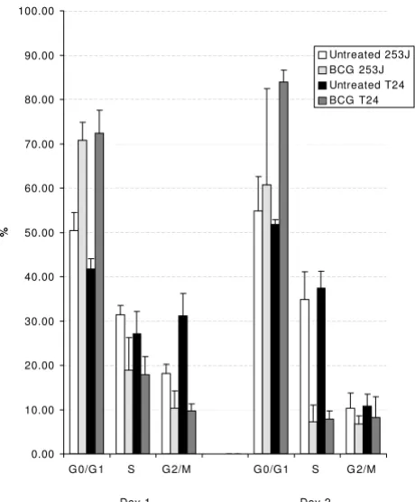
BCG induces cell cycle arrest at the G1/S interface
Figure 5 demonstrates the effect of BCG exposure on cell cycle compartmentalization in 253J and T24 cells. Follow-ing BCG exposure, the percentage of cells in G0/G1 increased 40% and 75% in the respective cell lines. The increase in G phase population was mirrored by a decrease in the percent of cells in S phase. The S phase cell
fraction in BCG treated cells represented 60% and 65% of the control groups in 253J and T24 cells respectively.
Antibody mediated α5β1 integrin cross-linking induces cell cycle arrest at the G1/S interface
Figure 6 demonstrates the effect of antibody mediated α5β1 cross-linking on cell cycle compartmentalization in 253J and T24 cells. Following cross-linking, the percent-age of cells in G0/G1 increased 29% and 48% in the respective cell lines. The increase in G phase population was mirrored by a decrease in the percent of cells in S phase. The S phase cell fraction in cross-linked cells repre-sented 71% and 60% of the control groups in 253J and T24 cells respectively.
BCG decreases the proliferation of human urothelial carci-noma cell lines as measured by the MTT assay
Figure 1
BCG decreases the proliferation of human urothelial carci-noma cell lines as measured by the MTT assay. Exposure of either 253J or T24 cells to BCG resulted in a decrease in via-ble cell number as measured by the optical density of the metabolic product. The day represents the duration of expo-sure to BCG
0
0.1
0.2
0.3
0.4
0.5
0.6
0.7
0.8
0.9
1
Day1
Day3
Day6
Dayspost-BCG
OD
Untreated 253J
BCG 253J
Untreated T24
BCG T24
BCG decreases the proliferation of human urothelial carci-noma cell lines as measured by Colony Forming assay
Figure 2
BCG decreases the proliferation of human urothelial carci-noma cell lines as measured by Colony Forming assay. Expo-sure of either 253J or T24 cells to BCG results in a time dependent decrease in viable cell number as measured by colony outgrowth. The day represents the duration of expo-sure to BCG.
0 100 200 300 400 500 600 700 800 900
Day1 Day2 Day3 Day6
Co
lony
Nu
mber
Antibody mediated cross-linking of a5b1 integrin decreases the proliferation of human urothelial carcinoma cell lines as meas-ured by the MTT assay
Figure 3
Antibody mediated cross-linking of a5b1 integrin decreases the proliferation of human urothelial carcinoma cell lines as meas-ured by the MTT assay. Results are shown for a5 cross-linking in both 253J cells (figure 3a) and T24 cells (Figure 3b). Identical results were obtained when cross-linking was performed using antibodies to b1 (data not shown).
3a
3b
0 0.2 0.4 0.6 0.8 1 1.2 1.4 1.6day 3 day 6
O.
D
.
Untreated Integrin a5 only GAM only Integrin a5 Crosslinking
0.000 0.400 0.800 1.200 1.600 2.000 2.400
day 3 day 6
O.
D.
Peptide blockade of integrin fibronectin binding sites using RGDS reverses the effect of BCG on proliferation and cell cycle arrest
BCG attaches to the cell surface via a fibronectin bridge linking mycobacterial receptors to cell surface integrins.[12-14] Mycobacterial binding of multiple integrin bound fibronectin molecules can affect crosslink-age of integrin receptors.[8] Blocking the ability of FN to bind to either cellular or mycobacterial receptors pre-cludes BCG attachment to the cell surface. [8,14] This series of experiments employed a competitive inhibitor of FN binding to integrins (RGDS) to assess the effect of inhibiting BCG binding to integrin receptors on the bio-logic response to BCG. While RGDS competitively inhib-its the ability of FN to function as an opsonin for BCG binding (effectively inhibiting BCG/integrin interaction), prior studies have demonstrated that RGDS does not effect integrin mediated signaling initiated in response to antibody mediated cross linking of α5β1.[8] 253J cells were pre-incubated with RGDS or the control (non-block-ing) peptide RGES for 1 hour prior to BCG exposure. The MTT and flow cytometric assays were then carried out as described above. Controls included each peptide alone, BCG alone, and untreated cells. In the MTT assay 2 con-centrations of both the blocking and control peptides were employed (100 or 250 ug/ml). The 250 ug/ml con-centration was used in the flow cytometry assay.
Figure 7 demonstrates the effect of integrin blockade on the anti-proliferative effect of BCG as measured by MTT assay. RGDS reduced the BCG effect in a "dose-response" manner with a maximal 4 fold inhibition at the 250 ug/ ml concentration. RGES had no effect. In the flow cyto-metric assay, RGDS inhibited G1 arrest. The BCG induced increase in the G1 fraction was reduced by 60% in RGDS treated cells. RGES pretreatment had no effect on G1 or S phase fraction compared to controls (Figure 8).
BCG does not induce an apoptotic response in urothelial carcinoma cell lines
A series of experiments were conducted to determine if BCG exposure induced apoptosis in either the 253J or T24 cell lines. Gel electrophoresis of cellular DNA obtained at intervals following BCG exposure failed to demonstrate the laddering associated with apoptosis. Flow cytometry for the apoptotic pathway enzyme caspase 3 failed to
Antibody mediated cross-linking of α5β1 integrin decreases the proliferation of human urothelial carcinoma cell lines as measured by Colony Forming assay
Figure 4
Antibody mediated cross-linking of α5β1 integrin decreases the proliferation of human urothelial carcinoma cell lines as measured by Colony Forming assay.
0 50 100 150 200 250 300 350 400 450 500
253J T24
Co
lo
ny
Nu
m
be
r
control alpha 5 GAM alpha 5 +GAM
Human urothelial carcinoma cells exposed to BCG undergo cell cycle arrest at the G1/S interface
Figure 5
Human urothelial carcinoma cells exposed to BCG undergo cell cycle arrest at the G1/S interface. Both 253J and T24 cell lines increased the percentage of cells in G1, with a concom-itant decrease in the S phase fraction, following BCG exposure.
0.00 10.00 20.00 30.00 40.00 50.00 60.00 70.00 80.00 90.00 100.00
G 0/G 1 S G 2/M G 0/G 1 S G 2/M
%
Untreated 253J BCG 253J Untreated T24 BCG T24
6a and 6b. Cell cycle compartmentalization in 253J (Figure 6a) and T24 (Figure 6b) cells following antibody mediated cross-link-ing of α5β1 integrin
Figure 6
6a and 6b. Cell cycle compartmentalization in 253J (Figure 6a) and T24 (Figure 6b) cells following antibody mediated cross-link-ing of α5β1 integrin. Similar to the effect observed in response to BCG, antibody mediated cross-linking resulted in an accumu-lation of cells in G0-G1 with a concomitant decrease in the S-phase fraction.
6a
6b
0.00 10.00 20.00 30.00 40.00 50.00 60.00 70.00 80.00
G0/G1 S G2/M G0/G1 S G2/M
Day 1 Day 2
%
Untreated a5 only(5ug/ml) GAM only(10ug/ml) a5+GAM
0.00 10.00 20.00 30.00 40.00 50.00 60.00 70.00 80.00
G0/G1 S G2/M G0/G1 S G2/M
Day 1 Day 2
%
Untreated
a5 only(5ug/ml)
GAM only(10ug/ml)
demonstrate activation of this enzyme in response to BCG. (Data not shown)
Discussion
The anti-tumor effect of BCG is well established. Clinical experience, the results of animal models, as well as in vitro experiments provide data in clear support of this effect. While an anti-tumor effect of BCG is certain, the mecha-nism through which this occurs is less clear. In vivo ani-mal models of BCG's anti-tumor effect suggest a critical role for a cell mediated immune response. At the same time, in vitro studies using human urothelial cell lines identify a direct effect of BCG. The relative contribution of these two possible mechanisms to BCG's clinical anti-tumor activity is unknown.
A number of studies suggest that a direct anti-tumor effect is at least in part responsible for BCG's activity. Work by Pryor, Ciao, and Sasaki all have reported a direct anti-pro-liferative effect of BCG.[5,6,15] Liu et al demonstrated that BCG, as a consequence of its interaction with cell sur-face bound fibronectin, abrogates invasion and motility
of urothelial carcinoma cell lines.[7] Cellular internaliza-tion of BCG, with resultant alterainternaliza-tions in reactive oxygen species and nitric oxide, has been proposed as a mecha-nism contributing to direct BCG mediated cytotoxicity. [16,17]
Fibronectin functions as an opsonin, linking BCG to cell surface fibronectin receptors of which α5β1 integrin is the predominate FN receptor on urothelial cells. This linkage is a requisite step for BCG's antitumor activity.[14] Rather than constituting a passive interaction, our prior reports have shown that FN mediated BCG adherence to the urothelial carcinoma surface has a pharmacogenetic effect as exemplified by transactivation of IL-6.[9] This effect is mediated through signal transduction pathways involving NF-κB and AP-1. BCG induced signaling and gene trans-activation pathways are identical to those observed in response to antibody mediated cross linking of α5β1.[8] Strategies that prevent the ability of FN to function as an opsonin, including competitive inhibition of FN receptors using RGDS and simultaneous saturation of mycobacte-rial and cell surface FN receptors using excess exogenous FN, inhibit the signaling and transactivation responses to
The effect of BCG on 253J Cellular Proliferation as measured by MTT assay
Figure 7
The effect of BCG on 253J Cellular Proliferation as measured by MTT assay. Peptide mediate integrin blockade (RGDS) reversed the anti-proliferative effect of BCG in a dose-response manner. The control peptide RGES had no effect.
0 0.2 0.4 0.6 0.8 1 1.2 1.4 1.6 1.8 2
day 1 day 2 day 3
O.D.
Untreated RGDS(250 ug/ml) RGES(250 ug/ml) BCG BCG-RGDS(100 ug/ml) BCG-RGDS(250 ug/ml) BCG-RGES(100 ug/ml) BCG-RGES(250 ug/ml)
The effect of BCG on 253J cell cycle compartmentalization as measured by flow cytometry
Figure 8
The effect of BCG on 253J cell cycle compartmentalization as measured by flow cytometry. Peptide mediate integrin block-ade (RGDS) reversed the G1 cell cycle arrest effect of BCG. The control peptide RGES had no effect.
G0/G1 S G2/M G0/G1 S G2/M
%
Untreated
RGDS(250 ug/ml)
RGES(250 ug/ml)
BCG
BCG-RGDS(250ug/ml)
BCG-RGES(250ug/ml)
BCG.[8,18] The current report moves beyond the details of the signaling pathway to examine the biologic effect of BCG induced signaling on a tumor biologic end point with clear treatment relevance.
Consistent with what has been reported by others, our results demonstrate that BCG exerts a direct anti-prolifer-ative effect on human urothelial carcinoma cell lines. The observed anti-proliferative effect does not appear to occur as a consequence of apoptosis. To our knowledge this report is the first to identify cell cycle arrest at the G1/S interface as a mechanism by which BCG exerts an anti-proliferative effect. As was the case for BCG induced sign-aling and gene transactivation, this effect was reproduced using antibody mediated cross-linking of α5β1 integrin. Elimination of the opsonin function of fibronectin using the peptide competitive inhibitor RGDS, effectively block-ing the ability of BCG to bind integrin receptors, reversed the biologic effects of BCG. These findings, together with the results of prior studies, demonstrate that a portion of the biologic response to BCG occurs as a consequence of FN mediated binding of BCG to integrin receptors present on the urothelial cell surface. The failure of simple integrin ligation to simulate the BCG response, the ability of antibody mediated crosslinking of α5β1 integrin to duplicate the BCG response, the central role of FN in the biologic response to BCG, together with the predomi-nance of α5β1 as the principal FN binding integrin on urothelial cells, strongly supports a model in which the biologic response of the tumor cell to BCG occurs as a consequence of BCG crosslinking of α5β1 integrin receptors.
The results of this study have direct clinical relevance. The demonstration that BCG has a direct anti-tumor effect opens this pathway for potential manipulation to improve treatment outcome. This is important given the fact that 30% of patients are BCG refractory, and that the long-term durability of response to BCG is limited. Finally, and equally important, the dissection of the path-way through which BCG exerts an anti-tumor effect will allow us to move further towards our ultimate goal of eliminating the need to expose patients to the risk of a via-ble biologic organism and perhaps allow the develop-ment of less toxic equally effective treatdevelop-ment approaches.
Conclusion
BCG, as a consequence of integrin cross-linking, exerts direct anti-tumor effect against human urothelial carci-noma cell lines. Cell cycle arrest at the G1/S interface, rather than apoptosis, is a mechanism contributing to BCG's anti-proliferative effect. Additional studies will be required to define the molecular pathways that contribute to this response.
Competing interests
The author(s) declare that they have no competing interests.
Authors' contributions
FC carried out the flow cytometric experiments for cell cycle arrest and apoptosis and participated in drafting the manuscript. GZ performed the cell viability and colony forming assays and aided in drafting the manuscript. YI developed the protocols for flow cytometric experiments, aided in the design of the studies and drafted the manu-script. WAS conceived of the study, participated in its design and coordination, and drafted the manuscript. All authors read and approved the final manuscript.
Acknowledgements
This work was supported by a grant from the Department of Veterans Affairs and the Milwaukee Veterans Affairs Medical Center.
References
1. Malkowicz SB: Intravesical therapy for superficial bladder cancer. Semin Urol Oncol 2000, 18(4):280-8.
2. Bohle A, Jocham D, Bock PR: Intravesical bacillus Calmette-Guerin versus mitomycin C for superficial bladder cancer: a formal meta-analysis of comparative studies on recurrence and toxicity. J Urol 2003, 169(1):90-5.
3. Sylvester RJ, van der MEIJDEN AP, Lamm DL: Intravesical bacillus Calmette-Guerin reduces the risk of progression in patients with superficial bladder cancer: a meta-analysis of the pub-lished results of randomized clinical trials. J Urol 2002, 168(5):1964-70.
4. Bohle A, Brandau S: Immune mechanisms in bacillus Calmette-Guerin immunotherapy for superficial bladder cancer. J Urol
2003, 170(3):964-9.
5. Pryor K, Stricker P, Russell P, et al.: Antiproliferative effects of bacillus Calmette-Guerin and interferon alpha 2b on human bladder cancer cells in vitro. Cancer Immunol Immunother 1995, 41(5):309-16.
6. Seow SW, Rahmat JN, Mohamed AA, et al.: Lactobacillus species is more cytotoxic to human bladder cancer cells than Myco-bacterium Bovis (bacillus Calmette-Guerin). J Urol 2002, 168(5):2236-9.
7. Liu BC, Redwood SM, Weiss RE, et al.: Bacillus Calmette-Guerin abrogates in vitro invasion and motility of human bladder tumor cells via fibronectin interaction. J Urol 1992, 148(3):900-5.
8. Chen F, Zhang G, Iwamoto Y, See WA: Bacillus Calmette-Guerin initiates intracellular signaling in a transitional carcinoma cell line by cross-linking α5β1 integrin. J Urol 2003, 170(2 Pt 1):605-10.
9. Chen F-H, Crist SA, Zhang G-J: Interleukin-6 production by human bladder tumor cell lines is upregulated by BCG through NF-κb and AP-1 via an immediate early pathway. J Urol 2002, 168:786-797.
10. Mosmann T: Rapid colorimetric assay for cellular growth and survival: application to proliferation and cytotoxicity assays.
J Immunol Methods 1983, 65:55-63.
11. Schultz DR, Harrington WJ Jr: Apoptosis: programmed cell death at a molecular level. Semin Arthritis Rheum 2003, 32(6):345-69.
12. Cheng DL, Shu WP, Choi JC, Margolis EJ, Droller MJ, Liu BC: Bacillus Calmette-Guerin interacts with the carboxyl-terminal heparin-binding domain of fibronectin: implications for
BCG-mediated anti-tumor activity. J Urol 1994,
152(4):1275-80.
Publish with BioMed Central and every scientist can read your work free of charge
"BioMed Central will be the most significant development for disseminating the results of biomedical researc h in our lifetime."
Sir Paul Nurse, Cancer Research UK
Your research papers will be:
available free of charge to the entire biomedical community
peer reviewed and published immediately upon acceptance
cited in PubMed and archived on PubMed Central
yours — you keep the copyright
Submit your manuscript here:
http://www.biomedcentral.com/info/publishing_adv.asp
BioMedcentral
14. Kavoussi LR, Brown EJ, Ritchey JK, Ratliff TL: Fibronectin-medi-ated Calmette-Guerin bacillus attachment to murine blad-der mucosa. Requirement for the expression of an anti-tumor response. J Clin Invest 1990, 85(1):62-7.
15. Sasaki A, Kudoh S, Mori K, et al.: Are BCG effects against urinary bladder carcinoma cell line T24 correlated with apoptosis in vitro. Urologia Internationalis 1997, 59(3):142-8.
16. Pook SH, Esuvaranathan K, Mahendran R: N-acetylcysteine aug-ments the cellular redox changes and cytotoxic activity of internalized mycobacterium bovis in human bladder cancer cells. J Urol 2002, 168(2):780-5.
17. Jansson OT, Morcos E, Brundin L, Lundberg JO, Adolfsson J, Soderhall M, Wiklund NP: The role of nitric oxide in bacillus Calmette-Guerin mediated anti-tumour effects in human bladder cancer. Br J Cancer 1998, 78(5):588-92.
18. Zhang GJ, Chen FH, Xu Y, Cao Y, Crist S, McKerrow A, Iwamoto Y, See WA: Autocrine overexpression of fibronectin by human transitional carcinoma cells impairs BCG adherence and signaling. J Urol 2004, 172(4):1496-1500.
Pre-publication history
The pre-publication history for this paper can be accessed here: