R E S E A R C H A R T I C L E
Open Access
Presenting symptoms predict local staging
of anal cancer: a retrospective analysis of
86 patients
Matthias Sauter
1,2†, Georg Keilholz
3†, Helmut Kranzbühler
3, Norbert Lombriser
3, Meher Prakash
2,
Stephan R. Vavricka
2,4and Benjamin Misselwitz
2*Abstract
Background:Incidence of anal carcinoma (AC) is increasing and timely diagnosis is critical for efficient therapy. However, there is a paucity of recent studies addressing clinical symptoms and physical findings of anal carcinoma. Methods:We performed a retrospective study reviewing history, symptoms and physical findings from 86 patients with newly diagnosed AC. We analyzed frequency of symptoms and physical findings according to T and TNM stage and their predictive value regarding tumor stage.
Results:Most patients presented with T2 (37 %) or T3 (29 %) cancer. 85 of 86 patients were symptomatic with anal bleeding (78 %), anal/perianal pain (63 %), weight loss (31 %) and foreign body sensation (22 %). 95 % of patients had≥1 finding on physical examination including a visible tumor, palpable resistance and pain/blood during digital rectal examination. Patients with locally advanced disease (T3/T4) presented with more symptoms (p< 0.01) and more physical findings (p= 0.04) than patients with T1/T2 disease. On multivariate regression analysis perianal pain, painful defecation and weight loss were significantly associated with T3/T4 disease.
Conclusion:Clinical symptoms and physical findings are present in nearly all AC patients. Pain referred to the perianal region, painful defecation and weight loss have predictive value for locally advanced disease.
Keywords:Anal carcinoma, Symptoms, Physical examination, Tumor staging
Background
Anal carcinoma is an uncommon malignancy with an in-cidence of 2 new cases per 100,000 per year in the USA [1–6], comprising approximately 0.4 % of all tumors and 2.5 % of all gastrointestinal malignancies. Within the last decades the incidence of anal carcinoma has steadily in-creased and anal carcinoma incidence is now 2-fold higher than 30 years ago and 4-fold higher in selected subgroups of patients [3, 6, 7]. Risk factors associated with anal carcinoma are the lifetime number of sexual partners, receptive anal intercourse, cigarette smoking, genital warts and viral infections especially human papil-lomavirus (HPV), and human immunodeficiency virus
(HIV) [8]. The increasing incidence of anal carcinoma might reflect changes in one or more of the risk factors mentioned above and might follow an increase in infec-tion rates of HPV and HIV.
Timely diagnosis of anal carcinoma is critical, since for the treatment of early cancer highly effective and function preserving radio-(chemo) therapy is available. In contrast, the options for advanced and metastasized carcinoma are limited [1, 4, 9]. Clinical symptoms in the assessment anal cancer have only been insufficiently studied. Previous studies with a limited number of patients identified rectal bleeding (45 %), anorectal pain (20–35 %) and the sensa-tion of a rectal mass (20–35 %) as the most prevalent complaints [10–14] with approximately 20 % of patients being free of symptoms in the largest study [10]. Clinical findings of anal carcinoma have not been summarized in detail. An association of symptoms or findings with early or an advanced disease has to the best of our knowledge
* Correspondence:benjamin.misselwitz@usz.ch
†Equal contributors
2Division of Gastroenterology and Hepatology, University Hospital Zurich,
Rämistr 100, 8091 Zürich, Switzerland
Full list of author information is available at the end of the article
not been tested. All studies were published between 1976 and 1986 and no recent data are available. In the interven-ing time medical practice has seen important changes including a massive increase in endoscopic evaluations of the colon. Following current guidelines, staging procedure of anal carcinoma now includes MRI and/or endosono-graphy [1, 9], techniques unavailable 30 years ago. Con-sidering these changes and the dramatic increase of anal carcinoma incidence, clinical presentation might differ now compared to the 1970s and the 1980s.
We therefore performed a systematic study of present-ing symptoms and physical findpresent-ings in patients with newly diagnosed anal cancer. Signs, symptoms and find-ings that differed for early and advanced anal carcinoma were identified.
Methods
We performed a retrospective analysis of all patients referred to Triemli-Hospital, a tertiary care center and teaching hospital in Zürich, Switzerland from 1999 until 2013 for treatment of anal carcinoma. Patients were identified by an automated search within the internal clinical information system. Histological evidence of anal carcinoma was a requirement for inclusion and all pa-tients with rectal carcinoma were excluded. We only considered symptoms and findings from the first presen-tation of anal carcinoma and recurrent anal carcinoma cases were excluded.
The study protocol was approved by the local ethics com-mittee of Zurich county (Registration KEK-ZH 2010–0555). The study was performed according to the Declaration of Helsinki.
Data collection
All relevant parameters regarding the initial clinical assess-ment including patient demographics, a detailed personal history, local and systemic symptoms, findings on physical examinations, and results of investigations (endoscopy with histology and staging investigations including CT, MRI and endosonography) were extracted from the documented pa-tient history as well as from referral letters.
Tumor classification followed the 7th edition of the American joint Committee on Cancer TNM staging [15] with T1 referring to a tumor size <2 cm, T2: a tumor between 2 and 5 cm, T3: a tumor >5 cm and T4 a tumor invading adjacent organs. N1 refers to lymph node metastasis in perirectal lymph nodes; N2 to metastasis in unilateral internal iliac and/or inguinal lymph node(s); and N3 to metastasis in perirectal and inguinal lymph nodes and/or bilateral internal iliac and/or inguinal lymph nodes. Stage I refers to a T1 tumor without lymph node involvement or distant metastasis. Stage II refers to a T2 or T3 tumor without lymph node or distant metastasis. Stage III refers to either i) T1-3 with
N1 without metastasis or ii) any N2 or N3 positivity without metastasis independent of T-Stage or iii) T4 with N0 or N1 without distant metastasis. Stage IV refers to a tumor with advanced (N3) lymph node involvement (bilateral inguinal or bilateral internal iliac) or a tumor with distant metastasis. During our chart review all find-ings from all original investigations (CT, MRI and EUS) were evaluated and the appropriate tumor stage noted.
We also differentiated between distal, middle and prox-imal location according to i) digital rectal exam; ii) findings reported by CT, MRI or endosonography. Distal tumors in-cluded the anal rim/perianal region and proximal tumors included local invasion into the rectum.
Data analysis
For each symptom and each physical finding we calculated the frequency for patients of each T-stage and TNM-stage. To identify differences for early and advanced carcinoma, χ2
significance of a trend (T1–T4 or stage I–stage IV, re-spectively) was calculated using a generalized linear model.
To study the effect of the age at diagnosis on tumor characteristics our cohort was divided according to the me-dian age (62 years) into a younger age group (≤62 years) and older age group (>62 years) and independent samples test was used for the comparison of means. A similar com-parison was also performed to evaluate differences between the subgroups with proximal and distal anal carcinomas.
Multivariate logistic regression was used to analyze the predictability of early and advanced T-stage (or TNM stage) cancers. For this purpose T1-2 cancers (or TNM stage I and II) were categorized as early, T3-4 (or TNM stage III and IV) as advanced cancers. We tested clinical symptoms, physical findings and patient demographics such as gender, body mass index (BMI) and age at diag-nosis as potential predictors. Potential predictors were sorted according to theirp-values. In a step-wise proced-ure all descriptors with p-values <0.1 were eliminated and the regression analysis was repeated. This procedure eliminated several general descriptors including gender, BMI and age at diagnosis and the remaining variables with p< 0.1 and their corresponding odds ratios (OR) are reported. Similar analyses were also performed for the prediction of the TNM stage. All statistical analyses were performed using SPSS software.
Results
at our institution during the study period. No PCR data for HPV was performed.
Staging was done by MRI in 29 patients, CT scan in 71 patients and anorectal endosonography in 56 patients and most patients presented with T2 or T3 cancers (TNM stage II or III), respectively. Demographic data, T stage and TNM stage of the cohort are presented in Table 1. Anal cancer of the outer margin was signifi-cantly more frequent in men (p= 0.05). In contrast, can-cer involving the anal channel tended to be more frequent in women (n.s.).
Clinical symptoms
Almost all patients reported symptoms due to anal car-cinoma: 85 of 86 patients described at least one clinical complaint. The mean duration of symptoms before diag-nosis was 8 months (median 3, range 0–62 months). The duration of the symptoms did not differ according to any of the symptoms, the total number of symptoms or tumor stage (not shown).
Clinical symptoms are summarized in Table 2. The most frequent symptoms were anal bleeding (78 %), anal/perianal pain (63 %, including 29 % with anal pain, 24 % with perianal pain and 38 % of patients with painful defecation), weight loss (31 %), tumor on self-palpation (26 %) and foreign body sensation (22 %). On average, patients described 3.3 symptoms.
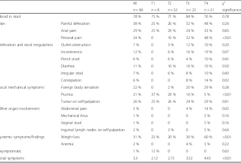
Clinical presentation differed according to the T-stage of the tumor; patients with advanced disease described significantly more symptoms (2.1 for T1 and 4.4 for T2;
p< 0.01, compare Table 2 and Fig. 1a). In addition, peri-anal pain, constipation, abdominal pain and weight loss were significantly more frequent in patients with locally advanced disease. Pruritus was more prevalent in pa-tients with early T-stages. The most common symptoms anal bleeding and anal pain occurred with similar fre-quency in early and advanced tumors.
Findings on physical examination
The vast majority of patients (82 out of 86) had patho-logical findings on physical examination (Table 3). Patients with locally advanced disease (T3 and T4) were likely to have more findings on physical examination than those with early disease (T1 and T2; Fig. 1b;p= 0.04). The fre-quency of all individual physical findings was similar in all tumor stages and no significant differences could be de-tected. Digital rectal examination was not possible due to pain in 2 patients, both of which presented with locally advanced carcinomas.
Regression analysis
Multivariate logistic regression analysis was used to distin-guish early carcinoma (T1 or T2) from locally advanced tumor (T3 or T4) using clinical symptoms and physical findings. We step-wise eliminated all variables which did not significantly contribute to T-stage prediction, using
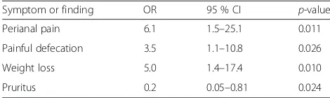
p= 0.1 as a cut-off. Our final model used 4 variables for T-stage prediction (Table 4). Thereby,perianal pain was the single symptom with the strongest predictive value regarding the presence of locally advanced disease (odds ratio 6.1, 95 %-CI 1.5–25.1,p= 0.011). In addition, painful defecation and weight loss were able to predict locally advanced disease.
We also tested predictability of TNM-stage by symp-toms and findings. According to our analysis TNM-stage could also be predicted, but with less precision (Additional file 1: Table S1). Perianal pain was the strongest predictor of TNM-stage, to a similar extent as described for T-stage.
Age at diagnosis
To analyze effects of the age at diagnosis we divided our cohort into a younger (≤62 years, n= 42) and an older age group (>62 years,n= 44; Additional file 1: Table S2). Younger patients had a higher TNM-stage (2.7 vs. 2.3,
Table 1Epidemiological characteristics of our patients with anal carcinoma
Description Numbers
Mean ± standard deviation
Gender Male: 30 (35 %)
Female: 56 (65 %)
Age 62 ± 13 years, range: 32–91 years
Diagnostic delay 8 ± 12 months, range: 0–62 months
BMI 24.7 ± 4.3 kg/m2, range: 17.3–34.7 kg/m2
Histology Squamous carcinoma: 85 (99 %)
Neuroendocrine carcinoma: 1 (1 %)
HIV Positive: 5, Negative: 1
Unknown: 80
T Stage T1: 8 (9 %)
T2: 32 (37 %)
T3: 25 (29 %)
T4: 21 (24 %)
TNM Stage Stage I: 8 (9 %)
Stage II: 27 (31 %)
Stage III: 50 (58 %)
Stage IV: 1 (1 %)
Tumor site involved Distal anal channel: 43 (50 %)
Middle anal channel: 37 (43 %)
Proximal anal channel: 54 (63 %)
Table 2Clinical symptoms of patients with various tumor stages (expressed as percent of total number of patients with respective tumor stage). Statistical analysis: Generalized linear model
All T1 T2 T3 T4 χ2
n= 86 n= 8 n= 32 n= 25 n= 21 significance
Blood in stool 78 % 75 % 77 % 84 % 76 % 0.78
Pain Painful defecation 38 % 25 % 26 % 52 % 48 % 0.26
Anal pain 29 % 25 % 29 % 24 % 33 % 0.83
Perianal pain 24 % 0 10 % 32 % 48 % <0.01
Defecation and stool irregularities Outlet obstruction 7 % 0 3 % 12 % 10 % 0.20
Incontinence 12 % 0 6 % 16 % 19 % 0.07
Pencil stool 6 % 0 6 % 4 % 10 % 0.45
Diarrhea 11 % 0 10 % 16 % 10 % 0.50
Irregular stool 7 % 0 6 % 8 % 10 % 0.40
Constipation 6 % 0 0 8 % 14 % 0.02
Local mechanical symptoms Foreign body sensation 22 % 0 2 % 20 % 29 % 0.28
Pruritus 21 % 37 % 29 % 16 % 5 % <0.01
Tumor on self-palpation 26 % 25 % 26 % 24 % 29 % 0.81
Other organ involvement Abdominal pain 5 % 0 0 4 % 14 % 0.02
Mechanical ileus 1 % 0 0 0 5 % 0.16
Vaginal stool 1 % 0 0 0 5 % 0.16
Inguinal lymph nodes on self-palpation 2 % 0 3 % 0 5 % 0.64
Systemic symptoms/findings Weight loss 31 % 25 % 20 % 30 % 60 % <0.01
Anemia 2 % 0 0 4 % 5 % 0.22
Asymptomatic 1 % 12 % 0 0 0 0.63
Total symptoms 3.3 2.12 2.75 3.52 4.43 <0.01
p= 0.026) and a higher likelihood of proximal cancer than the older subgroup (chance of involvement of the proximal anal channel: 91 vs. 72 %, p= 0.044), but no further significant differences were detected and age at diagnosis is unlikely to confound our analysis.
Location of anal carcinoma
Combining information available from imaging (MRI, CT-scan and endosonography) and the physical examination we determined for each patient whether the proximal anal channel (close to the rectum) and/or distal anal channel (bordering the skin) was involved by the carcinoma. Patients with proximal anal carcinoma had a higher T-stage than patients with distal cancer (mean T-T-stage 2.6 vs. 1.9, p= 0.03 for distal vs. proximal anal cancer, Additional file 1: Table S3). In addition, a description of distal carcinoma without proximal involvement was more frequent for localized cancers (OR for T-stage: 0.13, 95 % CI 0.03–0.64,p= 0.012) and proximal carcinoma was more common in locally advanced cancer (OR for T-stage: 3.0, 95 % CI 1.2–7.6,p= 0.019). However, multivariate logistic analysis did not result in any variables with significant con-tributions for prediction of early and advanced cancer.
Discussion
In this study we provide a comprehensive clinical characterization of a cohort of patients with newly diag-nosed anal carcinoma. According to our analyses, clin-ical symptoms have predictive value for local staging of anal carcinoma.
Our study fills a gap in our knowledge since no system-atic study regarding physical findings in anal carcinoma has been performed. Furthermore, no contemporary study describing the clinical presentation of anal carcinoma is available and the incidence of anal carcinoma, the preva-lence of risk factors and medical practice has tremendously changed since the publication of the last paper characteriz-ing anal carcinoma almost 30 years ago [10–14]. In agree-ment with previous studies, bleeding, anal pain and sensation of an anal mass remain the most frequent symp-toms of anal carcinoma. However, the presence of anal pain including painful defecation and perianal pain (63 vs. 20–35 %) as well as anal bleeding (77 vs. 45 %) were more frequent than in historical studies [10–14]. In addition we found that nearly all patients had at least one symptom likely associated with anal carcinoma. Truly asymptomatic anal carcinoma was exceedingly rare in our cohort (1 vs. 20 % in a previous study [10]). Pathological physical find-ings were also almost universal and only 6 % of our patients had a normal rectal examination.
Symptoms and findings of anal carcinoma were signifi-cantly correlated to the T-stage of the tumor. This pre-dictive value of the clinical presentation has not been described previously and might be unique among gastro-intestinal carcinomas (e.g. colon cancer, stomach cancer) for which early symptoms or findings are rare and symp-toms do not reflect T stage or TNM stage [16–19]. For anal carcinoma, patients with advanced disease had a greater number of symptoms than those with early dis-ease. They frequently reported perianal pain, changes in stool consistency and sometimes symptoms suggesting involvement of neighboring organs. Thereby, the predict-ive value of perianal pain for advanced tumor remained strong even in a multivariate regression analysis. This indicates that patients are able to“sense”local progression of this tumor. The ability of a significant fraction of patients with anal carcinoma to perceive locally advanced disease is likely related to the somatic sensation of the anal region, frequent mechanical stress during defecation and functional importance of the anal region for suc-cessful defecation. Similar to stomach and colon cancer, duration of symptoms does not predict tumor stage [16–18] but our study might be underpowered to detect subtle differences.
Table 3Physical findings in our patients during digital rectal examination. Statistical analysis: Generalized linear model
All T1 T2 T3 T4 χ2
n= 86 n= 8 n= 32 n= 25 n= 21 significance
Painful palpation 39 % 25 % 30 % 40 % 65 % 0.15
Resistance on palpation 88 % 50 % 90 % 100 % 90 % 0.32
Blood on palpation 31 % 14 % 29 % 36 % 40 % 0.44
No clinical findings (or normal rectal examination) 7 % 37 % 9 % 0 0 0.02
Total number of physical findings 1.60 0.87 1.68 1.76 2.09 0.04
Table 4Predictive value of symptoms for T-stage of anal carcinoma. The final model of our multivariate logistic regression analysis had significant predictive value to distinguish a localized vs. locally advanced tumor (R2= 0.37;p= 0.03). In the table the odds ratio (OR) of a symptom or physical finding for prediction of locally advanced cancer (T3/T4 vs. T1/T2) are shown. Only variables withp< 0.100 are indicated
Symptom or finding OR 95 % CI p-value
Perianal pain 6.1 1.5–25.1 0.011
Painful defecation 3.5 1.1–10.8 0.026
Weight loss 5.0 1.4–17.4 0.010
As a clinical message our data highlight the importance of a profound careful clinical history of patients with anal carcinoma. Attention should be paid to the localization of anal pain: According to our analysis, local anal pain or pain at defecation was compatible with both, early and advanced anal carcinoma. However, perianal pain (outside the anus) was almost exclusive found in patients with higher T-stages. Furthermore, the abundance of positive physical findings (in 99 % of our patients) underscores the importance of a careful examination of all patients.
Our study has limitations: i) data were collected retro-spectively and 86 patients may be too small to detect all relevant associations. However, a prospective study of such a rare tumor would be difficult and the size of our study compares favorably to historic analyses and is the only study regarding this subject in almost 30 years. ii) No disease control group is available and all symptoms under discussion are not specific for anorectal cancer and might also be caused by hemorrhoids, fissure, infec-tious proctitis, rectal abscess, fistula or other common conditions [3, 20–23]. Therefore, our results cannot be used to estimate the diagnostic accuracy of individual symptoms for anal cancer. iii) Our analyses tested the predictive power of symptoms for T and TNM stage; however, we did not test the predictive power of symp-toms and findings for overall survival or disease specific survival as hard clinical end points. Since in our follow-up analyses only 5 patients had died of anal cancer (not shown), our study is insufficiently powered for a survival analysis. iv) For the majority of our patients no result of an HIV test is available. Triemli Hospital is not a major referral center for HIV patients. We as-sume that for most of these patients HIV testing or at least a risk assessment has been performed with a negative result or a perceived low risk. Therefore, our results might not reflect the typical presentation of an HIV positive patient with anal carcinoma. v) No PCR testing for HPV was performed; with this method an HPV prevalence of 80–90 % has been described in other cohorts [24, 25]. vi) We did not perform a population-based study and therefore referral bias cannot be com-pletely excluded. However, we estimate that the majority of patients with anal cancer of the 300,000–500,000 pa-tients of the catchment area of our hospital were referred to the division of radiation oncology of this hospital for palliative or definite treatment. The number of 86 patients over a 10-year study period is in reasonable agreement with near-complete coverage if an incidence of 1–2 cases per 100,000 individuals is assumed [1–6].
Conclusion
In summary, our study provides a contemporary summary of the clinical presentation of anal carcinoma. Nearly all patients displayed symptoms including pain, bleeding and
foreign body sensation and/or physical findings such as pain on palpation, resistance and blood. Symptoms, es-pecially pain perceived outside the anal channel, painful defecation and weight loss had predictive value for ad-vanced disease. Due to the high sensitivity, functional importance and good accessibility of the anal and peri-anal region, clinical history and physical examination can identify patients with a high likelihood of advanced anal cancer.
Additional file
Additional file 1: Table S1.Predictive value of symptoms for TNM-stage of anal carcinoma (similar to T- in Table 4). The final model of our multivariate logistic regression analysis had significant predictive value to distinguish early from advanced cancer (R2=0.40;p=0.040).The odds ratio of a symptom or physical finding for prediction of advanced cancer (stage III/ IV vs. stage I/ II). Only variables with p-values less than 0.10 are shown.Table S2.Characteristics of the older and younger subgroup of our anal carcinoma cohort. Statistical analysis: Mann-Whitney U test.Table S3.Characteristics of patients with involvement of the proximal (close to rectum) and distal (close to skin) anal channel. Statistical analysis: Mann Whitney U test. (PDF 233 kb)
Abbreviations
AC:anal carcinoma; CI: confidence interval; CT: computer tomography; HIV: human immunodeficiency virus; HPV: human papillomavirus; MRI: magnetic resonance imaging; OR: odds ratio.
Competing interests
The authors declare that they have no competing interests.
Authors’contributions
BM, SV, HK, NL: study design; MS, GK, MP and BM: data acquisition, data analysis, drafting of manuscript; All authors: critical review and final approval of manuscript.
Funding
This study was supported by 1) the Division of Gastroenterology and 2) Radiation Oncology, Triemli Hospital Zurich; 3) Grant of the Swiss Cancer League to BM 4) Grant of the Horten Foundation to BM.
Author details
1Department of Medicine and Specialities, Triemli Hospital, Zurich,
Switzerland.2Division of Gastroenterology and Hepatology, University
Hospital Zurich, Rämistr 100, 8091 Zürich, Switzerland.3Division of
Radiation-Oncology, Triemli Hospital, Zürich, Switzerland.4Department of
Medicine and Specialities, Division of Gastroenterology, Triemli Hospital, Zurich, Switzerland.
Received: 16 September 2015 Accepted: 29 March 2016
References
1. Glynne-Jones R, Nilsson PJ, Aschele C, Goh V, Peiffert D, Cervantes A, Arnold D. Anal cancer: ESMO-ESSO-ESTRO Clinical Practice Guidelines for diagnosis, treatment and follow-up. Ann Oncol. 2014;25 Suppl 3:iii10–20.
2. Jemal A, Simard EP, Dorell C, Noone AM, Markowitz LE, Kohler B, Eheman C, Saraiya M, Bandi P, Saslow D, et al. Annual Report to the Nation on the Status of Cancer, 1975–2009, featuring the burden and trends in human papillomavirus(HPV)-associated cancers and HPV vaccination coverage levels. J Natl Cancer Inst. 2013;105(3):175–201.
3. Johnson LG, Madeleine MM, Newcomer LM, Schwartz SM, Daling JR. Anal cancer incidence and survival: the surveillance, epidemiology, and end results experience, 1973–2000. Cancer. 2004;101(2):281–8.
5. Siegel R, Ma J, Zou Z, Jemal A. Cancer statistics, 2014. CA Cancer J Clin. 2014;64(1):9–29.
6. Simard EP, Ward EM, Siegel R, Jemal A. Cancers with increasing incidence trends in the United States: 1999 through 2008. CA Cancer J Clin. 2012; 62(2):118–28.
7. Wise J. Anal cancer rates quadruple among UK women in past 40 years. BMJ. 2014;348:g3747.
8. Palefsky JM. Anal human papillomavirus infection and anal cancer in HIV-positive individuals: an emerging problem. AIDS. 1994;8(3):283–95. 9. NCCN Clinical Practice Guidelines in Oncology on Anal Carcinoma, Version
2.2015, http://www.nccn.org.
10. Beahrs OH, Wilson SM. Carcinoma of the anus. Ann Surg. 1976;184(4):422–8. 11. Clark J, Petrelli N, Herrera L, Mittelman A. Epidermoid carcinoma of the anal
canal. Cancer. 1986;57(2):400–6.
12. Schneider TC, Schulte WJ. Management of carcinoma of anal canal. Surgery. 1981;90(4):729–34.
13. Schraut WH, Wang CH, Dawson PJ, Block GE. Depth of invasion, location, and size of cancer of the anus dictate operative treatment. Cancer. 1983; 51(7):1291–6.
14. Singh R, Nime F, Mittelman A. Malignant epithelial tumors of the anal canal. Cancer. 1981;48(2):411–5.
15. Edge SBBD, Compton CC. AJCC Cancer Staging Handbook. 7thed. New
York: Springer; 2010.
16. Hamilton W, Round A, Sharp D, Peters TJ. Clinical features of colorectal cancer before diagnosis: a population-based case-control study. Br J Cancer. 2005;93(4):399–405.
17. Majumdar SR, Fletcher RH, Evans AT. How does colorectal cancer present? Symptoms, duration, and clues to location. Am J Gastroenterol. 1999;94(10): 3039–45.
18. Steinberg SM, Barkin JS, Kaplan RS, Stablein DM. Prognostic indicators of colon tumors. The Gastrointestinal Tumor Study Group experience. Cancer. 1986;57(9):1866–70.
19. Wanebo HJ, Kennedy BJ, Chmiel J, Steele Jr G, Winchester D, Osteen R. Cancer of the stomach. A patient care study by the American College of Surgeons. Ann Surg. 1993;218(5):583–92.
20. Abcarian H. Anorectal infection: abscess-fistula. Clin Colon Rectal Surg. 2011;24(1):14–21.
21. Daniel GL, Longo WE, Vernava 3rd AM. Pruritus ani. Causes and concerns. Dis Colon Rectum. 1994;37(7):670–4.
22. Nelson RL, Abcarian H, Davis FG, Persky V. Prevalence of benign anorectal disease in a randomly selected population. Dis Colon Rectum. 1995;38(4):341–4. 23. Zaghiyan KN, Fleshner P. Anal fissure. Clin Colon Rectal Surg. 2011;24(1):22–30. 24. Daling JR, Madeleine MM, Johnson LG, Schwartz SM, Shera KA, Wurscher
MA, Carter JJ, Porter PL, Galloway DA, McDougall JK. Human papillomavirus, smoking, and sexual practices in the etiology of anal cancer. Cancer. 2004; 101(2):270–80.
25. Frisch M, Glimelius B, van den Brule AJ, Wohlfahrt J, Meijer CJ, Walboomers JM, Goldman S, Svensson C, Adami HO, Melbye M. Sexually transmitted infection as a cause of anal cancer. N Engl J Med. 1997;337(19):1350–8.
• We accept pre-submission inquiries
• Our selector tool helps you to find the most relevant journal
• We provide round the clock customer support
• Convenient online submission
• Thorough peer review
• Inclusion in PubMed and all major indexing services
• Maximum visibility for your research
Submit your manuscript at www.biomedcentral.com/submit