Comparison of Screening
Colposcopy with Diagnostic Colposcopy
SETAREH FATEHI
1, MALIHEH ARAB
2*, MARYAM SADAT HOSSEINI
2,
FARAH FARZANEH
3, TAHEREH ASHRAFGANJOEI
3and ATEFEH MORIDI
11Gyneco-oncology Fellowship Assistant, Imam Hossein Medical Center, Shahid Beheshti University of Medical Siences, Tehran, Iran. 2Professor of Gyneco-oncology, Imam Hossein Medical Center,
Shahid Beheshti University of Medical Siences, Tehran, Iran. 3Associate Professor of Gyneco-oncology, Imam Hossein Medical Center,
Shahid Beheshti University of Medical Siences, Tehran, Iran. *Corresponding author E-mail:drmarab@yahoo.com
http://dx.doi.org/10.13005/bpj/961
(Received: April 01, 2016; accepted: May 19, 2016) ABSTRACT
Cervical cancer is among the common gynecologic malignancies effective and well developed cytologic screening programs are not available in developing countries. This study is to compare screening and diagnostic culposcopy. In a prospective cross-sectional study 2235 screening and 2318 diagnostic colposcopies were compared. Diagnostic colposcopy indications included chronic vaginal discharge, abnormal cervix, positive VIA test, abnormal cytology, postcoital bleeding and intermenstrual bleeding. CIN2 as more pathology findings were compared in two groups. High score colposcopy were found in 1084 out of 2235 (48.5%) screening cases and 1476 out of 2318 (63.7%) diagnostic group. Pathologic result of CIN2 as more was observed in 45 (2.3%) of screening and 110 (5.3%) of diagnostic colposcopies. Cases finding of diagnostic colposcopy was significantly more in diagnostic group in comparision to screening cases. Decision regarding screening colposcopy needs more studies in different aspects specially cast-benefit of alternative methods.
Key words:Colposcopy, Screening: diagnosis, Uterine Cervical neoplasms, Cervical Cancer.
INTRODUCTION
Epidemiology of cervical cancer in developing countries is different from developed regions. For instance, the most common Gynecologic malignancy In India is cervical cancer1,2. In Indonesia most new cases are diagnosed in late stage. About 80% new cases of cervical cancer in the world are diagnosed in developing countries1. Papanicolau test (pap test) has deeply influenced the cervical cancer incidence. Cytologic examination of cervical transformation zone followed by colposcopic biopsy if indicated have resulted in significant reduction of disease burden in developed countries3.
These effective and well developed cytologic screening programs are not available in developing countries. Besides, low sensitivity of pap test perse, in developing countries additional limitations exist. Ideal cervical cancer screening require trained personal, quality control, acceptable laboratories, proper referral pathways4,5.
In the present study, evaluation of colposcopy as screening in comparision to diagnostic colposcopy is considered.
MATERIAL AND METHODS
In a prospective cross-sectional study all married 25-65 years old women reffered for routine screening between 2011-2014 in a charitable free of charge setting to Imam Hossein medical center included in screening colposcopy if accepted written informed consent.
Post- hysterectomy, pregnant, diagnosed cervical neoplasia, disabling physical as mental disease impossible to cooperate excluded from the study. About 2235 cases underwent screening colposcopy after demographic data entry. All colposcopies were done by one expert gyneco-oncologist by D.F.vasconcellos machine made in Brazil. Colposcopy was performed via 10-40 magnification with both white and green filter with and without acetic acid (3%). Logul solution was not used in these patients. Colposcopic reports were recorded in a sheet including satisfactory/ unsatisfactory, luko plakia, acetowhite regions, punctation, mosaism and abnormal vessel items. In abnormal findings colposcopic biopsy was done and samples were send to one university-linked laboratory. Pathologic results were reported by 2 pathologists. Data of this group were compared to diagnostic colposcopy data of the same years of the same centre. Diagnostic colposcopy group included patients reffered to gyneco-oncology outpatient ward of the same centre. Colposcopic indications of this group included postcoital bleeding, inter menstrual bleeding, hypermenorrhea, positive HPV test, chronic vaginal discharge, VIA positive test, abnormal cervix in exam, genital wart, history of genital wart, history of genital wart in sexual partner, post menopausal bleeding and abnor mal cervical cytology. All diagnostic colposcopies were done under supervision of 4 expert Gyneco-oncologists by the same machine in the same centere. Colposcopy report sheet and exclusion criteria was the same as screening group. Data of diagnostic group was retrospective, extracted from existing data base. Outcome of CIN2 or more studied and compared in two groups. Data analysis was done by SPSS V 20
and related-appropriate tests were used. RESULTS
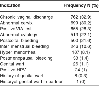
Screening and diagnostic groups included 2235 and 2318 cases, respectively. Mean age of diagnostic group was (45.9 ± 10) and Median age was 46 years with Range of 18-86 years. In screening group Mean age was (39.5 ± 10) and median age was 39 years with range of 18-88 years. The most common Colposcopic indications in diagnostic group were chronic vaginal discharge (32.9%). Abnormal cervix (30.2%), positive VIA test (28.3%), abnor mal cytology (22.1%), post coital bleeding (21.6%) and inter menstrual bleeding (10.6%). In 1030 out of 2318 (44.5%) of cases more than one Colposcopic indication existed (Table 1).
Colposcopy was satisfactory in 623 out of 2235 (30%) screening colposcopy cases and 1336 out of 2318 (58%) diagnostic colposcopies. Results of colposcopy in both groups are presented in table 2.
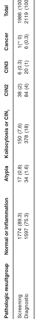
Pathologic results of Colposcopic biopsies in both groups are presented in table 3.
In screening colposcopy group 45 (2.3%) and in diagnostic group 110 (5.3%) revealed CIN2 or more (P= 0.000). Case finding was significantly
Table 1: Colposcopic indications frequency in diagnostic group
Indication Frequency N (%)
Chronic vaginal discharge 762 (32.9)
Abnormal cervix 699 (30.2)
Positive VIA test 655 (28.3) Abnormal cytology 513 (22.1) Postcoital bleeding 500 (21.6) Inter menstrual bleeding 246 (10.6)
Hyper menorrhea 187 (8.1)
Postmenopausal bleeding 33 (1.4)
Genital wart 26 (1.1)
Positive HPV 24 (1)
T
a
ble 2:
Colposcopic results of screening and dia
gnostic Colposcopic gr
oups.
Group
Satisfactory
Unsatisfactory
T
otal
Normal or low score
**High
score
T
otal
Screening
623
(27.9)
1612 (72.1)
2235 (100)
1151 (51.5)
1084 (48.5)
2235 (100)
Diagnostic
1336
(57.6)
982 (42.4)
2318
(100)
842 (63.7)
1476
(63.7)
2318
(100)
T
otal
1959 (43)
2594 (57)
1993 (43.8)
2560 (56.2)
2560 (56.2)
4
5
5
3
*sums are presented as absolute number and percent N (%) **high score colposcopy: lesions with dense acetowhite or pealing or punctation or mosaism or abnormal vessels or large lesions
.
T
a
b
le 3:
P
athologic Colposcopic biopsy results in screening and dia
gnostic gr
oups (if biopsy is done).
Pathologic resultgroup
Normal or inflammation
Atypia
K
oilocytosis or CIN
1
CIN2
CIN3
Cancer
T
otal
Screening
1774 (89.3)
17
(0.8)
150
(7.6)
38
(2)
6
(0.3)
1(‘“
0)
1986 (100)
Diagnostic
1597
(75.3)
34 (1.6)
378
(18)
84 (4)
20 (1)
6
(0.3)
2119
(100)
more in diagnostic colposcopy in comparison to screening colposcopy (Table 4).
DISCUSSION
In the present study , disease finding in screening colposcopy group (2%) was significantly less than Diagnostic group (4%). Consideration of significantly more unsatisfactory colposcopy reports in screening group in comparison to diagnostic group (72.1% versus 42.2%, P<0.001) in explication of this difference is necessary. In the other hand diagnostic group were selected among referral oncology patients with probably more positive cases.
In a similar study in USA and Canada 797 diagnostic colposcopy following abnormal pap test were compared to 971 screening colposcopy cases. High risk HPV frequency of screening group cases was 9%, besides 41% in diagnostic group patients. In screening group 21 out af 971(2%) and in diagnostic group, 231 out of 797 (29%) CIN2 as higher were detected. Disease finding in screening group was similar to our study. Probable cause of significant different disease finding of diagnostic group in USA study compared to our study might be due to abnormal pap test indication which is highly specific besides 41% positive rate of high risk HPV test. It means higher pretest (pre colposcopy) probability of CIN2 ad more in their
diagnostic group in contrast to our study with versatile (table 1) indications (6).
In another Indian study in diagnostic colposcopy including 78 cases including abnormal pap test (48) as positive HPV test (2) as abnormal pap test and positive HPV (28), CIN2 as more as outcome was confirmed in 18(23%) (192).
This study might confirm abnormal pap test and positive HPV test indications harboring high probability of disease finding in colposcopy.
It seems reasonable to consider high risk screening groups (pre colposcopy high probabiligy), rate of satisfactory colposcopies, cost of colposcopy-biopsy in contrast to alternative screening tests to make decision regarding colposcopy screening in a specific society.
For instance in our society cost of HPV plus pap test is 2/800/000 Rials vesus 1/600/000 Rials for colposcopy (that is 1/75 times for HPV plus pap test)
CONCLUSION
In the present study diagnostic colposcopy is significantly more effective in disease finding. Decision regarding screening colposcopy need more studies in different aspects specially cost-benefit of alternative methods.
REFERENCES
1. Indar ti.J, Farid Aziz.M, Sur yawati. B, Fer nanda.D. Scor ing system and management algorithm Assessing the role of survivin Expression in predicting progressivity of HPV infections in precancerous cervical lesions. Asian pacific J Cancer prev, 14(3): 1643-1647 (2013). 2. Surabhi.K, Ragini.M. Complementary
procedures in cervical cancer screening in low resource settings federation of obstetric & Gynecological societies of India (2011). 3. Zhao.Y.g, Chang.I.J, Zhao.F.h, Huosoy,
Smith.J.S, Zhang.X, et.al. Distribution of
cervical intraepithelial neoplusia on the cervix in Chinese women: pooled analysis of 19 population based screening studies Biomed central 15: 485 (2015).
4. Zong.L.j, Zhang.Y.Z, Yang.X.S, Jiang.J, Cui.B.X, Qiao.Y.B, et.al. Evaluation of several screening Approaches for Detection of cervical lesions in Rural Shandong, China, Asian Pacific Journal Cancer prevention 16: 1907-1912 (2015).
screening test and cancer precursors obstetrics Gynecol, 121: 829-46 (2013). 6. Contor.S.B, Taranzas.M.C, Cox.D.D,
Atkinson.G.N, Gonzalez.G.M.N, Bech.J.K, et.al. Accuracy of colposcopy in the Diagnostic setting compared with the screening setting. American College of Obstetricians and Gynecologists. 111(1): 7-14 (2008).
7. Syrjanen.K, NAUD.P, Derchain.S, Roteli-Martins.C, Longatto-Filho.A, TATTI.S, et al. comparing papsmear cytology, Aided visual inspection, screening colposcopy, cervicography and HPV testing as optional screening tools in Latin America. Study Design and Baseline Data of the LAMS
Study. Anticancer Research. 25: 3469-80 (2005).
8. Syrjanen.F.E, de-wolf.K, Pamiek.G, Ferenczy. J, MC Googan.E, Bosch.X, et al. New developments in cervical cancer screening and prevention work shop cancer Epidemiol Biomarkers Prev. 5: 853-56 (1996).
9. Ponten.J, Adami.H.O, Bergstnom.R, Dillner.J, Friberg.L.G, Gustafsson.L. Strategies for global control of cervical cancer. Int J Cancer. 60: 1-26 (1995). 10. Syrjanen.K, Erzen.M and Syrjanen.S.