Effect of Shenqin biochemical extract on
hypoxia-inducible factor-1α expression in
ultraviolet B-irradiated HaCaT cells
X. Chen1, J. Bai1, Y. Li1, X.-G. Li1, G.-F. Lv2 and H.-Ch. Zhang2
1School of Pharmacy, Changchun University of Chinese Medicine, Changchun, China 2Department of Pharmacology Center of Traditional Chinese Medicine and Bioengineering Changchun University of Chinese Medicine, Changchun, China Corresponding author: H.-C. Zhang
E-mail: hongczhangdr@126.com Genet. Mol. Res. 16 (2): gmr16029430 Received October 13, 2016
Accepted May 2, 2017 Published May 31, 2017
DOI http://dx.doi.org/10.4238/gmr16029430
Copyright © 2017 The Authors. This is an open-access article distributed under the terms of the Creative Commons Attribution ShareAlike (CC BY-SA) 4.0 License.
ABSTRACT. Hypoxia-inducible factor-1α (HIF-1α) is considered the main transcriptional regulator of the hypoxia-specific cellular and
developmental response. This study was performed to investigate the
effect of Shenqin biochemical extract (SQBE) on HIF-1α expression
in ultraviolet B (UVB)-irradiated HaCaT cells and the possible action mechanisms of SQBE against UVB-induced skin cancer. HaCaT cells
in logarithmic growth phase were seeded in Dulbecco’s modified
Eagle’s medium with 10% fetal bovine serum, and conventionally cultured at 37°C with 5% CO2. Cells were divided into control group (administered the same amounts of dimethyl sulfoxide), SQBE1 group (12.5 μg/mL SQBE), SQBE2 group (25.0 μg/mL SQBE), and SQBE3 group (50.0 μg/mL SQBE). Four hours post administration,
the control and treatment groups were irradiated with UVB (0, 20, 40, and 60 mJ/cm2). After 24 h, cell survival rate was detected by
3-(4,5-dimethylthiazol-2-yl)-2,5-diphenyltetrazolium bromide (MTT)
by polymerase chain reaction and western blotting, respectively. SQBE-treated, UVB-irradiated cells had improved survival rates. This
increase was most significant in SQBE3 group (P < 0.01), which also had effectively reduced expression of intracellular HIF-1α mRNA
and protein. Hence, SQBE had a protective effect on UVB-irradiated HaCaT cells and inhibited the UVB irradiation-induced expression
of HIF-1α. This indicates that SQBE could prevent the occurrence of
UVB radiation-induced skin cancer.
Key words: Shenqin biochemical extract; Ultraviolet B;
Immortalized human keratinocytes; Hypoxia-inducible factor-1α
INTRODUCTION
Ultraviolet B (UVB) radiation can cause acute skin damage (such as sunburn) and
chronic cumulative damage (such as skin cancer and photoaging) (Sliney, 2001; Fisher et al., 2002). Studies have shown that hypoxia inducible factor 1α (HIF-1α), a part of HIF
transcription complexes, has a close association with cellular aging and cancer. UVB radiation
can enhance HIF-1α protein expression in HaCaT cells (Li and Bi, 2009).
HIF-1α is highly expressed in many human cancers such as skin, breast, and ovarian cancers. HIF-1α plays an important role in hypoxia. Moreover, increased HIF-1α is believed
to be associated with tumor development. Therefore, it is considered an effective target for
cancer treatment. HIF-1α can be activated by UVB radiation. UVB radiation contributes to the development and metastasis of tumors by enhancing the expression of HIF-1α and promoting
the transcription of its target genes.
Ginseng not only has good anti-radiation and anticancer effects but can also inhibit cell mutation and apoptosis (Song et al., 2003; Zhang et al., 2013; Zhu et al., 2014). Scutellaria has anti-radiation, anticancer, free radical scavenging, and antioxidant effects (Mei et al., 2004; Lei, 2010). In this study, a bionic extraction technology (Chen et al., 2012; Chen et al., 2013; Tang et al., 2014) was used to obtain an extract of the active ingredients of ginseng and Scutellaria.
Furthermore, the effect of different doses of UVB radiation on HIF-1α expression in HaCaT
cells was observed to clarify the possible mechanisms of action of Shenqin biochemical extract (SQBE) against UVB radiation-induced skin cancer.
MATERIAL AND METHODS
Materials
Cell lines and drugs
HaCaT cells were purchased from Cell Bank of Xiangya central laboratory, Central South University. SQBE contained the active ingredients of ginseng and Scutellaria extracted using bionic extraction technology. Scutellaria was purchased from Hebei KangPai Company
(Shijiazhuang, China). Ginseng was purchased from Fusong medicine market in Jilin Province
Reagents
Fetal bovine serum (FBS) was purchased from Thermo, USA (Chicago, CA, USA). Dulbecco’s Modified Eagle’s medium (DMEM) was purchased from Gibco, USA. Dimethyl
sulfoxide (DMSO) and 3-(4,5-dimethylthiazol-2-yl)-2,5-diphenyltetrazolium bromide (MTT)
were purchased from Beijing Dingguo Biological Technology Co. (Beijing, China). Total RNA extraction kit, BCA-200 protein quantification kit, and radioimmunoprecipitation assay (RIPA) cell lysis buffer were purchased from Nanjing Jiancheng Bioengineering Institute (Nanjing, China). HIF-1α and β-actin primers were synthesized by Shanghai Sangon Biological Ltd. (Shanghai, China). Mouse anti-human HIF-1α and mouse anti-human β-actin monoclonal antibodies were purchased from Novus, USA while a goat anti-mouse secondary antibody
was purchased from Beijing CW company (Beijing, China).
Main instruments
Ultraviolet (UV) light source (Matsushita National Germicidal Lamp); 550 microplate reader, Bio-Rad, USA; ABI7500PCR amplification instrument, Biometra, Germany; DYY40B
transfer electrophoresis tank, Beijing Liuyi Instrument factory (Beijing, China); and Chemi Imager 5500 gel electrophoresis image analysis system, Alpha Innotech, USA.
Methods
SQBE preparation
Electronic scales were used to weigh 1 mg bionic extract of ginseng and Astragalus.
The extract was dissolved in 1 mL of DMSO to formulate 1 mg/mL SQBE, filtered through a 0.22-μm micropore membrane, and stored at 4°C.
Treatment groups and intervention protocol
HaCaT cells in logarithmic growth phase were seeded in petri dishes containing
DMEM and 10% FBS, and conventionally cultured at 37°C under 5% CO2. Cells were divided into control group (administered the same amount of DMSO), SQBE1 group (12.5 μg/mL SQBE), SQBE2 group (25.0 μg/mL SQBE), and SQBE3 group (50.0 μg/mL SQBE). After 4 h post administration, the control and treatment groups were irradiated with UVB (0, 20, 40, and 60 mJ/cm2). After 24 h, cell survival rate was detected by MTT assay. Exposure dose (mJ/
cm2) = the irradiation intensity (W/cm2) × irradiation time (s).
Observation methods
MTT assay to detect cell viability
HaCaT cells in the logarithmic growth phase were seeded in 96-well plates with concentration of 5 × 10⁵ /mL, 200 μL/well. Cells were cultured according to “1.2.2” method.
MTT (5 mg/mL; 20 μL/well) was added. After 4 h, the medium was removed and DMSO (150
570 nm was measured by a microplate reader and cell survival calculated. Cell survival = (A value of experimental group / A value of non-irradiation control group) x 100%.
Quantitative polymerase chain reaction (PCR) to detect HIF-1α mRNA expression
HaCaT cells in the logarithmic growth phase were seeded in 6-well plates with 5 × 10⁵/ mL. Cells were cultured according to “1.2.2” method. Total RNA was extracted using Trizol method. cDNA was synthesized and the expression of HIF-1α mRNA was determined by
real-time PCR method. β-Actin was used as internal control. PCR conditions: 94°C denaturation for 5 min, (94°C, 30 s; 60°C, 30 s; and 72°C, 30 s) × 35 cycles, and 72°C extension for 5 min.
HIF-1α upstream primer: 5'-TCCATGTGACCATGAGGAAA-3'; and downstream primer 5'-TATCCAGGCTGTGTCGACTG-3'. β-actin upstream primer 5'-AGTTGCGTTAC ACCCTTTC-3'; and ownstream primer 5'-CCTTCACCGTTCCAGTTT-3'.
PCR fragments of HIF-1α and β-actin were 338 and 152 bp, respectively. The amplification product was electrophoresed on 1.5% agarose gel and the gel-image analysis
system was used for image acquisition and analysis. The ratio of gray values of the target gene
and β-actin mRNA bands represented the relative expression of the target gene.
Western blotting to detect HIF-1α protein expression
HaCaT cells in logarithmic growth phase were seeded in 6-well plates at 5 x 10⁵ /mL.
Cells were cultured according to “1.2.2” method. After being rinsed twice with
phosphate-buffered saline, RIPA cell lysis buffer was added to the cells. Next, the cells were allowed to
stand on ice for 15 min. The lysate was collected and stored at -20°C.
Repeated freezing and thawing of the frozen lysate was done three times. After centrifugation at 12,000 g for 30 min, protein quantification was performed according to the BCA-200 protein quantification kit instructions. After sodium dodecyl sulfate-polyacrylamide
gel electrophoresis, 50 μg protein was transferred to a polyvinylidene difluoride membrane. The membrane was blocked with blocking solution and incubated for 2 h with HIF-1α monoclonal
antibody (1:250) at 37°C. After washing with Tris-HCl plus Tween (TBST) (3 times for 10 min each), the membrane was incubated for 1 h with diluted (1:5000) goat anti-mouse IgG secondary antibody at 25°C. After washing the membrane, enhanced chemiluminescence (ECL) was added and a minute later, the membrane was exposed, washed and scanned. The gradation ratio of protein
bands of the target protein and β-actin represented the relative expression of the target protein.
Statistical methods
SPSS 13.0 software was used for statistical analysis. Data are reported as means ± standard deviation. Samples among multiple groups were compared using analysis of variance
(ANOVA), and pairwise comparisons were performed using the Student t-test.
RESULTS
Rate of HaCaT cell survival
increased, the survival rate of cells gradually decreased, compared to that observed at the
previous dose of the same group (P < 0.05). However, compared to the results for the control group, the survival rates of cells in each treatment group increased (P < 0.05). Cell survival rate increased most significantly in the SQBE3 group (P < 0.01). Thus, all subsequent experiments
used this SQBE concentration. The data and comparative analysis are shown in Table 1.
Table 1. HaCaT cell survival after treatment with Shenqin biochemical extract and ultraviolet B irradiation (N
= 6; means ± SD; %).
Comparison with the same group before a dose, *P < 0.05; comparison with the control group of the same dose,
#P < 0.01; and comparison between SQBE1 and SQBE2 group, △P < 0.05.
Group 0 mJ/cm2 20 mJ/cm2 40 mJ/cm2 60 mJ/cm2
Control group 100 89.8 ± 1.2* 68.2 ± 0.5* 51.8 ± 1.0* SQBE1 group 100 92.6 ± 1.4* 71.6 ± 1.3* 57.5 ± 1.7* SQBE2 group 100 92.8 ± 1.5* 75.9 ± 0.8* 62.9 ± 1.1* SQBE3 group 100 95.7 ± 2.2*#Δ 81.7 ± 1.2*#Δ 70.2 ± 0.7*#Δ
Quantitative PCR to detect HIF-1α mRNA expression
Compared to the results reported for the control group, the expression of HIF-1α mRNA significantly decreased after treatment with 50 μg/mL SQBE 24 h post UVB (0, 20,
40, and 60 mJ/cm2) irradiation (P < 0.01), as shown in Table 2.
Table 2. mRNA expression of hypoxia-inducible factor-1α in the control and Shenqin biochemical
extract-treated groups after ultraviolet B irradiation (N = 6, means ± SD).
*P < 0.01 vs control group.
Group HIF-1α mRNA/β-actin mRNA
0 mJ/cm2 20 mJ/cm2 40 mJ/cm2 60 mJ/cm2
Control group 0.12 ± 0.02 0.24 ± 0.03 0.42 ± 0.02 0.64 ± 0.01 SQBE 0.12 ± 0.01 0.16 ± 0.01* 0.24 ± 0.01* 0.31 ± 0.02*
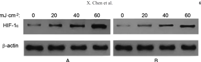
Western blotting to detect HIF-1α protein expression
The treatment group (50 μg/mL SQBE) was irradiated and cultured for 24 h. Western
blot analysis showed that in the control group, HIF-1α protein was highly expressed, and with
increasing irradiation dose (20, 40, and 60 mJ/cm2), HIF-1α protein expression increased. However, in the SQBE treatment group, HIF-1α protein expression significantly decreased compared to that reported for the control group. Gradation analysis showed that the HIF-1α protein expression level in the control group was significantly higher than that in the SQBE treatment group (P < 0.01), as shown in Table 3 and Figure 1.
Table 3. Protein expression of hypoxia-inducible factor-1α in the control and Shenqin biochemical
extract-treated groups after ultraviolet B irradiation (N = 6, means ± SD).
*P < 0.01 vs control group.
Group HIF-1α/β-actin
0 mJ/cm2 20 mJ/cm2 40 mJ/cm2 60 mJ/cm2
DISCUSSION
According to wavelengths, UV radiation in sunlight can be divided into ultraviolet C (UVC), UVB, and ultraviolet A (UVA), of which almost all the UVC is absorbed by the ozone layer when reaching the earth’s surface. The UV radiation affecting the human body is mainly UVB. UVB radiation can cause skin damage, aging, and even cancer. It can lead to
point mutations in the tumor-suppressor gene p53, and p53 and HIF-1α can affect each other
(Kiselyov et al., 2007; Heidenreich et al., 2009).
HIF-1α is mainly expressed in hypoxic cells. Hypoxia can cause increased accumulation and activity of HIF-1α, and regulate the expression of vascular endothelial growth factor (VEGF) (Li et al., 2010). High expression of VEGF can promote angiogenesis
to create conditions for the invasion and metastasis of tumor cells. After being activated by
UVB radiation, HIF-1α and the downstream VEGF, through regulation of transcription and
expression of downstream genes, can stimulate angiogenesis, inhibit tumor cell apoptosis, and promote tumor cell metastasis, which results in tumor cells that adapt to the hypoxic microenvironment and continue to proliferate, invade, and metastasize (Liu et al., 2011).
Studies have shown that HIF-1α activity is closely related to tumorigenesis, and it is considered a target for cancer therapy (Hoffman et al., 1991; Forsythe et al., 1996).
UVB radiation promotes tumorigenesis by enhancing the expression of HIF-1α that
promotes transcription of target genes, and subsequently, participates in tumorigenesis and metastasis. UVB radiation can also promote tumor growth by enhancing the expression of
transferrin receptor (TFR), increasing the absorption of iron (Jang, 2013).
Ginseng and Scutellaria have beneficial anti-radiation, anticancer, and antioxidant
effects. In this study, the active ingredients of ginseng and Scutellaria were extracted using bionic extraction technology, and the effects of different doses of UVB radiation and SQBE on
intracellular expression of HIF-1α and viability of HaCaT cells were investigated. The results showed that SQBE had a significant protective effect on HaCaT cells after UVB irradiation. It also significantly reduced UVB irradiation-induced expression of HIF-1α mRNA and protein,
indicating that SQBE could play a role in preventing UVB-induced skin carcinogenesis by
modulating intracellular expression of HIF-1α. The results of this study provide a starting
point for the development of this Chinese medicine as an agent for the prevention of skin cancer, and is worthy of further research.
This study has two limitations. In the present study, we only observed the effect of SQBE
on HIF-1α expression in UVB-irradiated HaCaT cells. The entire molecular pathway was not
examined. Moreover, we did not investigate the effect of SQBE on an appropriate animal model.
Figure 1. Protein expression of hypoxia-inducible factor-1α in the control and Shenqin biochemical extract-treated
In conclusion, the present study showed that SQBE has protective effect on
UVB-irradiated HaCaT cells, and it inhibited the UVB irradiation-induced expression of HIF-1α at both the mRNA and protein levels, which indicates that SQBE could prevent the occurrence
of UVB radiation-induced skin cancer.
Conflicts of interest
The authors declare no conflict of interest.
ACKNOWLEDGMENTS
Research supported by the Jilin Province Science and Technology Department of Science and Technology Development Program funded project (#20140414051GH), and Jilin Province Pharmaceutical Administration funded project (#2014-ZD25).
REFERENCES
Chen X, Yan QX and Zhang XE (2013). Extract with water security comparative study of ginseng extract bionic. Chang Chun Zhong Yi Yao Da Xue Xue Bao 29: 393-394.
Chen X, Hu ZQ, Zhang HC and Sun Y (2012). A preliminary study bionic extraction of ginsenosides. Zhong Guo Yao Fang 23: 1752-1754.
Fisher GJ, Kang S, Varani J, Bata-Csorgo Z, et al. (2002). Mechanisms of photoaging and chronological skin aging. Arch. Dermatol. 138: 1462-1470. https://doi.org/10.1001/archderm.138.11.1462
Forsythe JA, Jiang BH, Iyer NV, Agani F, et al. (1996). Activation of vascular endothelial growth factor gene transcription
by hypoxia-inducible factor 1. Mol. Cell. Biol. 16: 4604-4613. https://doi.org/10.1128/MCB.16.9.4604
Heidenreich R, Röcken M and Ghoreschi K (2009). Angiogenesis drives psoriasis pathogenesis. Int. J. Exp. Pathol. 90: 232-248. https://doi.org/10.1111/j.1365-2613.2009.00669.x
Hoffman EC, Reyes H, Chu FF, Sander F, et al. (1991). Cloning of a factor required for activity of the Ah (dioxin) receptor. Science 252: 954-958. https://doi.org/10.1126/science.1852076
Kiselyov A, Balakin KV and Tkachenko SE (2007). VEGF/VEGFR signalling as a target for inhibiting angiogenesis. Expert Opin. Investig. Drugs 16: 83-107. https://doi.org/10.1517/13543784.16.1.83
Lei F (2010). Advances in the pharmacological effects of baicalin. Zhong Guo Yao Ye 19: 87-88.
Li W, Xiong ZW, Li HW, Hu HX, et al. (2010). Expression of COX-2, VEGF and E-cad in breast cancer and their clinicopathologic significance. Zhong Guo Xian Dai Pu Tong Wai Ke Jin Zhan 13: 766-771.
Li YH and Bi ZG (2009). A study on effects of UVB-induced HIF1α expression of HaCaT. Shi Yong Lin Chuang Yi Yao Za Zhi 13: 36-38.
Jang Y, Jeong SH, Park YH, Bae HC, et al. (2013). UVB induces HIF-1α-dependent TSLP expression via the JNK and
ERK pathways. J. Invest. Dermatol. 133: 2601-2608. https://doi.org/10.1038/jid.2013.203
Liu Y, Yang FB, Diao ZW, Liu WP, et al. (2011). Expressions of glucose transporter-3 and hypoxia-inducible factor-1α in
astrocytoma. Zhong Hua Shen Jng Yi Xue Za Zhi 10: 29-32.
Mei W, Luo D and Lin XF (2004). Experimental study on the photo-protection of baikal skullcap root of human skin cells
from ultraviolet radiation damage. Xu Zhou Yi Xue Yuan Xue Bao 24: 76-79.
Sliney DH (2001). Photoprotection of the eye - UV radiation and sunglasses. J. Photochem. Photobiol. B 64: 166-175.
https://doi.org/10.1016/S1011-1344(01)00229-9
Song JY, Han SK, Bae KG, Lim DS, et al. (2003). Radioprotective effects of ginsan, an immunomodulator. Radiat. Res.
159: 768-774. https://doi.org/10.1667/0033-7587(2003)159[0768:REOGAI]2.0.CO;2
Tang Y, Liu MX, Xu X, Zhang HC, et al. (2014). The protective effect of imitation extracts from ginseng baikal skullcap root on the injury of organisms caused by UVB irradiation. Chang Chun Zhong Yi Yao Da Xue Xue Bao 30: 208-210. Zhang HC, Wang EP, Chen X, Tang Y, et al. (2013). Protective effect of ginsenoside Re on damaged cells irradiated by
UVC. Ji Lin Da Xue Xue Bao: Yi Xue Ban 39: 507-511.