832 | 2018 | ISSUE 4 | VOLUME 13 © 2018 University of Alicante
Knee kinematics of ACL-deficient patients: A
development of a portable motion analysis system
PUI WA FUNG, KAM MING MOK 1 , RUEN SHAN LEOW, SAI CHUEN FU, PATRICK SHU HANG YUNG,
KAI MING CHAN
Department of Orthopaedic and Traumatology, Faculty of Medicine, The Chinese University of Hong Kong, Hong Kong
ABSTRACT
This study is to compare the knee kinematic measurements between the novel portable skin marker-based motion analysis system (Opti-Knee®) and a conventional system (Vicon®). Nineteen subjects were recruited and asked to perform stair descent with lower limb placed with skin markers. Knee kinematic data was
computed from the trajectories of the skin markers. Pearson’s correlation coefficient and Root-mean-square deviation (RMSD) were used to analyse the data. For the waveform, sagittal plane rotation is strongly positive correlated between systems, while for axial and coronal plane rotation, it was moderately to strongly positive correlated in both normal (ACLN) and ACL-deficient (ACLD) group. Substantial difference between two groups was found in correlation of abduction/adduction in both stance and swing phase, as well as in external/internal rotation in all selected regions of interest. Moreover, the RMSD was larger in ACLN than in ACLD in three planes of rotation. The capability of Opti-Knee® in tracking lower limb sagittal plane rotation was comparable to Vicon®. However, for coronal and axial plane rotation, although the correlation to Vicon® in kinematic waveforms was moderately high, their ROM and peak values substantially deviated from the values in Vicon®. Keywords: Anterior cruciate ligament; Dynamic knee stability; Sports injury; Stair descent; Tibiofemoral kinematics.
1Corresponding author. Department of Orthopaedic and Traumatology, Faculty of Medicine, The Chinese University of Hong
Kong, Hong Kong. http://orcid.org/0000-0003-2185-6468 E-mail: [email protected]
Submitted for publication January 2018 Accepted for publication May 2018 Published in press July 2018
JOURNAL OF HUMAN SPORT & EXERCISE ISSN 1988-5202 © Faculty of Education. University of Alicante
doi:10.14198/jhse.2018.134.11 Cite this article as:
Fung, P., Mok, K., Leow, R., Fu, S., Yung, P., & Chan, K. (2018). Knee kinematics of ACL-deficient patients: A development of a portable motion analysis system. Journal of Human Sport and Exercise,
VOLUME 13 | ISSUE 4 | 2018 | 833 INTRODUCTION
Three-dimensional motion analysis systems for functional knee stability assessment
Skin marker-based three-dimensional (3D) motion analysis systems have been widely used to investigate knee joint kinematic changes following different injuries. Particularly in anterior cruciate ligament (ACL) injury, the change of kinematics of running, stair negotiation, other daily activities and exercise are of concern after ACL reconstruction (ACLR) (Salem, et al. 2003; von Porat, et al. 2006; Waite, et al. 2005; Webster and Feller. 2011). However, space occupying and time required and expertise to interpret the data are the main drawbacks that render the conventional systems difficult to be implemented in the clinical setting. Currently, there are no well-accepted use of quantitative data regarding the functional tests in the rehabilitation progress after ACLR.
A portable motion analysis system (Opti-Knee®, Shanghai Innomotion Inc., China), which was originally designed specifically for tibiofemoral kinematic analysis, has been developed recently. It is a compact motion capture system, which includes a portable workstation consisting of an integrated 2-head stereo-infrared camera which sampled at 60Hz and a high-speed camera. The accuracy of the optical tracking system was 0.3mm RMS as reported by the original equipment manufacturer (Northern Digital Inc., Ontario, Canada). The system consists of a marker cluster set of 8 retroreflective markers.
Recently, we have conducted a systematic review regarding the motion tasks that can assess dynamic stability after ACL reconstruction, which showed that simple and uniplanar movement like stair descent or single leg hop can be one of the available options (Chua, et al. 2016). This indicated that a sophisticated motion system might not be required in clinical setting but a simplified one, as long as its capability can cater for the detection of uniplanar movements. In this study, stair descent was chosen to be the motion task as it is less challenging for the subjects who have ACL injury.
Before further application of the portable motion system, it is necessary to understand the capability of the novel motion analysis system. Thereby, this study is to compare the portable motion system with a conventional motion analysis system. In this study, we also included ACL injured patients because change in kinematic following ACL injury was evidential (von Porat, et al. 2006; Waite, et al. 2005): increased or decreased range-of-motion (ROM) . We hypothesized that there were no significant differences on the kinematics data between Opti-Knee® and Vicon®, which serves as a conventional standard.
MATERIALS AND METHODS
Study design
834 | 2018 | ISSUE 4 | VOLUME 13 © 2018 University of Alicante
Part 1: The capability of detecting the retroreflective markers’ positions using the two systems
A brief comparison study between the motion capturing volume of two motion systems was conducted using a digital caliper (accuracy close to 0.01mm) as the gold standard. The accuracy of capturing systems of Vicon® and Opti-Knee® was compared using the electronic caliper (Digimatic caliper, Mitutoyo, Japan) as the gold standard.
Experimental procedure
Two retroreflective markers were placed on the caliper: one at the fixed outside measuring step; another at the slider. The caliper was fixed on a flat platform. Translations were performed by moving the slider with the marker attached. Four planes of translations were randomly selected and 5 translations (6 spatial positions of the marker) were made per plane. The Trajectories of the marker on the slider of the caliper were captured simultaneously by both Vicon® and Opti-Knee®. In the process, only 12 out of 15 Vicon cameras were operating to avoid interference. The sampling frequency of Vicon® was adjusted to 60 Hz (same as Opti-Knee®). And, the readings from the electronic caliper were recorded. Background noise was greatly reduced when only two retroreflective markers were used.
Data analysis
Marker movement trajectories were extracted and relative distances between two retroreflective markers were calculated based on the spatial positions. Dependent t-test was used to compute the difference between (A) the readings from the calipers and the measurements from Vicon® and (B) the readings from the caliper
and the measurements from Opti-Knee®. Statistical significance level is reached when p-value is smaller
than 0.05. The mean errors from the gold standard were 0.055 ± 0.053mm (Vicon®) and 0.040 ± 0.057mm
(Opti-Knee®). Paired t-test showed no significant difference (p=0.233) between Vicon® and Opti-Knee® of
their deviation of calculated translations from the measured values of the electronic caliper.
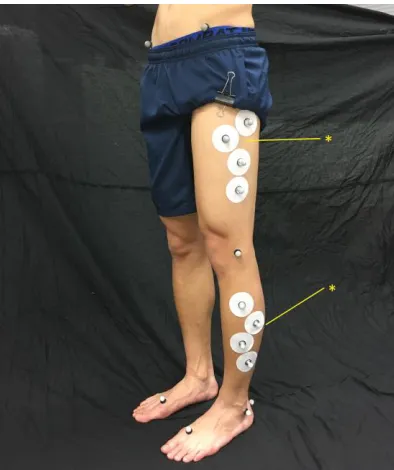
Without significant difference in detection of spatial positions of markers, in order to optimize the experimental procedures and reduce the time cost of data collection, the marker movement trajectories were captured only by the cameras of Vicon® system in the remaining body of the study. And the data were processed in Vicon® Nexus 1.7.1 and Opti-Knee® separately. Movement trajectories were recorded using a Vicon® (MX-T40) motion capture system (Vicon®, Oxford, UK), sampled at 200 Hz. The knee kinematic data was computed by 2 sets of marker sets from 2 systems: the lower extremity version of plug-in-gait (Vicon®) and the Opti-Knee® (Figure 1).
Part 2:To compare the difference in knee kinematic measurements between Opti-Knee® and Vicon® Subject recruitment
Nineteen subjects were recruited from the community (14 males, 5 females, age 27.0 ± 4.7 years; body height 170.7 ± 9.2cm; body weight 64.5 ± 9.8 kg). Nine of them were confirmed of ACL rupture by magnetic resonance imaging (6 males, 3 females, age 27 ± 5.6 years; body height 172.7 ± 9.3cm; body weight 67.4 ± 12.7 kg). All of the subjects have signed and kept an informed consent form. The study protocol was
approved by The Joint Chinese University of Hong Kong – New Territories East Cluster Clinical Research
Ethics Committee. All experimental procedures were performed in accordance with the approved procedures.
Experimental procedure
VOLUME 13 | ISSUE 4 | 2018 | 835
should be performed with the use of a probe to locate anatomical landmarks and three randomly selected points at the ground level to generate lower limb joint coordinate systems and neutral joint positions. In the calibration process, the subject was asked to stand in a neutral position in which the foot was 10 cm apart and the arms were crossed at the chest to avoid blocking the infrared signals. All kinematic meanings were defined relatively to this neutral starting position but not anatomical bony landmarks.
Figure 1. A hybrid protocol consisting plug-in-gait (lower extremity) and the skin marker cluster of Opti-Knee®
was placed on the lower extremity of subjects. Two skin markers of Opti-Knee®’s protocol were chosen to
be the sharing points of two marker cluster sets as indicated by asterisks.
In the part of the motion task, subjects would perform stair descent on a staircase which consists of two steps with each step of 20 cm height, with arms crossed at chest to avoid blocking signals, and in a pace of 80 steps per minute guided by a metronome, which is set to simulate the stair descent rhythm in real life. Subjects would stand on the top step of the staircase, and follow the command from the instructor to begin stair descending with the tested leg in a step-over-step manner. No unnecessary contact between the foot and the stair platform during any swing phase or hesitation was considered a successful trial. Five successful trials were collected. The movement trajectories were captured by the cameras of Vicon® system.
Data analysis and statistical treatment
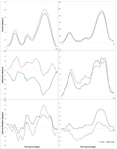
The tibiofemoral kinematics of the tested leg were extracted for analysis. Extracted kinematics were then normalized into 100 data points in each trial of a subject (Figure 2). Three tibiofemoral degrees of freedom (DOF) were included: axial rotation (external and internal rotation), sagittal rotation (flexion and extension)
and coronal rotation (abduction and adduction).Difference in ROM and in peak value of three DOF between
Vicon® and Opti-Knee® (calculated values of Opti-Knee® minus calculated values of Vicon®) were calculated from the mean value of the five trials. They were also expressed in percentage over full ROM which were taken from calculations of Vicon® as shown below. All joint rotations were expressed in degree (°).
𝐕𝐚𝐥𝐮𝐞 𝐨𝐟 𝐎𝐩𝐭𝐢𝐊𝐧𝐞𝐞 − 𝐕𝐚𝐥𝐮𝐞 𝐨𝐟 𝐕𝐢𝐜𝐨𝐧
836 | 2018 | ISSUE 4 | VOLUME 13 © 2018 University of Alicante For the comparison of the kinematic waveform, Pearson’s correlation coefficient (r) was used to analyse the similarity. The significance level of correlation coefficient was obtained by computing the p-value. Root-mean-square deviation (RMSD) was to compare the deviation of movement trajectories (Thies, et al. 2007). Statistical significance level is reached when p-value is smaller than 0.05. Different regions of interest were taken to do correlation separately: (A) a whole gait cycle (includes a stance phase and a swing phase); (B) stance phase in the gait cycle; and (C) swing phase in the gait cycle.
VOLUME 13 | ISSUE 4 | 2018 | 837 RESULTS
Difference in full range-of-motion
In group ACLN, the mean difference in flexion/extension was 1.79° ± 6.22° (in percentage of full ROM: 1.69% ± 6.47%). Mean difference in Abduction/adduction was 10.09° ± 13.01° (in percentage of full ROM: 29.89% ± 37.18%). Mean difference in External/internal rotation was 16.51° ± 11.11° (in percentage of full ROM: 123.48% ± 96.81%) (Table 1).
In group ACLD, the mean difference in flexion/extension was 3.74° ± 4.14° (in percentage of full ROM: 4.33% ± 4.57%). Mean difference in Abduction/adduction was 5.63° ± 9.44° (in percentage of full ROM: 11.19% ± 57.54%). Mean difference in External/internal rotation was 10.09° ± 10.65° (in percentage of full ROM: 62.35% ± 71.48%) (Table 1).
Table 1. Difference in range of motion and difference in peak value of three degrees of motion
Mean ± SD Mean/ROM ± SD
ACLN
Difference in ROM
Flex/Ext -1.79 ± 6.22 -1.69% ± 6.47%
Abd/Add -10.09 ±13.01 -29.89% ± 37.18%
Ext/Int Rot 16.51 ±11.11 123.48% ± 96.81%
Difference in peak value
Flexion -1.34 ± 5.92 -1.22% ± 6.20%
Abduction 14.73 ± 13.17 46.96% ± 37.12%
Internal Rotation 13.30 ± 11.77 100.54% ± 99.13%
Extension 0.45 ± 1.04 0.47% ± 1.09%
Adduction 4.64 ± 4.24 17.08% ± 17.30%
External Rotation -3.20 ± 3.08 -22.94% ± 20.79%
ACLD
Difference in ROM
Flex/Ext 3.74 ± 4.14 4.33% ± 4.57%
Abd/Add -5.63 ± 9.44 -11.19% ± 57.54%
Ext/Int Rot 10.09 ± 10.65 62.35% ± 71.48%
Difference in peak value
Flexion 3.50 ± 4.20 4.07% ± 4.64%
Abduction 5.56 ± 11.88 12.49% ± 63.95%
Internal Rotation 5.11 ± 12.59 34.72% ± 71.45%
Extension -0.24 ± 0.37 -0.27% ± 0.42%
Adduction -0.07 ± 6.82 1.30% ± 24.33%
External Rotation -4.99 ± 4.05 -27.63% ± 21.11%
Abd: Abduction; Add: Adduction; Ext: Extension; Ext/Int Rot: External and internal rotation; Flex: Flexion; ROM: Range of motion *Value measured by Opti-knee minus value measured by Vicon
838 | 2018 | ISSUE 4 | VOLUME 13 © 2018 University of Alicante
Difference in peak values
In group ACLN, the mean difference in flexion was -1.34° ± 5.92° (in percentage of full ROM: 1.22% ± 6.20%). For extension, it was 0.45° ± 1.04° (in percentage of full ROM: 0.47% ± 1.09%). In Abduction and adduction, it was 14.73° ± 13.17° (in percentage of full ROM: 46.96% ± 37.12%) and 4.64° ± 4.24° (in percentage of full ROM: 17.08% ± 17.30%) respectively. For axial rotation, it was 13.30° ± 11.77° (in percentage of full ROM: 100.54% ± 99.13%) in internal rotation; and -3.20° ± 3.08° (in percentage of full ROM: -22.94% ± 20.79%) (Table 1).
In group ACLD, the mean difference in flexion was -3.50° ± 4.20° (in percentage of full ROM: 4.07% ± 4.64%). For extension, it was -0.24° ± 0.37° (in percentage of full ROM: -0.27% ± 0.42%). In Abduction and adduction, it was 5.56° ± 11.88° (in percentage of full ROM: 12.49% ± 63.95%) and -0.07° ± 6.82° (in percentage of full ROM: 1.30% ± 24.33%) respectively. For axial rotation, it was 5.11° ± 12.59° (in percentage of full ROM: 34.72% ± 71.45%) in internal rotation; and -4.99° ± 4.05° (in percentage of full ROM: -27.63% ± 21.11%) (Table 1).
Correlations of kinematic waveforms
Pearson’s correlation coefficient and RMSD were used to analyse the kinematic waveforms from two systems (Table 2a & 2b).
Table 2a. Pearson’s correlation coefficient in three degrees of freedom
Pearson’s correlation coefficient
One gait cycle Stance Swing
ACLN ACLD ACLN ACLD ACLN ACLD
F/E 0.984** 0.994** 0.979** 0.992** 0.975** 0.991**
Ab/Ad 0.506** 0.487** 0.509** 0.382** 0.326** 0.584**
Ext/Int 0.466** 0.651* 0.549 (p=0.90) 0.654** 0.365** 0.602**
Note: Ab/Ad: abduction/adduction; Ext/Int: External/internal rotation; F/E: flexion/extension *p<0.01
**p<0.001
Table 2b. Root-mean-square deviation in three degrees of freedom
Root-mean-square deviation
One gait cycle Stance Swing
ACLN ACLD ACLN ACLD ACLN ACLD
F/E 6.29 4.92 6.33 4.91 6.22 4.93
Ab/Ad 17.28 11.65 15.82 10.89 19.48 12.91
Ext/Int 11.54 9.07 9.08 7.98 14.75 10.75
Note: Ab/Ad: abduction/adduction; Ext/Int: External/internal rotation; F/E: flexion/extension; RMSD: Root-mean-square deviation
For one gait cycle of the tested leg, the correlation of flexion/extension was strongly positive (ACLN: r=0.984,
p <.001; ACLD: r=0.994, p<0.001); and it was moderately positive in abduction/adduction (ACLN: r=0.506, p
<.001; ACLD: r=0.487, p<0.001) and in external/internal rotation (ACLN: r=0.466, p <.001; ACLD: r=0.651,
p<0.001).
VOLUME 13 | ISSUE 4 | 2018 | 839
When the stance phase and swing stance in the gait cycle were isolated to do correlation, for the stance phase, the correlation of flexion/extension was strongly positive (ACLN: r=0.979, p <.001; ACLD: r=0.992,
p<0.001); and it was moderately positive in abduction/adduction (ACLN: r=0.509, p <.001; ACLD: r=0.382,
p<0.001) and in external/internal rotation (ACLN: r=0.549, p =0.900; ACLD: r=0.654, p<0.001). RMSD was used to measure the deviation of kinematics. It was 6.33° and 4.91° in flexion/extension; 15.82° and 10.89 ° in abduction/adduction; 9.08° and 7.98° in external/internal rotation for ACLN and ACLD respectively.
For the swing phase, the correlation of flexion/extension was strongly positive (ACLN: r=0.975, p <.001; ACLD: r=0.991, p <.001); and it was moderately positive in abduction/adduction (ACLN: r=0.326, p <.001; ACLD: r=0.584, p <.001) and in external/internal rotation (ACLN: r=0.365, p <.001; ACLD: r=0.602, p <.001). RMSD was used to measure the deviation of kinematics. It was 6.22° and 4.93° in flexion/extension; 19.48° and 12.91° in abduction/adduction; 14.75° and 10.75° in external/internal rotation for ACLN and ACLD respectively.
DISCUSSION
This study aimed to compare the novel portable motion system to the conventional one. The capability of Opti-Knee® in tracking tibiofemoral sagittal plane rotation was comparable to Vicon®, with small difference
in ROM and peak values and positive Pearson’s correlation. However, for coronal and axial plane rotation,
their ROM and peak values substantially deviated from the values of Vicon®. And, the difference in axial plane rotation was generally high when the error percentage in ROM was considered.
Between group ACLN and ACLD, we found substantial difference in correlation in abduction/adduction in both stance and swing phase (Table 2a), as well as in external/internal rotation in all selected regions of interest. Moreover, the RMSD was larger in ACLN than in ACLD in three planes of rotation.
Vicon® was chosen as a model to compare because of its prevailing use in the field of biomechanics. Recently, different kinds of commercialized 3D motion analysis systems have been introduced and investigated the accuracy and capability to capture joint kinetics and kinematics in different motion tasks. There were studies comparing their capacity with the conventional motion analysis systems like Vicon® using Plug-in-gait model marker set and other marker protocols (Carse, et al. 2013; Pfister, et al. 2014). Clinically, recent studies have investigated different gait and specific motion pattern through the use of the portable system (Mok, et al. 2016; Yeung, et al. 2016; Zhang, et al. 2015; Zhang, et al. 2016). As the current study reported differences between two systems, the findings from the above-mentioned studies may not be reproducible through the use of Vicon® system.
840 | 2018 | ISSUE 4 | VOLUME 13 © 2018 University of Alicante To clarify, this study was only to compare a novel model of 3D motion system to a conventional one, but it should not be considered a validation study. In fact, the model plug-in-gait used in this study had been criticised for the relatively small number of markers that were placed on the lower extremity, while the novel system had eight markers on the thigh and shank. A validation study of plug-in-gait model and Vicon® using a dynamic stereo-radiographic system showed that substantial errors of the plug-in-gait model in tibiofemoral kinematics were found in different motion tasks (Li, et al. 2012), which can be greater than the difference between the injured group and the control group in stair ascent. The intrinsic error may lead to misinterpretation of results.
In short, this study indicated that kinematic data from the novel system and the conventional system were not totally consistent. Opposite trend was observed frequently in axial and coronal plane rotations. However, due to the intrinsic deficit of the conventional system, the absolute accuracy of the novel system is inconclusive. Therefore, validation of the novel motion analysis system using dynamic stereo-radiographic system is needed in the future.
Though comprehensive knowledge regarding the kinematics and dynamic stability of knee joint following ACLR is yet to establish, Tashman et al. found that there is abnormal knee axial plane rotation under dynamic loading using a stereo-radiographic system with a high frame rate (Tashman, et al. 2004; Tashman, et al. 2007). All evidence urges the objective assessment on the rotational stability. Effort should be made to increase the accuracy of 3D motion systems especially in axial plane rotation.
CONCLUSIONS
In conclusion, the portable motion analysis system (Opti-Knee®) is a satisfactory substitute for the conventional lab-based motion analysis system (Vicon®) in terms of kinematics regarding sagittal plane rotation. The portable system has the advantage of enabling clinicians to perform knee kinematics assessment in clinical setting outside the gait laboratory. Future technical advancement is needed to reduce the difference in the knee frontal and transverse plane kinematics between two systems.
ACKNOWLEDGEMENTS
This study is supported by the Innovation and Technology Support Program, Hong Kong (Ref. No.: ITS/289/14FX).
CONFLICT OF INTEREST DISCLOSURE
This study is supported by the Innovation and Technology Support Program, Hong Kong (Ref. No.: ITS/289/14FX). The Shanghai InnoMotion Inc. financially supports the program but they do not influence any part of the current project.
REFERENCES
Carse, B., Meadows, B., Bowers, R., & Rowe, P. (2013). Affordable clinical gait analysis: An assessment of the marker tracking accuracy of a new low-cost optical 3D motion analysis system. Physiotherapy, 99(4), 347-351. https://doi.org/10.1016/j.physio.2013.03.001
VOLUME 13 | ISSUE 4 | 2018 | 841
review. Arthroscopy: The Journal of Arthroscopic & Related Surgery, 32(7), 1453-1465. https://doi.org/10.1016/j.arthro.2016.01.057
Li, K., Zheng, L., Tashman, S., & Zhang, X. (2012). The inaccuracy of surface-measured model-derived
tibiofemoral kinematics. Journal of Biomechanics, 45(15), 2719-2723.
https://doi.org/10.1016/j.jbiomech.2012.08.007
Mok, K. M., Chua, E. N., Leow, R. S., Yeung, M. Y., Fu, S. C., Yung, P. S. H., et al. (2016). Utilization of portable motion capture system for knee stability assessment in ACL-deficiency during stair descent. Asia-Pacific Journal of Sports Medicine, Arthroscopy, Rehabilitation and Technology, 6, 24-25. https://doi.org/10.1016/j.asmart.2016.07.047
Muneta, T., Ezura, Y., Sekiya, I., & Yamamoto, H. (1996). Anterior knee laxity and loss of extension after anterior cruciate ligament injury. The American Journal of Sports Medicine, 24(5), 603-607. https://doi.org/10.1177/036354659602400507
Pfister, A., West, A. M., Bronner, S., & Noah, J. A. (2014). Comparative abilities of microsoft kinect and vicon 3D motion capture for gait analysis. Journal of Medical Engineering & Technology, 38(5), 274-280. https://doi.org/10.3109/03091902.2014.909540
Salem, G. J., Salinas, R., & Harding, F. V. (2003). Bilateral kinematic and kinetic analysis of the squat exercise after anterior cruciate ligament reconstruction. Archives of Physical Medicine and Rehabilitation, 84(8), 1211-1216. https://doi.org/10.1016/S0003-9993(03)00034-0
Tashman, S., Collon, D., Anderson, K., Kolowich, P., & Anderst, W. (2004). Abnormal rotational knee motion during running after anterior cruciate ligament reconstruction. The American Journal of Sports Medicine, 32(4), 975-983. https://doi.org/10.1177/0363546503261709
Tashman, S., Kolowich, P., Collon, D., Anderson, K., & Anderst, W. (2007). Dynamic function of the ACL-reconstructed knee during running. Clinical Orthopaedics and Related Research, 454, 66-73. https://doi.org/10.1097/BLO.0b013e31802bab3e
Thies, S., Tresadern, P., Kenney, L., Howard, D., Goulermas, J., Smith, C., et al. (2007). Comparison of linear accelerations from three measurement systems during "reach & grasp". Medical Engineering & Physics, 29(9), 967-972. https://doi.org/10.1016/j.medengphy.2006.10.012
Torry, M. R., Decker, M. J., Ellis, H. B., Shelburne, K. B., Sterett, W. I., & Steadman, J. R. (2004). Mechanisms of compensating for anterior cruciate ligament deficiency during gait. Medicine and
Science in Sports and Exercise, 36(8), 1403-1412.
https://doi.org/10.1249/01.MSS.0000135797.09291.71
von Porat, A., Henriksson, M., Holmström, E., Thorstensson, C., Mattsson, L., & Roos, E. (2006). Knee kinematics and kinetics during gait, step and hop in males with a 16 years old ACL injury compared with matched controls. Knee Surgery, Sports Traumatology, Arthroscopy, 14(6), 546-554. https://doi.org/10.1007/s00167-006-0071-4
Waite, J., Beard, D., Dodd, C., Murray, D., & Gill, H. (2005). In vivo kinematics of the ACL-deficient limb during running and cutting. Knee Surgery, Sports Traumatology, Arthroscopy, 13(5), 377-384. https://doi.org/10.1007/s00167-004-0569-6
Webster, K. E., & Feller, J. A. (2011). Alterations in joint kinematics during walking following hamstring and patellar tendon anterior cruciate ligament reconstruction surgery. Clinical Biomechanics, 26(2), 175-180. https://doi.org/10.1016/j.clinbiomech.2010.09.011
842 | 2018 | ISSUE 4 | VOLUME 13 © 2018 University of Alicante Zhang, Y., Huang, W., Yao, Z., Ma, L., Lin, Z., Wang, S., et al. (2016). Anterior cruciate ligament injuries
alter the kinematics of knees with or without meniscal deficiency. The American Journal of Sports Medicine, 44(12), 3132-3139. https://doi.org/10.1177/0363546516658026
Zhang, Y., Yao, Z., Wang, S., Huang, W., Ma, L., Huang, H., et al. (2015). Motion analysis of chinese normal knees during gait based on a novel portable system. Gait & Posture, 41(3), 763-768. https://doi.org/10.1016/j.gaitpost.2015.01.020