The Theoretical Study on a Nano Biosystem Consisting of
Nano Tube-Catalytic Site of Bacillus Subtilis α-Amylase,
PDB: 1UA7
Z. Eslamifar
*Department of Biology, Payamnoor University, Qom, I. R. Iran
(*) Corresponding author: [email protected]
(Received:28 Sep. 2013 and Accepted: 12 Jan. 2013)
Abstract:
α-Amylase has been studied extensively from various sides. This enzyme is used in many industries .Many applications of this enzyme have encouraged us for greater attempts on the study of α-amylase and to search for more effective processes. In this investigation, the structure of nanotube - catalytic site of bacillus subtilis α- amylase was optimized by hyperchem 7.0 and then it was investigated with ab initio/hartree fock and density functional theory /B3LYP methods using the STO-3G, 3-21G and 6-31G basis sets for a physicochemical explanation of interactions within these nano biosystem. Then nuclear Magnetic Resonance (NMR) parameters and so charge, dipole moment, and stability energy were calculated on the optimized structure. We have found each of active atoms that indeed play an important role in imparting extra stability. In the current study, we have reported the NMR parameters of 8 atoms of catalytic site of bacillus subtilis alpha-amylasethe. Interesting finding of the present study is that in NMR shielding for each of active atoms, O8 and O14 had maximal shift in all of levels. In catalytic mechanism of this enzyme, O14 is adopting a chair structure leading to the easy cleavage of the glucoside bond (fixer for catalysis). This investigation suggests that nanotube interactions in this nano biosystems indeed play an important role in imparting extra stability of the catalytic site of the enzyme .Energy parameters in B3LYP level in different basis sets have more negative values than HF and have indicated the most stability in B3LYP6-31/G level and so dipole moment in this structure have observed that in HF3-21/G is maximum. The aim of this work was to discuss the aspects of the electronic structure of this nano biosystem to increase their advantages in practical applications.
Keywords: Nanotube-catalytic site, Bacillus subtilis α-amylase, AB initio, DFT, NMR shielding, GIAO, CSGT, HF, B3LYP.
Int. J. Nanosci. Nanotechnol., Vol. 10, No. 1, Mar. 2014, pp. 35-44
1. INTRODUCTION
α -Amylase (α-1, 4-glucan-4-glucanohydrolase,
EC 3.2.1.1) catalyzes the hydrolysis of D-(1, 4)-glycosidic linkages in polysaccharides such as starch, glycogen, and malto oligosaccharides, so that produces anomeric products [1]. This enzyme has been studied extensively from various sides, including structure, function, secretion, and
industrial application. α-Amylases are the most
widely studied members of the glycosyl hydrolase family 13 [1,2].
Microorganisms such as bacteria and fungi have
been extensively screened for suitable α-amylase production. Several extracellular α -amylases are of
prominent industrial importance [3].
study α-amylases and to search for more effective
processes [4-5].
Today NMR spectroscopy is a powerful tool in chemistry and bio chemistry [6-7]. In quantum mechanics, the quantity is directly related with the
NMR chemical shift. The shielding is defined as the
mixed second derivative of the energy with respect to magnetic moment of the nucleus and the strength
of the applied magnetic field. It is solved through the
second-order perturbation theory with the Zeeman Hamiltonian, treated as a perturbing term [8-9]. The methods currently employed for calculating of the NMR shielding are done by ab initio method consist of the Hartree Fock (HF) [10] and so density functional theory (B3LYP) levels. [11-13]. The term “AB initio” refers to calculations that are considered directly from theoretical principles, without inclusion of experimental data [14].
Gaussian basis sets are normally employed as
the basis functions to fit the electronic orbital
in a molecule [15]. The NMR shielding at very accurate levels of approximation are available in literature .The widely used methods for calculating of chemical shifts are as follows: GIAO and CSGT approximations [16-19]. The GIAO approach is known to give satisfactory shielding for different nuclei with larger molecules [17-19].
This study have surveyed the active atoms which play in stability by examining the NMR shielding. Also optimum energy and dipole moment were evaluated. The aim of this work is to discuss the aspects of the electronic structure of this nano
biosystem and to increase their advantages in practical applications.
1.2. Computational methods
The zigzag single-walled carbon nanotubes (SWCNT) with (5, 0) structure were made using the implement in HyperChem7.0. The nanotube symmetry is D5d [20, 21].
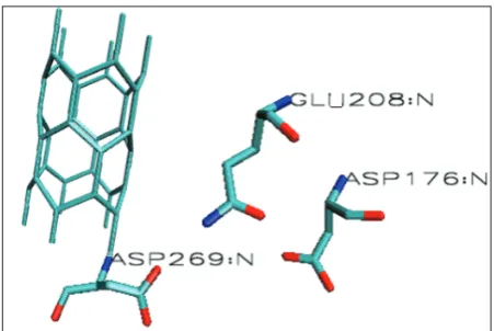
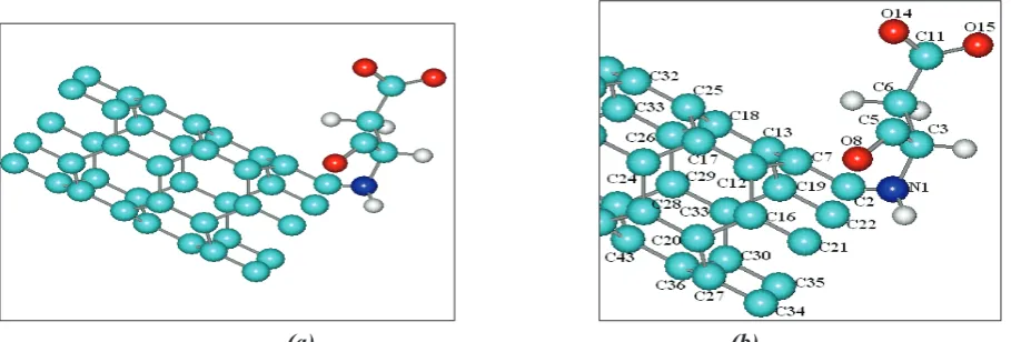
In this study, a structure of nanotube –catalytic
site of bacillus subtilis α amylase was made by
HyperChem7.0 (Figures 1, 2). The structure of this enzyme is deposited in Protein Data Bank as
1UA7. At first, nanotube –catalytic site of enzyme
was optimized. The Gaussian 98 was used for all calculations [22] and then Nuclear Magnetic Resonance (NMR) parameters by AB initio (hartree fock) and density Functional Theory ( B3LYP) methods was calculated on the optimized structure (Figure 2.) by GIAO and CSGT methods using the STO-3G ,3-21G and 6-31G basis sets .The results of the hartree fock and B3LYP methods of NMR
shielding values (ppm) including isotropic (σiso )
and anisotropic (σaniso) effects and so charge , energy
(kcal/mol) and dipole moment (debye) parameters for 8 number of active atoms of catalytic site were reported.
2. RESULTS AND DISCUSSION
In the current study, we performed the structural computations on the nanostructure of the
nanotube-catalytic site system of bacillus subtilis α amylase.
In this investigation, we analyze and report the NMR shielding values of 8 atoms of catalytic site at HF and B3LYP levels by STO-3G, 3-21G and 6-31G basis sets. We have studied principle calculations on this nanostructure.
The NMR parameters are given in Table 1 and 2.
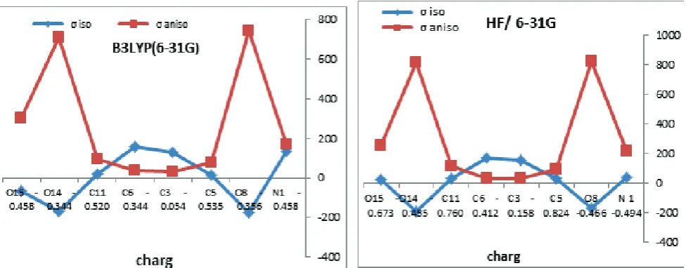
Also, parameters of σiso and σaniso of mentioned
atoms versus atom number and charge is shown in Figure 3, 4.
The results of Table 1 are shown in Figure 3 and 4, where we plot the NMR shielding isotropy (σ iso) and NMR shielding anisotropy (σaniso) of the nanotube-catalytic site for each of active atoms.
We found that O8 and O14 had maximal shift. Other
indicated atoms had the similar shifts in different positions therefore some atoms on nanotube-catalytic site of bacillus subtilis alpha amylase indeed play an important role in imparting extra stability and functionality and the best interaction in the catalytic site of the enzyme.
It should be noted that Calculations at the B3LYP and HF levels in CSGT and GIAO methods have shown that shielding properties in6-31 G is better than the other basis sets, (Figures 3a, 3b, 3c, 3d). In catalytic mechanism of this enzyme, one hydrogen-bonding is between O14 of carboxyl group of ASP269 and sugar in the catalytic subsite -1 in the enzyme/ acarbose. This atom is adopting a chair structure leading to the easy cleavage of the glucoside bond) fixer for catalysis. [23,1] The interesting finding of the present study is that O8 and O14 have maximal shift in B3LYP levels. In the course of parameters deliberation and relating them to the fundamental electronic structure of
the considered system, σaniso and σiso parameters
effects provided beneficial information on the
interaction characteristics.
The graphs of the charge value versus the isotropic and anisotropic parameters at B3LYP /6-31G and HF /6-31G Levels in Figure 4 shows charge Density is high on O8 and O14.
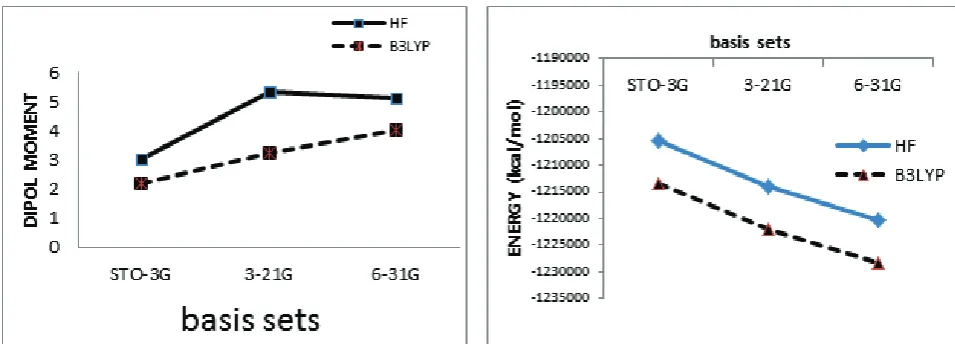
Also, we have detected the electromagnetic nature of this system by calculating the parameters consisting energy and dipole moment which provide valuable information on the interaction characteristics. By plotting the energy values versus basis sets, it is observed that stability energies decrease linearly in
basis sets consisting STO – 3 G, 3 - 21 G and 6 - 31 G in HF and B3LYP levels, respectively (Figure 5). Also, B3LYP/6-31G (5b) is better than the other level/basis sets (Table 2).
The factors such as dipole moment values are very important quantities for determining and estimating polarity of component [24]. The dipole moment values of this system are reported in Table 2 and Figure 5a.
In this nano biosystem, it was observed that
dipole moment value in HF/3-21G was maximum. Therefore maximum polarity was observed in this level.
Enzymes are big structures that cannot directly be utilized by QM methods (quantum mechanics methods). Only the substrate and residues in the catalytic site are treated quantum mechanically and the rest of the enzyme is explained at the MM method (molecular mechanics methods). This decreased the computational dispersion and made it possible to investigate large enzyme structures [25]. The QM method is increasingly applied and is preferred to traditional methods due to their versatility and ability to describe the same system at different levels of detail. [14, 26, 27].The NMR shielding quantity has been applied to a broad range of researches in chemistry and biochemistry and has revealed to be in valuable investigations. It has
played a significant role in the structural studying
of protein [28-30]. AB initio calculation of NMR shielding has become an indispensable tool in the analysis of molecular structure and accurate assignment of NMR spectra of systems [7].
(a) (b)
Figure 3: The curves of nanotube- catalytic site atoms via atom number versus σiso and σaniso at HF and B3LYP levels.
(a)
(b)
(c)
Table
1:
Parameters of active atoms of nanotube- catalytic site at the B3L
YP/ST
O-3G,3-21G,6-31G and HF/
ST
Table
2:
Parameters of active atoms consisting char
ge , dipole moment(DEBYE) and stability ener
gy
(kcal/mol)of nanotube- catalytic site of bacillus
subtilis α-amylase at the B3L
YP/ST
O-3G , 3-21G,6-31G and HF/ ST
The studying of nanometer sized structures will lead to products which are multi-functional.
Therefore, the studying of qualities of structures at the nano scale with the assistance of computational
calculations is important to plan the specific material
properties [7, 31, 32].
3. CONCLUSION
1. In the current study, we analyzed the NMR parameters of 8 atoms of catalytic site of bacillus subtitles alpha-amylase at HF and B3LYP levels with STO-3G, 3-21G and 6-31G basis sets. The results of NMR shielding for each of active atoms, showed that O8 and O14 had maximal shift. This investigation suggests that some interactions on this nano biosystems indeed play an important role in imparting extra stability and functionality of enzyme.
2. In catalytic mechanism of this enzyme, O14 of carboxyl group of ASP269 is adopting the chair structure that leading to easy cleavage of the glycoside bond (During hydrolysis), the results of this study indicated that O8 and O14 had maximum shift in B3LYP levels and played an important role in extra stability and the best interactions.
3. The charge value versus the isotropic and anisotropic values showed that charge Density was on O8 and O14.
4. The stability energy was reported and shown the most stability in B3LYP/6-31G level. 5. The dipole moment was observed that
HF/3-21G was maximum.
3.1. Future objectives
1. A number of α-amylase structures are available
therefore we can study the interesting results of this study on these structures.These structures including pig pancreatic amylase I [33,34], pig pancreatic amylase II [35,36], Aspergillus
oryzae α-Amylases [37], barley α-amylase
[38], Bacillus circulans cyclodextrin glycosyl transferase [39-41], Pseudoalteromonas
haloplanctis α-amylase [42], and Bacillus stearo thermophilus “maltogenic” α-amylase [21].
In the overall fold of these structures, despite differences in their amino acid sequences, they have similar three dimensional structures, suggesting a similar catalytic mechanism. Therefore we can suggest the interesting results of this study on these structures. We suggest some of atoms that are similarly at nanotube-catalytic site of these alpha amylases and that
play an important role in extra stability.
2. These theoretical calculations have become a competitive alternative to successful assumptions and interpretation for nano-biochemical investigations.
REFERENCES
1. M. Kagawa, Z.Fujimoto, M. Momma, K.Takase, and H.Mizuno, Crystal Structure of Bacillus subtilis α-Amylase in Complex with Acarbose. Journal of bacteriology., Vol. 185, No. 23, (2003), pp. 6981– 6984.
2. M. Monajjemi, Z. Eslamifar. Density functional theory outlook (DFT) of biological active site junction to single walled carbon nano tube. African. J. of Microbiology., Vol. 6, No. 10, (2010), pp. 2338-2345.
3. V. N. Ivanona, E. P. Dobreva and E. I. Emanuilova, Purification and charaterization of a thermostable alpha–amylase from Bacillus licheniformis, J. Biotechnol., Vol. 28, (1993), pp. 277–289.
4. K.Ohdan, T. Kuriki, H. Takata, H. Kaneko and S. Okada, Introduction of raw starch–binding domains into Bacillus subtilis alpha-amylase by fusion with the starch–binding domain of Bacillus cyclomaltodextrin glucanotramferase. Appl. Environ. Microbiol., Vol. 66, (2000), pp. 3058–64.
5. N. Riaz, I.-Ul-Haq and M.A. Qadeer, Characterization of α-Amylase by Bacillus subtilis, International Journal of Agriculture & Biology., Vol. 5, No. 3, (2003), pp. 249-252.
6. Seyed N. Azizi and C.Esmaili , Quantum Mechanical Calculation of 13C NMR Chemical Shift of a Lienear and Cyclo a, b Unsaturated Ketones. World Applied Sciences Journal., Vol. 7, No. 5, (2009), pp. 559-566.
7. L. Pishkar, M. Monajjemi, A.R. Ilkhani and F. Mollaamin*., Theoretical investigation of dielectric effects on Nanostructure of β- amyloid peptide. International Journal of the Physical Sciences.,Vol. 5, No. 9, (2010), pp. 1450-1464.
8. B. Friedrich, J.D.Weinstein, R. Decarvalho and J.M. Doyle., Zeeman Spectroscopy of CaH Molecules in a Magnetic Trap. J. Chem. Phys., Vol. 110, (1999), pp. 2376-2383.
9. Ramsay, N.E., Magnetic Shilding of Nuclei. J. Phys. Rev., Vol. 78, (1950), pp. 699-703.
10. M. J. Frischend and J. B. Foresman., Gaussian 94 user reference, Gaussian, Inc., Pittsburgh, (1995). 11. J.R. Cheeseman, M. J. Frisch, F. J. Devlin and P.
J. Stephens., Ab initio calculation of atomic axial tensors and vibrational rotational strengths using density functional theory. Chemical Physics Letters., Vol. 252, (1996), pp. 211-220.
12. C. Pisani, S. Casassa and P. Ugliengo., Proton-ordered ice structures at zero pressure. A quantum-Figure 5: Graphs of dipole moment (a) and stability energy (b) of nanotube-catalytic site in different basis
mechanical investigation. Chemical Physics Letter., Vol. 253 , (1996), pp. 201-208.
13. V. Subramanian, K. Venkatesh, D. Mary Prabha and T. Ramasami., Density functional calculations of VanderWaals clusters: NeN2+ as a case study. Chemical physics letter., Vol. 276, (1997), pp. 9-12. 14. M. Monajjemi, M. Heshmat, H. Aghaei, R. Ahmadi
and K. Zare., Solvent effect on 14N nmr shielding of glycine, serine, leucine, and threonine: comparison between chemical shifts and energyversus dielectric constant. Fullerenes, Nanotubes, and Carbon Nanostructures . Bull. Chem. Soc. Ethiop., Vol. 21, No. 1, (2007), pp. 111-116.
15. R. Poirer, R. Kari and I.G. Csizmadia., Handbook of Gaussian Basis Sets: A compendium for Ab-initio Molecular Orbital Calculations. Elsevier Science Publishing: New York., (1985).
16. P.V.R. Schleyer, N.L. Allinger, T. Clark, J. Gasteiger, P.A. Kolmann, H.F. Schaefer and P.R. Schreiner, The Encyclopedia of Computational Chemistry., (1998). 17. R. Ditchfield, Self-consistent perturbation theory of diamagnetism. Mol. Phys., Vol. 27, (1974), pp. 789-807.
18. M. Barfiled and P. Fagerness., Density Functional Theory/GIAO Studies of the 13C, 15N and1H NMR chemical shifts in aminopyrimidines and aminobenzenes: Relationships to electron densities and amine group orientations. J. Am. Chem. Soc., Vol. 119, (1977), pp. 8699-8711.
19. J. M. Manaj, D. Maciewska and I. Waver., 1H, 13C and 15N NMR and GIAO CPHF calculations on two quinoacridinium salts. Magn. Reson. Chem., Vol. 38, (2000), pp. 482-485.
20. Hyper Chem 7.0, Hypecube Inc., Gainesville, FL, USA., (2001).
21. I.Przylas, Y. Terada, K. Fujii, T. Takaha, W. Saenger, and N. Stra¨ter, X-ray structure of acarbose bound to amylomaltase from Thermus aquaticus: implications for the synthesis of large cyclic glucans. Eur. J. Biochem., Vol. 267, (2000), pp. 6903–6913.
22. M. J. Frisch, G. W.Trucks, H. B. Schlegel, G. E.Scusera, M. A. Robb, J. R. Cheeseman, V. G. Zakrzewski, J. A.Montgomery, R. E., Jr.Stratmann, J. C. Burant, S. Dapprich, J. M. Millam, A. D. Daniels, K. N.Kudin, M. C. Strain, O.Farkas, J.Tomasi, V. Barone, M. Cossi, R. Cammi, B. Mennucci, C. Pomelli, C. Adamo, S. Clifford, J. Ochterski, G.
A. Petersson, P. Y. Cui, Q.Ayala, K.Morokuma, D. K. Malick, A. D. Rabuck, K. Raghavachari, J. B. Foresman, J. Cioslowski, J. V. Ortiz, A. G. Baboul, B. B. Stefanov, G. Liu, A. Liashenko, P. Piskorz, I. Komaromi, R. Gomperts, R. L. Martin, D. J. Fox, T. Keith, M. A. Al-Laham, C. Y. Peng, A. Nanayakkara, C.Gonzalez, M. Challacombe, P. M. W. Gill, B.Johnson, W. Chen, M. W. Wong, J. L. Andres, C. Gonzalez, M. Head-Gordon, E. S. Replogle, J. A.Pople, Gaussian 98, revision A.7; Gaussian, Inc.: Pittsburgh, PA., (1998).
23. R. Mosi, H. Sham, J. C. Uitdehaag, R. Ruiterkamp, B. W. Dijkstra, and S. G. Withers., Reassessment of acarbose as a transition state analogue inhibitor of cyclodextrin glycosyltransferase. Biochemistry., Vol. 37, (1998), pp. 17192–17198.
24. M. Monajjemi, A. Abedi, and H. Passdar., The comparison of structure and property of azt conformers and its analogue cs-87 using density functional theory calculations: a study of anti-aids. Bull. Chem. Soc. Ethiop., Vol. 20, No. 1, (2006), pp. 133-142.
25. F. Claeyssens et.al., High-Accuracy Computation of Reaction Barriers in Enzymes . Angewandte Chemie., Vol. 45, (2006), pp. 6856–6859.
26. HM. Senn, W. Thiel., QM/MM methods for biomolecular systems. Angew Chem Int Ed Engl., Vol. 48, No. 7, (2009), pp. 1198-1229.
27. M. Monajjemi, M. Heshmata ,HH. Haeria ., QM/ MM model study on properties and structure of some antibiotics in gas phase: Comparison of energy and NMR chemical shift. Biochemistry., Vol. 1, (2006), pp. 113-22.
28. M. Monajjemi, L.Saedi, F. Najafi and F. Mollaamin., Physical properties of active site of tubulin–binding as anticancer nanotechnology investigation. International Journal of the Physical Sciences., Vol. 5, No. 10, (2010), pp. 1609-1621.
29. F. Mollaamin, I. Layali, A. R. Ilkhani and M. Monajjemi., Nanomolecular simulation of the voltage–gated potassium channel protein by gyration radius study. African Journal of Microbiology Research., Vol. 4, No. 24, (2010), pp. 2795-2803. 30. F. Mollaamin,K. Shahani poor, T. Nejadsattari and
31. M. Monajjemi, M . Khaleghian, N. Tadayonpour and F. Mollaamin, The effect of different solvents and temperature s on stability of singlewalled car bon nano tube: A QM/MD study. International Journal of Nanoscience., Vol. 9, (2010), pp. 517-529.
32. M. Monajjemi, V. S. Lee, M. Khaleghian, B. Honarparvar, and F. Mollaamin., Theoretical Description of Electromagnetic Nonbonded Interactions of Radical, Cationic, and Anionic NH2BHNBHNH2 Inside of the B18N18 Nanoring . J.
Phys. Chem. C., Vol. 114, (2010), pp. 15315–15330. 33. M. Qian, R. Haser, G. Buisson, E. Du´ee, and
F. Payan., The active center of a mammalian α-amylase. Structure of the complex of a pancreatic α-amylase with a carbohydrate inhibitor refined to 2.2-A˚ resolution. Biochemistry, Vol. 33, (1994), pp. 6284–6294.
34. M. Qian, V. Nahoum, J. Bonicel, H. Bischoff, B. Henrissat, and F. Payan., Enzyme-catalyzed condensation reaction in a mammalian α-amylase. High-resolution structural analysis of an enzyme-inhibitor complex. Biochemistry., Vol. 40, (2001), pp. 7700–7709.
35. C. Gilles, J. P. Astier, G. Marchis-Mouren, C. Cambillau, and F. Payan., Crystal structure of pig pancreatic α-amylase isoenzyme II, in complex with the carbohydrate inhibitor acarbose. Eur. J. Biochem., Vol. 238, (1996), pp. 561–569.
36. M. Machius, L. Vertesy, R. Huber, and G. Wiegand., Carbohydrate and protein-based inhibitors of porcine pancreatic α-amylase: structure analysis and comparison of their binding characteristics. J. Mol. Biol., Vol. 260, (1996), pp. 409–421.
37. A. M. Brzozowski, and G. J. Davies., Structure of the Aspergillus oryzae α-amylase complexed with the inhibitor acarbose at 2.0 A˚resolution. Biochemistry., Vol. 36, (1997), pp. 10837– 10845.
38. A. Kadziola, M. Sgaard, B. Svensson, and R. Haser., Molecular structure of a barley α-amylase-inhibitor complex: implications for starch binding and catalysis. J. Mol. Biol., Vol. 278, (1998), pp. 205–217.
39. R. Mosi, H. Sham, J. C. Uitdehaag, R. Ruiterkamp, B. W. Dijkstra, and S. G. Withers., Reassessment of acarbose as a transition state analogue inhibitor of cyclodextrin glycosyltransferase. Biochemistry., Vol. 37, (1998), pp. 17192–17198.
40. B. Strokopytov, R. M. Knegtel, D. Penninga, H. J. Rozeboom, K. H. Kalk, L. Dijkhuizen, and B. W. Dijkstra., Structure of cyclodextrin glycosyltransferase complexed with a maltononaose inhibitor at 2.6 A˚resolution. Implications for product specificity. Biochemistry., Vol. 35, (1996), pp. 4241–4249.
41. B.Strokopytov, D. Penninga, H. J. Rozeboom, K. H. Kalk, L. Dijkhuizen, and B. W. Dijkstra., X-ray structure of cyclodextrin glycosyltransferase complexed with acarbose. Implications for the catalytic mechanism of glycosidases. Biochemistry., Vol. 34, (1995), pp. 2234–2240.