Asian J. Med. Biol. Res. 2016, 2 (1), 1-8; doi: 10.3329/ajmbr.v2i1.27561
Asian Journal of
Medical and Biological Research
ISSN 2411-4472 (Print) 2412-5571 (Online)www.ebupress.com/journal/ajmbr
Review
Omega-3 fatty acids transport through the placenta
Ariful Islam1,2, Takanori Kodama2,4, Yui Yamamoto2, Majid Ebrahimi3, Hirofumi Miyazaki3, Yuki Yasumoto3, Yoshiteru Kagawa3, Tomoo Sawada2, Yuji Owada3 and Nobuko Tokuda2, 4*
1
Department of Pharmacy, University of Rajshahi, Rajshahi-6205, Bangladesh
2
Department of Organ Anatomy, Yamaguchi University Graduate School of Medicine, Yamaguchi 755-8505, Japan
3
Department of Organ Anatomy, Tohoku University Graduate School of Medicine, Miyagi 980-8575, Japan
4
Faculty of Health Sciences, Yamaguchi University Graduate School of Medicine, Yamaguchi 755-8505, Japan
*
Corresponding author: Professor Dr. Nobuko Tokuda, Faculty of Health Sciences, Yamaguchi University Graduate School of Medicine, Yamaguchi 755-8505, Japan. Tel.: +81-836-22-2805; E-mail: toku@yamaguchi-u.ac.jp
Received: 21 December 2015/Accepted: 02 February 2016/Published: 31 March 2016
Abstract: The placenta is a temporary vital organ for sustaining the development of the fetus throughout gestation. Although the fatty acid composition delivered to the fetus is largely determined by maternal circulating levels, the placenta preferentially transfers physiologically important long-chain polyunsaturated fatty acids (LC-PUFAs), particularly omega-3 (n-3) FAs. The precise mechanisms governing these transfers were covered in a veil, but have started to be revealed gradually. Several evidences suggest fatty acid transport proteins (FATPs), placental specific membrane bound fatty acid binding proteins (pFABPpm) and fatty acid translocases (FAT/CD36) involved in LC-PUFAs uptake. Our studies have shown that the placental transfer of omega-3 FAs through the trophoblast cells is largely contributed by fatty acid binding protein 3 (FABP3). Recently there are considerable interests in the potential for dietary omega-3 FAs as a therapeutic intervention for fetal disorders. In fact, prenatal supply of omega-3 FAs is essential for brain and retinal development. Recent findings suggest a potential opportunity of omega-3 FA interventions to decrease the incidence of type 2 diabetes in future generations. In this review, we discuss the molecular mechanism of transportation of omega-3 FAs through the placenta and how omega-3 FAs deficiency/supplementation impact on fetal development.
Keywords: Omega-3 fatty acids; FABP3; placenta; fetal development; trophoblast
1. Introduction
The placenta is the principal site of nutrient exchange between the mother and the fetus. Survival and growth of the fetus are critically dependent on the placenta. Trophoblast cells fuse to form a syncytium, resulting in a two layered structure of multinucleated syncytiotrophoblast (SCTB) and cellular cytotrophoblast. Protusions of SCTB interdigitate into the decidualised endometrium, forming contacts with the maternal blood supply (Frost and Moore, 2010). Nutrients must cross through the multilayer trophoblast cells which separates the fetal capillary from the maternal sinusoids (Watson and Cross, 2005). Placental nutrient uptake and transfer have a unique role in the fetal development, as changes in nutrient-dependent signaling pathways in placental trophoblast leads to the alteration of fetal cell metabolism and may affect the fetal growth and the health programming after birth (Jansson et al., 2013; Jansson and Powell, 2013).
and cellular proliferation. For this rapidly growing infant, there is a high demand for complex lipids, such as docosahexaenoic acid (DHA, 22:6n-3) to form vital cell membrane structures. Human fetuses have a limited ability to synthesize omega-3 LCPUFA de novo and have to be supplied via maternal sources (Joffre et al, 2014). Other studies evaluating the early exposition to PUFAs, in particular omega-3 PUFAs, showed benefits in the offspring development and epigenetic regulation, which seem to prevent obesity, insulin resistance and cardiovascular diseases onset (Mennitti et al, 2015). Therefore, elucidating the pathways of placental omega-3 FAs transport and the regulatory processes governing these pathways are critical for advancing our understanding about the relationships between maternal omega-3 FAs metabolism and the placental supply of metabolites to the developing fetus. In this review, we would summarize recent development in the biochemical processes involved in placental omega-3 FAs delivery to the fetus. In accordance with this, we would show the impact of deficiency/supplementation of omega-3 FAs on the fetus.
2. Structure of the human placenta
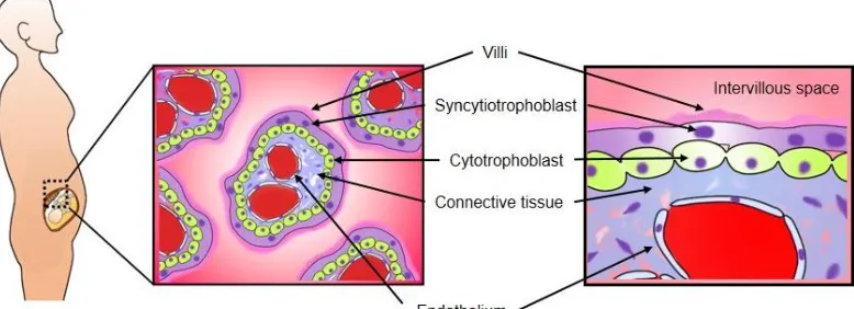
Placenta is directly responsible for bringing maternal and fetal blood supplies into contact, facilitating nutrient exchange and determining resource allocation (Frost and Moore, 2010). Human have an invasive hemochorial placenta resulting in close opposition of fetal and maternal blood and cells (Krishnan et al., 2013). The human placenta is a villous organ, whereby maternal blood comes into direct contact with placental trophoblast cell. The intervillous space is completely lined with a multinucleated syncytium called SCTB (Figure 1). Circulating maternal blood enters the intervillous space via spiral endometrial arteries, bathes the villi and drains back through endometrial veins. (Gude et al., 2004). Protrusions of SCTB interdigitate into the endometrium, forming contacts with the maternal blood supply. Interstitial trophoblast cells invade to expand the placenta from its edge outwards (Frost and Moore, 2010). Initially, the placental membrane is made up of four layers, the maternal facing SCTB, a layer of cytotrophoblast cells, connective tissue of the villus and the endothelium lining the fetal capillaries (Figure 1a). By approximately 20 weeks of gestation, the cytotrophoblast cell layer of many villi becomes attenuated and disappears gradually (Figure 1b). Subsequently, in most of the chorionic villi, the membrane consists of three layers and, in some areas, becomes extremely thin such that the SCTB comes in direct contact with the fetal capillary endothelium (Gude et al., 2004). The SCTB constitute the transporting epithelium of the placenta, with two polarized membranes, the microvillous membrane (MVM) facing maternal circulation and the basal plasma membrane (BM) facing the fetal capillary (Figure 1b). After passage across the SCTB membranes, substrates must cross the second layer of cells, the fetal capillary epithelium, before entry into the fetal circulation is complete. Only smaller solutes are highly permeable through the MVM and BM, and thus the SCTB constitutes a barrier and rate-limiting step of the transport of nutrients into fetal circulation (Brett et al., 2014).
3. Molecular mechanism of omega-3 FAs transport
3.1. Transport of omega-3 and other FAs through the placenta
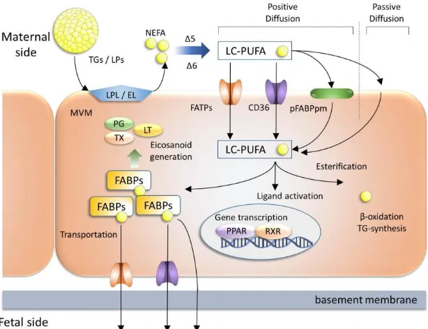
All of the PUFAs accumulated in the fetus must be obtained from the mother by placental transfer (Innis, 2005), predominantly originate from two sources in maternal circulation: nonesterified fatty acids (NEFAs) and esterified fatty acids in triglycerides (TGs) carried by lipoproteins (LPs) (Haggarty, 2010) (Figure 2). Fatty acids cannot cross the placenta in the form of TGs, therefore they must be converted to NEFAs by hydrolysis. This enzymatic process of TGs conversion is accomplished by several lipases expressed in the MVM (Lindegaard et al., 2006). Among the several lipases, lipoprotein lipase (LPL) and endothelial lipase (EL) have been well studied in the MVM. In addition to the LPL and EL activity, TG hydrolase expression and activity also exist in the MVM of human placenta (Waterman et al., 1998; Waterman et al., 2000) which is also involved with the conversion of TGs to NEFA. The hydrolysis of TGs by lipase or hydrolase appears to be of critical importance for the supply of LC-PUFA available for fetal transport (Benassayag et al., 1997). The placenta lacks the enzymes △5- and △6-desaturase which are involved in the conversion of NEFA or essential fatty acids (EFA) to LC-PUFA must be supplied from mother (Hanebutt et al., 2008; Wadhwani et al., 2013; Mennitti et al, 2015). TGs and NEFA after conversion into LC-PUFA are ready to cross the lipid bilayer of SCTB cells (Figure 2).
3.2. Transporters of fatty acids
(Haggarty et al., 1997), as well as from in vivo studies with DHA labeled with stable isotopes (Gil-Sanchez et al., 2010). Among the several membrane proteins, fatty acid transport proteins (FATPs, also known as SLC27A) are familiar for its cellular uptake of LC-PUFAs (Kazantzis and Stahl, 2012). Out of six family members of FATPs, FATP1-4 and FATP6 are expressed in both human and mouse placenta (Schaiff et al., 2005; Schaiff et al., 2007). FATP1 has been detected at protein level of both the MVM and the BM (Campbell et al., 1998; Duttaroy, 2009). Recently, FATP1 and FATP4 have been found to be involved in fatty acid uptake (Zhan et al.,
2012). FATP4 is found to be highly expressed by the epithelial cells of the visceral endoderm of the yolk sac and localizes at the brush-border membrane of extraembryonic endodermal cells (Gimeno et al., 2003). FATP4 gene deletion model shows early embryonic lethality (Gimeno et al., 2003), thus suggesting a critical role in materno-fetal fatty acid transport during early embryogenesis. In cultured trophoblast cells from term placenta, peroxisome proliferator-activated receptor (PPAR)γ/retinoid X receptor signaling and hypoxia regulate FATP4 mRNA expression (Mishima et al., 2011; Schaiff et al., 2005). Besides FATPs, two other membrane fatty acid transporters- namely, placental specific membrane bound fatty acid binding protein (pFABPpm) and fatty acid translocase (FAT/CD36) are involved in fatty acid uptake in the placenta (Campbell et al., 1998) (Figure 2). pFABPpm is exclusively expressed in the MVM (Campbell et al., 1998). Functionally, pFABPpm exhibits a high affinity for LC-PUFAs, suggesting this transporter to be involved in preferential uptake of these fatty acids for transfer across the placenta (Campbell et al., 1997). Recent study using an epithelial cell line has demonstrated that while comparing the efficacy to uptake fatty acids, FAT/CD36 is found to be 30 fold higher than that of FATP4. FAT/CD36 may directly facilitate fatty acid transport across the plasma membrane, whereas the intracellular FATP4 enhance fatty acid uptake indirectly by metabolic trapping in epithelial cells (Schneider et al., 2014). Since trophoblast cells are epithelial in character expressing both the FAT/CD36 and FATP4, similar mechanism of fatty acid uptake in the plasma membrane is possible.
After entering into the cytosol of SCTB cells, cytoplasmic fatty acid binding proteins (FABPs), traffic the fatty acids to sites for fatty acid uptake and metabolism, storage, beta-oxidation, signal transduction, ligand activation or transfer to the fetus (Owada, 2008) (Figure 2). Out of 12 members of FABPs family, FABP1, FABP3, FABP4, FABP5 and FABP7 have been observed to occur in both human and rodent placenta (Biron-Shental et al., 2007; Daoud et al., 2005; Das et al., 1993; Knipp et al., 2000; Larque et al., 2006; Masouye et al., 1997; Watanabe et al., 1991). In our study, FABP3, FABP4, FABP5 and FABP7 have been found to be spatially localized throughout rodent placenta, suggesting that functional properties may differ from each other depending on their localization (Islam et al., 2014). In our study, FABP3 gene deletion model shows significant down regulated transportation of omega-3 FA compared to wild type fetus. Similar observation has also been demonstrated by human trophoblast cell line. Our data suggest FABP3 regulates omega-3 FA transport in trophoblasts and plays a pivotal role in fetal development (Islam et al., 2014). In addition to FABPs and FATPs, exploring other molecules involved in fatty acid metabolism have been described, i.e. microsomal triglyceride transfer protein and apolipoprotein B (Farese et al., 1996; Raabe et al., 1998).
4. Impact of omega-3 FAs deficiency/supplementation on fetus
2011). These results suggest that the higher intake of omega-3 FAs during both maternal pregnancy and lactation may be beneficial for early brain development.
Gestational diabetes is a common pregnancy complication affecting 3.5% to 7.2% of pregnancies (Bardenheier
et al., 2013). LC-PUFA is fundamental to the functional integrity of pancreatic beta-cells (Dixon et al., 2004; Konard et al., 1996). A growing body of evidence suggests that fatty acid nutritional status in early life affects fetal tissue lipids as well as neuroendocrine and metabolic pathways relevant to metabolic ‘‘programming’’ (Korotkova et al., 2005). Babies of gestational diabetic mothers have reduced DHA in the plasma and erythrocyte cholinephosphoglycerides. The total omega-3 fatty acids of the erythrocyte cholinephosphoglycerides are significantly lower in these babies (Min et al., 2005). Recent studies have revealed that low circulating fetal DHA levels are associated with compromised fetal insulin sensitivity, and may be involved in ‘‘programming’’ the susceptibility to type 2 diabetes in the offspring of gestational diabetic women. These findings suggest a potential opportunity of early life nutritional interventions (e.g. DHA supplementation) to halt adverse metabolic programming to decrease the incidence of type 2 diabetes in future generations (Zhao et al., 2014).
Pregnancy has been suggested to be a critical period for developmental programming, which may influence the later development of allergic diseases in childhood (Prescott, 2010). Maternal diet, especially maternal PUFA intake, during pregnancy may modify neonatal immune response through epigenetic mechanisms and therefore alter disease susceptibility and predisposition (Calder et al., 2010; Prescott, 2010). The mechanism of anti-allergic reaction of omega-3 FAs in neonate is still not clear. However, it is proposed that a higher intake of omega-3 FA, for example, by fish oil supplementation, may have protective effects against the development of atopic diseases in the offspring although not all studies show conclusive results (Calder et al., 2010; Kremmyda
et al., 2011; Larque et al., 2012).
Figure 1a. Trophoblast layers in human placenta at 20 weeks of gestation. The placental membrane is made up of four layers, the maternal facing SCTB, a layer of cytotrophoblast cells, connective tissue of the villus and the endothelium lining the fetal capillaries.
Figure 2. Transport of fatty acids across the SCTB. A model of placental fatty acid transport through SCTB is shown. A complex interplay of different fatty acid transport proteins orchestrates fatty acid uptake mechanism. Within the cells LC-PUFAs are bound by different FABPs and have multiple functions like eicosanoid and TG synthesis, activation of nuclear transcription factors like PPAR/RXR and energy generation. Some LC-PUFAs are transported to fetal side through transporters. EL; endothelial lipase, FATP; fatty acid transport protein, FAT; fatty acid translocase, LC-PUFA; long chain polyunsaturated fatty acid, LP; lipoprotein, LPL; lipoprotein lipase, LT; leukotriene NEFA; non-esterified fatty acid, p-FABPpm; placental plasma membrane FABP, PG; prostaglandin, PPAR; peroxisome proliferator activated receptor, RXR; retinoid X receptor, TG; triglyceride, TX; thromboxane. [Figure was modified from Hanebutt et al., 2008].
5. Conclusions
Alterations in fetal development and growth have been closely associated with lifelong adverse health consequences. Since fetal growth and placental fatty acid transport are closely linked, a cohesive knowledge of placental nutrient transport mechanism will most certainly bring us closer to understanding those mechanisms underlying altered fetal growth. As reviewed in this paper, research has predominantly focused upon how the fatty acids, especially omega-3 FAs are transported through placental barrier (SCTB). Several recent studies resulted in the discovery of novel mechanisms involved in the regulation of placental nutrient transport. However, further work is needed to elucidate how fetal, maternal, and placental signals are integrated, regulate omega-3 FA transport from mother to fetus, and impact on fetal development ultimately.
Acknowledgments
This work was supported in part by grants from the Ministry of Education, Science and Culture of Japan, no. 23500956 (to N. T.)
Conflict of interest
None to declare.
References
Bardenheier BH, A Elixhauser, G Imperatore, HM Devlin, EV Kuklina, LS Geiss and A Correa, 2013. Variation in prevalence of gestational diabetes mellitus among hospital discharges for obstetric delivery across 23 states in the United States. Diabet. care., 36: 1209-1214.
Biron-Shental T, WT Schaiff, CK Ratajczak, I Bildirici, DM Nelson and Y Sadovsky, 2007. Hypoxia regulates the expression of fatty acid-binding proteins in primary term human trophoblasts. Am. J. Obstet. Gynecol., 197: 516 e511-516.
Brett KE, ZM Ferraro, J Yockell-Lelievre, A Gruslin and KB Adamo, 2014. Maternal-fetal nutrient transport in pregnancy pathologies: the role of the placenta. Int. J. of Mol. Sci., 15: 16153-16185.
Calder PC, 2012. The role of marine omega-3 (n-3) fatty acids in inflammatory processes, atherosclerosis and plaque stability. Mol. Nutr. & Food Res., 56: 1073-1080.
Calder PC, LS Kremmyda, M Vlachava, PS Noakes and EA Miles, 2010. Is there a role for fatty acids in early life programming of the immune system? The Proceedings of the Nutrition Society 69: 373-380.
Campbell FM, PG Bush, JH Veerkamp and AK Dutta-Roy, 1998. Detection and cellular localization of plasma membrane-associated and cytoplasmic fatty acid-binding proteins in human placenta. Placenta, 19: 409-415. Campbell FM, AM Clohessy, MJ Gordon, KR Page and AK Dutta-Roy, 1997. Uptake of long chain fatty acids
by human placental choriocarcinoma (BeWo) cells: role of plasma membrane fatty acid-binding protein. J. Lipid. Res., 38: 2558-2568.
Daoud G, L Simoneau, A Masse, E Rassart and J Lafond, 2005. Expression of cFABP and PPAR in trophoblast cells: effect of PPAR ligands on linoleic acid uptake and differentiation. Biochim. Biophys. Acta.,1687: 181-194.
Das T, G Sa and M Mukherjea, 1993. Characterization of cardiac fatty-acid-binding protein from human placenta. Comparison with placenta hepatic types. Eur. J. Biochem., 211: 725-730.
Dixon G, J Nolan, NH McClenaghan, PR Flatt and P Newsholme, 2004. Arachidonic acid, palmitic acid and glucose are important for the modulation of clonal pancreatic beta-cell insulin secretion, growth and functional integrity. Clin. Sci., 106: 191-199.
Duttaroy AK, 2009. Transport of fatty acids across the human placenta: a review. Prog. Lipid Res., 48: 52-61. Dziechciarz P, A Horvath and H Szajewska, 2010. Effects of n-3 long-chain polyunsaturated fatty acid
supplementation during pregnancy and/or lactation on neurodevelopment and visual function in children: a systematic review of randomized controlled trials. J.of the Am. College of Nutr., 29: 443-454.
Farese RV, Jr., S Cases, SL Ruland, HJ Kayden, JS Wong, SG Young and RL Hamilton, 1996. A novel function for apolipoprotein B: lipoprotein synthesis in the yolk sac is critical for maternal-fetal lipid transport in mice. J. Lipid Res., 37: 347-360.
Frost JM and GE Moore, 2010. The importance of imprinting in the human placenta. PLoS Genetics, 6: e1001015.
Gil-Sanchez A, E Larque, H Demmelmair, MI Acien, FL Faber, JJ Parrilla and B Koletzko, 2010. Maternal-fetal in vivo transfer of [13C]docosahexaenoic and other fatty acids across the human placenta 12 h after maternal oral intake. Am. J. Clin. Nutr., 92: 115-122.
Gimeno RE, DJ Hirsch, S Punreddy, Y Sun, AM Ortegon, H Wu, T Daniels, A Stricker-Krongrad, HF Lodish and A Stahl, 2003. Targeted deletion of fatty acid transport protein-4 results in early embryonic lethality. J. Biol. Chem., 278: 49512-49516.
Gould JF, LG Smithers and M Makrides, 2013. The effect of maternal omega-3 (n-3) LCPUFA supplementation during pregnancy on early childhood cognitive and visual development: a systematic review and meta-analysis of randomized controlled trials. Am. J. Clin. Nutr., 97: 531-544.
Gude NM, CT Roberts, B Kalionis and RG King, 2004. Growth and function of the normal human placenta. Thromb. Res., 114: 397-407.
Haggarty P, 2010. Fatty acid supply to the human fetus. Annu. Rev. Nutr., 30: 237-255.
Haggarty P, K Page, DR Abramovich, J Ashton and D Brown, 1997. Long-chain polyunsaturated fatty acid transport across the perfused human placenta. Placenta,18: 635-642.
Hanebutt FL, H Demmelmair, B Schiessl, E Larque and B Koletzko, 2008. Long-chain polyunsaturated fatty acid (LC-PUFA) transfer across the placenta. Clin. Nutr., 27: 685-693.
Innis SM, 2005. Essential fatty acid transfer and fetal development. Placenta, 26 Suppl A: S70-75.
Islam A, Y Kagawa, K Sharifi, M Ebrahimi, H Miyazaki, Y Yasumoto, S Kawamura, Y Yamamoto, S Sakaguti, T Sawada, N Tokuda, N Sugino, R Suzuki and Y Owada, 2014. Fatty Acid Binding Protein 3 Is Involved in n-3 and n-6 PUFA Transport in Mouse Trophoblasts. J. Nutr., 144: 1509-1516.
Jansson N, FJ Rosario, F Gaccioli, S Lager, HN Jones, S Roos, T Jansson and TL Powell, 2013. Activation of placental mTOR signaling and amino acid transporters in obese women giving birth to large babies. J. Clin. End. Metab., 98:105-113.
Joffre C, A Nadjar, M Lebbadi, F Calon and S Laye, 2014. n-3 LCPUFA improves cognition; The young, the old and the sick. Prost. Leukot. Essen. Fatt. Acids., 91: 1-20.
Kazantzis M and A Stahl, 2012. Fatty acid transport proteins, implications in physiology and disease. Biochim. Biophys. Acta., 1821: 852-857.
Knipp GT, B Liu, KL Audus, H Fujii, T Ono and MJ Soares, 2000. Fatty acid transport regulatory proteins in the developing rat placenta and in trophoblast cell culture models. Placenta, 21: 367-375.
Konard RJ, JZ Stoller, ZY Gao and BA Wolf, 1996. Eicosapentaenoic acid (C20:5) augments glucose-induced insulin secretion from beta-TC3 insulinoma cells. Pancreas, 13: 253-258.
Korotkova M, BG Gabrielsson, A Holmang, BM Larsson, LA Hanson and B Strandvik, 2005. Gender-related long-term effects in adult rats by perinatal dietary ratio of n-6/n-3 fatty acids. Am. J. Phys. Reg., Integ. and Comp. Phys., 288: R575-579.
Kremmyda LS, M Vlachava, PS Noakes, ND Diaper, EA Miles and PC Calder,2011. Atopy risk in infants and children in relation to early exposure to fish, oily fish, or long-chain omega-3 fatty acids: a systematic review. Clin. Rev. Aller. Immun., 41:36-66.
Krishnan L, T Nguyen and McComb S, 2013. From mice to women: the conundrum of immunity to infection during pregnancy. J. Repr. Immun., 97: 62-73.
Larque E, H Demmelmair, M Klingler, S De Jonge, B Bondy and B Koletzko, 2006. Expression pattern of fatty acid transport protein-1 (FATP-1), FATP-4 and heart-fatty acid binding protein (H-FABP) genes in human term placenta. Early Hum. Dev., 82: 697-701.
Larque E, A Gil-Sanchez, MT Prieto-Sanchez and B Koletzko, 2012. Omega 3 fatty acids, gestation and pregnancy outcomes. Br. J. Nutr., 107 Suppl 2: S77-84.
Lindegaard ML, P Damm, ER Mathiesen and LB Nielsen, 2006. Placental triglyceride accumulation in maternal type 1 diabetes is associated with increased lipase gene expression. J. Lipid Res., 47: 2581-2588.
Makrides M, CT Collins and RA Gibson, 2011. Impact of fatty acid status on growth and neurobehavioural development in humans. Matern. Child Nutr.,7 Suppl 2: 80-88.
Masouye I, G Hagens, TH Van Kuppevelt, P Madsen, JH Saurat, JH Veerkamp, MS Pepper and G Siegenthaler, 1997. Endothelial cells of the human microvasculature express epidermal fatty acid-binding protein. Circ. Res., 81: 297-303.
Mennitti LV, JL Oliveira, CA Morais, D Estadella, LM Oyama, CM Nascimento and LP Pisani, 2015. Types of fatty acids in maternal diets during pregnancy and/or lactation and metabolic consequence of the offspring. J. Nutr. Biochem. 26: 99-111.
Min Y, C Lowy, K Ghebremeskel, B Thomas, D Bitsanis and MA Crawford, 2005. Fetal erythrocyte membrane lipids modification: preliminary observation of an early sign of compromised insulin sensitivity in offspring of gestational diabetic women. Diabetic Medicine : J. Br. Diab. Assoc., 22: 914-920.
Mishima T, JH Miner, M Morizane, A Stahl and Y Sadovsky, 2011. The expression and function of fatty acid transport protein-2 and -4 in the murine placenta. PLoS One., 6: e25865.
Morse NL, 2012. Benefits of docosahexaenoic acid, folic acid, vitamin D and iodine on foetal and infant brain development and function following maternal supplementation during pregnancy and lactation. Nutrients., 4: 799-840.
Mozurkewich EL and C Klemens, 2012. Omega-3 fatty acids and pregnancy: current implications for practice. Curr. Opin. Obst. Gyn., 24: 72-77.
Owada Y, 2008. Fatty acid binding protein: localization and functional significance in the brain. Tohoku J. Exp. Med., 214: 213-220.
Prescott SL, 2010. Allergic disease: understanding how in utero events set the scene. Proc. Nutr. Soc., 69: 366-372.
Raabe M, LM Flynn, CH Zlot, JS Wong, MM Veniant, RL Hamilton and SG Young, 1998. Knockout of the abetalipoproteinemia gene in mice: reduced lipoprotein secretion in heterozygotes and embryonic lethality in homozygotes. Proc Natl Acad Sci USA., 95: 8686-8691.
Schaiff WT, I Bildirici, M Cheong, PL Chern, DM Nelson and Y Sadovsky, 2005. Peroxisome proliferator-activated receptor-gamma and retinoid X receptor signaling regulate fatty acid uptake by primary human placental trophoblasts. J. Clin. Endocrinol. Metab., 90: 4267-4275.
Schneider H, S Staudacher, M Poppelreuther, W Stremmel, R Ehehalt and J Fullekrug, 2014. Protein mediated fatty acid uptake: synergy between CD36/FAT-facilitated transport and acyl-CoA synthetase-driven metabolism. Arch. Biochem. Biophys., 546: 8-18.
Tian C, C Fan, X Liu, F Xu and K Qi, 2011. Brain histological changes in young mice submitted to diets with different ratios of n-6/n-3 polyunsaturated fatty acids during maternal pregnancy and lactation. Clin.Nutri., 30: 659-667.
Tobin KA, GM Johnsen, AC Staff and AK Duttaroy, 2009. Long-chain polyunsaturated fatty acid transport across human placental choriocarcinoma (BeWo) cells. Placenta, 30: 41-47.
Wadhwani NS, KD Dangat, AA Joshi and SR Joshi, 2013. Maternal micronutrients and omega 3 fatty acids affect placental fatty acid desaturases and transport proteins in Wistar rats. Prostaglandins Leukot Essent Fatty Acids, 88: 235-242.
Watanabe A, T Toyota, Y Owada, T Hayashi, Y Iwayama, M Matsumata, Y Ishitsuka, A Nakaya, M Maekawa, T Ohnishi, R Arai, K Sakurai, K Yamada, H Kondo, K Hashimoto, N Osumi and T Yoshikawa, 2007. Fabp7 maps to a quantitative trait locus for a schizophrenia endophenotype. PLoS Biol., 5: e297.
Watanabe M, T Ono and H Kondo, 1991. Immunohistochemical studies on the localisation and ontogeny of heart fatty acid binding protein in the rat. J. Anat., 174: 81-95.
Waterman IJ, N Emmison and AK Dutta-Roy, 1998. Characterisation of triacylglycerol hydrolase activities in human placenta. Biochim. Biophys. Acta., 1394: 169-176.
Waterman IJ, N Emmison, N Sattar and AK Dutta-Roy, 2000. Further characterization of a novel triacylglycerol hydrolase activity (pH 6.0 optimum) from microvillous membranes from human term placenta. Placenta, 21: 813-823.
Watson ED and JC Cross, 2005. Development of structures and transport functions in the mouse placenta. Physiology (Bethesda), 20: 180-193.
Zhan T, M Poppelreuther, R Ehehalt and J Fullekrug, 2012. Overexpressed FATP1, ACSVL4/FATP4 and ACSL1 increase the cellular fatty acid uptake of 3T3-L1 adipocytes but are localized on intracellular membranes. PLoS One, 7: e45087.