as a method of mutation detection.
Helene Cadiou
Department of Genetics and Biometry University College London
University of London
submitted October 1994
ProQuest Number: 10018526
All rights reserved
INFORMATION TO ALL USERS
The quality of this reproduction is dependent upon the quality of the copy submitted.
In the unlikely event that the author did not send a complete manuscript and there are missing pages, these will be noted. Also, if material had to be removed,
a note will indicate the deletion.
uest.
ProQuest 10018526
Published by ProQuest LLC(2016). Copyright of the Dissertation is held by the Author.
All rights reserved.
This work is protected against unauthorized copying under Title 17, United States Code. Microform Edition © ProQuest LLC.
ProQuest LLC
789 East Eisenhower Parkway P.O. Box 1346
responsible for genetic disease, the considerable m olecular heterogeneity at these loci has placed a growing emphasis on the importance of mutation detection techniques both in the coding and non-coding regions of human DNA.
This thesis is principally concerned with the evaluation of one m ethod of m utation a n alysis: D enaturing G ra d ie n t Gel Electrophoresis (DGGE), which has been used to identify seq ue nce v a ria tio n in thre e m odel system s: the w ell characterised alpha-1-antitrypsin (AAT) gene, with a range of known mutations; a segment of the Y chromosome where there is evidence for very little m utation and the Phenylalanine Hydroxylase (PAH) gene which mutated causes the autosomal re c e s s iv e d is o rd e r P h e n y lk e to n u ria (P KU ), a d ise a se characterised by severe mental retardation.
The AAT gene was used as a test locus to evaluate the general applicability of DGGE for the detection of single base pair polymorphisms. The sequence on the Y chromosome was used to set up the com puter program s associated with the technique. Subsequently the technique was used to screen for mutation in 5 exons and their splice site sequences of the PAH gene in 50 unrelated Phenylketonuria (PKU) patients.
the international data base. Two further sequence variants which caused no obvious alteration in the function of the PAH gene were also identified. A comparison of the efficiency of DGGE, in relation to single strand conformation polymorphism (SSCP) as a mutation detection system was also performed. Only four out of the twelve mutations identified using DGGE were resolved using SSCP analysis. Furtherm ore SSCP analysis did not identify any other mutations not observed in the initial DGGE screen.
Pages
TITLE PAGE 1
ABSTR ACT 2 - 3
CONTENTS 4 - 1 0
LIST OF FIGURES 1 1 - 1 7
LIST OF TABLES 1 8 - 1 9
ABBRE VIAT IONS 2 0 - 2 1
CONTRIBUTERS 22
ACKNOWLEDGMENTS 2 3
DEDICATION 24
CHAPTER 1: INTRODUCTION 2 5 - 5 7
CHAPTER 2: MATERIALS AND METHODS 5 8 - 7 8
CHAPTER 3: RESULTS 7 9 - 1 6 3
CHAPTER 4: DISCUSSION 1 6 4 - 1 8 8
CHAPTER 1: INTRODUCTION 2 5 - 5 7
1.1 Bakground 25
1.2 Mutation 25
1.2.1 Structural basis 25
1.2.2 Analysis of phenotypic variation 27
1.2.3 Analysis of genetic variation 31
1.2.3.1 Utilisation of restriction enzymes 32 1.2.3.2 M inisatellites and m icrosatellites 34
1.3 Mutation detection strategies 35
1.3.1 Denaturing Gradient Gel Electro phor esis 36
(DGGE) and related techniques
1.3.2 Single Strand Conformation Polymorphism 44 ( S S C P )
1.3.3 Heteroduplex analysis 44
1.3.4 Chemical Mismatch Cleavage (CMC) 46
1.3.5 RNase protection Assay 48
1.3.6 Direct sequencing (DS) 48
1.4.3 Phenylketonuria (PKU) 51
1.5 Aims and objectives 56
5 8 -7 8
CHAPTER 2 : MATERiALS AND METHODS
2.1 Materiais and suppiiers 58
2.2 Methods 62
2.2.1 Isoiation of DNA 62
2.2.1.1 DNA extracted from whole blood 62
2.2.1.2 plasmid DNA, extracted from bacterial cultures 62
2.2.2 Concentration of DNA 63
2.2.3 PCR primer sequences and PCR reaction 64
c o n d i t i o n s
2.2.3.1 Alpha-one-antitrypsin gene 64
2.2.3.2 Y sequence originated from the pY80 fragment 65
2.2.3.4 PCR protocol 67
2.2.4 Agarose gel electrophoresis 67
2.2.5 Denaturing Gradient Gel Electrophoresis 67
2.2.5.1 Gel preparation 67
2.2.5.2 Electrophoresis conditions 71
2.2.5.3 Visualisation of DGGE products 72
2.2.5.3.1 Ethidium bromide staining 71
2.2.5.3.2 Silver staining 71
2.2.6 Constant Dénaturant Gel Electrophoresis 73
(CDGE)
2.2.7 Single Strand Conformation Polymorphism 73 (S S C P )
2.2.8 Cioning of PCR product 74
2.2.9 Dideoxy sequencing 77
CHAPTER 3: RESULTS
3.1 Mutation analysis of the human AAT gene. 79
3 . 1 .4 S c r e e n i n g of i n d i v i d u a i e x o n s fo r 81 m u t a t i o n
3.1.5 Conciusion 91
3.2 S c r e e n i n g for m u t a t i o n in th e Y 92
ch ro m o s om e
3.2.1 Summary 92
3.2.2 DGGE anaiysis of pY-80 92
3.3 Mutation a n a ly s is of the P h e n y l a i a n i n e 105 Hydroxyiase (PAH) gene
3.3.1 Summary 105
3.3.2 Sample preparation 106
3.3.3 Generation of exon specific PCR products 106
3.3.4 Denaturing Gradient Gei Eiectrophoresis 107
3 . 3 . 5 S c r e e n i n g of i n d i v i d u a i e x o n s fo r 109 m u t a t i o n
3.3.5.1 Exon 3 109
3.3.5.2 Exon 10 127
3.5.5.4 Exon 12 146
3.3.5.5 Exon 5 154
3.3.6 Evaluation of SSCP as a method of 154
mutation detection
3.3.6.1 Evaluation of SSCP as a method of mutation 154 detection fo r resolving the 12 d iffe re n t m utation types identified by DGGE.
3.3.6.2 SSCP as a technique to identify mutation other 160 than those resolved by DGGE
CHAPTER 4: DiSCUSSiON
4.1 Summary 164
4.2 DGGE studies 168
4.2.1 A i p h a - o n e - a n t i t r y p s i n 168
4.2.2 Y chromosome 169
4.2.3 Studies on PKU 171
4.2.3.1 Family studies: DGGE analysis 171
4.2.3.2 Population studies 174
4.2.3.3 Clinical aspects of PKU mutation analysis 176
4.3.3 S tudies ev ai u a ting d iffe re nt s c r e e n in g 186 ap pro a c he s
4.4 Conclusion 1 88
LIST OF FIGURES 1 1 -1 7
CHAPTER 1
1.1 D iagram m atical re p rese ntatio n illu s tra tin g the 37 principle of DGGE
1.2 D iagram m atical re p rese ntatio n com paring the 39 melting properties of two different DNA duplex forms by Parallel Denaturing Gradient Gel Electrophoresis
1.3 Diagram m atical representation of the generation 4 0 of a high melting domain through the incorporation of a GC clamp at one end of the PCR product
1.4 Diagrammatical representation of a Perpendicular 42 Denaturing Gradient Gel Electrophoresis
1.5 Diagram m atical representation of Single Strand 4 5 Conformation Polymorphism (SSCP)
1.6 D ia g ra m m a tic a l re p re s e n ta tio n mismatch cleavage (CMC)
of ch e m ica l 4 7
1.7 D ia g ra m m a tic a l r e p re s e n ta tio n p h e n yla la n in e h yd ro xyla tio n pathw ay in described by Kaufman in 1976
the cloning site
CHAPTER 3
A A T
3.1.1 An ethidium bromide stained perpendicular DGG 82 of exon 3 from the AAT gene
3.1.2a An ethidium bromide stained parallel DGG of 84 exon 3 from the AAT gene resolving the M and S alleles at codon position 265 using a 50-70% denaturing g ra d ie n t
3.1.2b An ethidium bromide stained parallel DGG of 85 exon 3 from the AAT gene resolving the M lV a l and M l Ala alleles at codon position 213 using a 50-70% denaturing gradient
3.1.3a An ethidium bromide stained parallel DGG of 87 exon 5 from the AAT gene resolving the M3 allele from the M l allele at codon position 376 using a 55-70% denaturing gradient
3.1.4 An ethidium bromide stained parallel DGG of exon 90 2 from the AAT gene resolving the M2 from the M a lle le at codon p o sitio n 101 using a 56-76% denaturing gradient
p Y - 8 0
3.2.1 Sequence of the pY-80 taken from Tsukahara e t 93 a/., 1990.
3.2.2 Melting map of the pY-80 sequence as determined 95 by the MELT87 program
3.2.3 a and b Melting map of the pY-80 sequence, 97 determ ined by the MELT87 program follow ing the introduction of a 40 bp GC clamp at the 5' and 3' end of the sequence respectively
3.2.4 Melting map of the pY-80 sequence, position 1- 98 270 bp determined by the MELT87 program following the introduction of a 40 bp GC clamp at the 3' end of the sequence
3.2.5 Melting map of the pY-80 sequence, position 99 201-470 bp determ ined by the MELT87 program following the introduction of a 50 bp GC clamp at the 3' end of the sequence
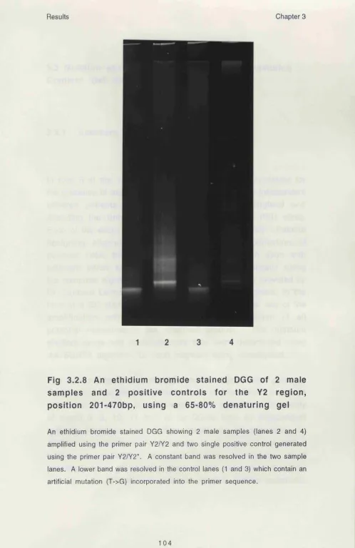
3.2.8 An ethidium bromide stained DGG of 2 male 104 samples and 2 positive controls for the Y2 region, position 201-470bp, using a 65-80% denaturing gel
PAH
3.3.1 PCR am plification of exon/intron sequences of 108 exon 3 ,5 ,1 0 , 11 and 12 of the PAH gene
3.3.2 Melting map of the sequence containing the exon 11 0 3 and splice site sequence of the PAH gene as determined by the MELT87 program
3.3.3 Melting map of the sequence containing the exon 111 3 and splice site sequence of the PAH gene determined
by the MELT87 program following the introduction of a 40 bp GC clamp at the 3' end of the sequence
3.3.4 Melting map of the sequence containing the exon 11 2 3 and splice site sequence of the PAH gene determined by the MELT87 program following the introduction of a 40 bp GC clamp at the 5'end of the sequence
3.3.5 A silver stained CDG of the two mutation types 113 identified in exon 3 using a 52% constant denaturing
3.3.6 A silver stained CDG of the two mutation types 115 identified in exon 3 using a 52% constant denaturing
gel
3.3.7 EcoRI digestion of a cloned exon 3 PCR product of 122 the PAH gene
3.3.8 A silver stained CDG of exon 3 mutation 1, 123 identified using a 52% constant dénaturant gel
3.3.9 Sequence comparison of mutated and normal 124 alleles of the exon 3 region containing mutation 1
3.3.10 Comparison of the coding region containing the 125 I65T mutation in the human, rat, mouse and drosophila PAH protein
3.3.11 Sequence comparison of mutated and normal 126 alleles of the exon 3 region containing mutation 2
3.3.12 A silver stained DGG of the three m utation 128 types identified in exon 10 using a 48-52% denaturing g ra d ie n t
3.3.13 An ethidium bromide stained CDG of the three 129 m utation types identified in exon 10 using a 49%
constant denaturing gel
3.3.14 Sequence comparison of mutated and normal 131 alleles of the exon 10 region containing mutation 1
K341R and L347F mutations in the human, rat, mouse and drosophila PAH protein
3.3.18a An ethidium bromide stained CDG of the five 136 m utation types identified in exon 11 using a 56% constant dénaturant gel
3 .3 .1 8 b A silver stained CDG of the five m utation 137 types identified in exon 11 using a 56% constant denaturing gel
3.3.19 Silver staining of a CDG of mutation 1 under 138 id e n tic a l d e n a tu rin g c o n d itio n s usin g d iffe re n t percentages of acrylamide
3 .3 .2 0 A silver stained CDG of exon 11 mutation 2 and 140 3
3.3.21 Sequence comparison of mutated and normal 141 alleles of the exon 11 region containing mutation 1
3.3.22 Sequence comparison of mutated and normal 142 alleles of the intron 10 splice site region containing mutation 2
3.3.24 Sequence comparison of mutated and normal 144 alleles of the exon 11 region containing mutation 4
3.3.25 Sequence comparison of mutated and normal 145 alleles of the exon 11 region containing mutation 5
3.3.26 A silver stained DGG of the two mutation types 147 identified in exon 12 using a 54-57% denaturing g ra d ie n t
3.3.27 CDGE analysis of the PAH exon 12 in a PKU 150 fam ily showing mendelian inheritance of m utationi
3.3.28 Sequence comparison of mutated and normal 151 alleles of the exon 12 region containing mutation 1
3.3.29 Sequence comparison of mutated and normal 152 alleles of the intron 12 splice site region containing mutation 2
3.3.30 Comparison of the coding region containing the 153 R408W m utation in the human, rat, m ouse and drosophila PAH protein
3.3.31 A silver stained CDG of exon 5 using a 55% 155 denaturing concentration
3.3.32 A silver stained SSCP gel corresponding to 1 5 7 /1 5 8 analysis of exon 3, 10, 11 and 12 of the PAH gene
1.1 Review of the historical approaches used to study 28 genetic variation in man
CHAPTER 2
2.1 Stock solutions for the preparation of 6.5% and 69 10% acrylamide DGG
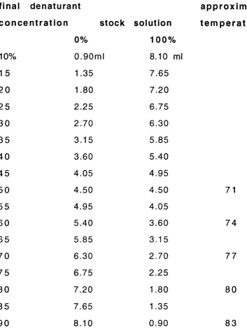
2.2 Volumes of stock solutions required to obtain the 70 appropriate gradient of chemical dénaturant in the DGG and tem p eratu res as fun ction of the d e na rura nt c o n ce n tra tio n
CHAPTER 3
3.3.1 The six common variant alleles of human AAT 80 studied in this project.
3.3.2 Distribution of the identified PAH mutations in 1 1 6 -8 50 unrelated PKU patients and close relatives
3.3.3 Distribution of 50 unrelated PKU patient samples 1 1 9 -
and their relatives 120
3.3.4 Summary of the results of the SSCP analysis for 161 the 12 different mutations identified by DGGE
CHAPTER 4
4.1 DGGE applications in Human Molecular Genetics 1 6 5 -7 4.2 A dvantages and disadvantages of scree ning 179 methods for detection of single base mutations.
4.3 D iagram m atical representation of the diffe re n t 182 types of DGGE banding patterns observed in the DGGE studies presented.
bp base pairs
°c degrees centigrade CDG Constant Denaturing Gel
CDGE Constant Denaturing Gel Electrophoresis cDNA complementary deoxyribonucleic acid DGG Denaturing Gradient Gel
DGGE Denaturing Gradient Gel Electrophoresis DNA deoxyribonucleic acid
EDTA ethylene-diam ine-tetra acetic acid FISH Fluorescent in situ hybridisation
g grams
g g ra v ity
Kb kilob ase
M m o la r
Mb Megabase
pg m icrogra m
m 1 m i l l i l i t r e p i m ic r o litr e Mins m in u te s
mRNA messenger ribonucleic acid CD optical density
PAH Phenylalanine hydroxylase PCR polymerase chain reaction PKU P henylketonuria
RFLP Restriction Fragment Length Polymorphism rpm revolutions per minute
SSCP Single strand conformation polymorphism TAE Tris acetate EDTA
TBE Tris borate EDTA
UV u ltra v io le t light
VNTR Variable number tandem repeat
provided the computer MELT87 and SQHTX algorithms
John Attwood
set up the Lerman computer program
Sue Povey, David Whitehouse
provided genomic DNA from a range of people with the most common AAT variants
Phillip Johnson
with whom the AAT studies were carried out
Michel Vidaud
offered the key to the understanding of the SQHTX algorithm
Dr D. Brenton and G. Gillett
collected the PKU blood samples
Dr L. Tyfield
A c k n o w l e d g m e n t s
Professors E. Robson and D. Hopkinson, Dr Phillip Johnson who helped me throughout the course of my studies.
I am very gratefull to Dr D. Brenton who was extrem ely supportive over the last year of my PhD, Margaret Lillburn for her dedication to the PKU patients in the Middlesex Hospital and her spontaneous help each time I needed some information Thanks also to Isabelle Chantret, Joseph Nahmias, Wendy and Twee for their support.
Introduction Chapter 1
Chapter 1 INTRODUCTION
1.1 Background
Techniques that allow the detection of genetic changes at the level of deoxyribonucleic acid (DMA) have had a major impact on our understanding of human genetic diseases, both by identifying specific mutations that result in disease, and by identifying DNA polym orphism s that are used as genetic markers in linkage stud ies.
This thesis is m ainly concerned with the evaluation of a technique. Denaturing Gradient Gel Electrophoresis (DGGE), for the analysis of mutation in DNA, for the detection of polymorphisms which might be used as genetic markers for gene mapping and also to analyse the relationships between genotype and phenotype. This general introduction will present the background relevant to these studies, thereafter the different studies will be considered sep ara tely.
1.2. Mutation
1.2.1. Structural basis
In coding sequence, single base pair changes (point m utations) can result in nonsense mutations (chain term inator m utations) and the generation of a stop codon in a translated sequence. They rep rese nt approxim ately 5% (H a rris ,1975) of all m utations. Synonymous mutations, where the nucleotide change does not result in a change in amino acid, due to degeneracy of the code, occur in approximately 25% of all mutations. Non synonymous m utations (missense mutations), where there is an amino acid substitution account for approxim ately 70% of all m utations in coding sequences. Point mutations can also occur in the region of splice site junctions or in the region of regulatory sequences of a gene.
Small deletions or insertions generally involve the loss or gain of a sm all number of nucleotides and may result in fram esh ift mutations or the generation of a mature protein containing or lacking a few amino acid residues if the mutation involves coding sequences. Such mutations can also affect the function of regulatory sequences and splicing mechanisms.
Large deletions/insertions or other rearrangments of greater than 1 m egabase (Mb) of DNA may be visualised cytog en etica lly (Grompe et al., 1993) while smaller submicroscopic changes are usually identified through the use of direct DNA analysis using restriction enzyme, gel electrophoresis, DNA transfer to filte r support (Southern blot analysis. Southern,1975) and hybridisation to specific cloned DNA segments used as probes (Vetrie at a!.,
Introduction Chapter 1
fluorescence in situ hybridization (FISH) have been used to detect m icro de letio ns, du plica tion s and rearrangem ents (C a rte r e t a l.,^ 992).
A further category of mutation, heritable unstable DNA, in v o lv e s the amplification and expansion of a simple trinucleotide repeat sequence. This expansion of a trinucleotide repeat has been observed as a mechanism underlying the seven disorders, spinal and bulbar muscular atrophy (SBMA) also known as Kennedy disease (La Spada et a/.,1991), fragile X (FX) (Yu et a/.,1 9 9 1 ), FRAXE fragile site (Knight et al., 1993), myotonic dystrophy (DM) (M a h a d e va n et a/.,1993), H u n tin g to n 's disease (HD) (The H u n tin g to n 's D isease C o lla b o ra tive R esearch G ro u p ,1993), S p in o c e re b e lla a ta x ia ( S e a l) (O rr e t a!., 1993) and de ntatorubropallidoluysian atrophy (DRPLA) (Nagafuchi et al.,
1 9 9 4 ). C linically these diseases exhibit a phenomenon called a n tic ip a tio n where increasing severity of the disease is observed in subsequent generations of an affected fam ily. M olecular analysis of the trinucleotide repeat sequences in these pedigrees p(CGG)n in FX, p(GCC)n in FRAXE, p(CTG)n in DM and p(CAG)n in HD, SBMA, DRPLA and SCA1 suggest a correlation between the size of the am plification element in the patient and the severity of the phenotype observed. These trinucleotide repeats are located in the 5' (FX) and 3' (DM) untranslated regions and are also present in the coding sequence (SBMA, HD and SC A).
1.2.2 Analysis of phenotypic variation.
bleeding disorders haemophilia A 1 9 5 3 Alexander, 1953
c e lls red cell ABO blood groups 1 9 0 0
Rhesus blood groups 1941
Landsteiner, 1900, 1901
Landsteiner and Wiener, 1941
other cells h istoco m p atibility
systems (HLA)
1 9 7 0 Teraski, 1970; Dausset, 1972
m e ta b o lite alkaptonuria
phenylalanine+related metabolites
1 9 0 0
1 9 3 5
Garrod, 1902
Penrose, 1935
p ro te in sickle cell anaemia
haemoglobin variants
serum protein and
isozyme polymorphism
1 9 4 9
1 9 6 0
Pauling, 1949
Harris and Hopkinson, 1972
genotype DNA/ molecular genetics 1 9 7 8 Kan and Dozy, 1978
Introduction Chapter 1
Mendel was the first to study the inheritance of these phenotypic changes. By creating "a rtificia l" crosses between d iffe re n t varieties of the common garden pea plant, he was able to follow the segregation of several traits in subsequent generations. From his observations, he subdivided these features into two distinct forms that he referred to as dominance and recessive. He defined dom inance by those characters which are transm itted in their entirety and therefore constitute the character of the hybrid. Those which become latent in the process, he termed recessive. In essence a recessive allele will manifest its phenotypic effect only when present in the homozygous state, in contrast to a dominant allele which need only be present as a single copy to exert its effect on the phenotype.
In human beings, obvious morphological variations for example facial features, skeletal abnormalities and some genetic diseases can be traced in families in order to determine the recessive or dom inant nature of a particular gene. Haem ophilia A in the English royal family is probably one of the best examples. In the case of these royals, the pattern of segregation suggested that haemophilia A was an X-linked recessive disease. This ability to follow a particular characteristic through a pedigree is the basis fo r linkage analysis (section 1.1 .1 .3 .) and has advanced considerably in recent years with the introduction of m olecular techniques which can lead to the identification of a disease locus from a knowledge of its chromosomal position, an application known as positional cloning or reverse genetics (Collins, 1992; Orkin, 1986).
termed A and B, the red cells could be classified into four groups A, B, AB and O. Therefore, O individuals which have no antigens, have both antibody-A and antibody-B, whilst AB persons were without such antibodies. This has become the basis for blood transfusion. Since these initial studies, about 22 blood group systems (Daniels et a/.,1993) have now been identified.
Landsteiner, suggested that the difficulties in matching for blood tra n s fu s io n w e re s im ila r to th o s e e n c o u n te re d fo r transplantation. Early work in the mouse had established that genetic differences between recipient and donor tissue affected the outcome of a graft. This led to the discovery of the antigens im portant for transplantation called the major histocom patibility system by Snell (Snell at a/.,1948). This initial work was carried out in the mouse and became an important model for studies of the Human Leucocyte Antigen (HLA) system.
G arrod in 1902 put forward the notion that there existed a relationship between the genetic constitution of an individual and his gene products, subsequently referred to as the well known one-gene-one enzyme (one polypeptide) theory. Through his work on the chemistry of the blood and other body fluids, a whole range of metabolic disorders have been identified. Among those, he de scribed alka p to n u ria which was the firs t exam ple of a recessive inherited Mendelian disorder in man. This gene has only recently been localised to human chromosome 3q (Janocha at a/., 1994). In recognition of these achievements Garrod is referred to as the father of Biochemical Genetics.
Introduction Chapter 1
of m any p la sm a p ro te in p o ly m o rp h is m s and en zym e polym orphism s in health and disease (H arris, 1975). The combination of electrophoresis and refinement strategies such as isoelectric focusing together with the developm ent of methods for the specific detection of enzymes and other proteins allow the analysis of very large range of genes in fam ilies. The detailed studies on the variant haemoglobin polypeptides and the analysis of the structural variation underlying the haptoglobin polym orphism s layed the fou nd atio n of know ledge on the structural basis of mutation which introduced this chapter. It also set the scene tow a rd the next m a jo r m e th o d ica l developments in the analysis of genetic variation at the level of the gene itself.
It is only relatively recently, that studies have reverted from the investigation of altered proteins to the development of methods for the direct analysis of the DNA. Moreover, these techniques have facilitated the search for nucleotide changes in non coding region of the genome. These new so called molecular techniques enabled DNA to be visualised directly, for example by sequencing (Sanger et a/., 1977). As a result, more accurate diagnosis and better assessments of prognosis has become possible.
The next section considers the progress of the last ten years, in particular the utilisation of genetic variation within coding and non-coding region to allow the chrom osom al assignm ent of disease loci and the range of approaches which have facilitated the identification of these genes and the characterisation of the m utations involved.
1.2.3 Analysis of genetic variation
mutations at the level of genomic DNA. At the present time changes responsible for genetic disease can be readily identified and the chrom osom al position of different traits localised by linkage analyses. The most direct and unequivocal way of identifying the m olecular changes underlying m utation is of course by DNA sequencing analysis. In the early days of molecular an alysis, direct sequencing was em ployed to confirm the predictions from protein phenotypes in human genes such as the a and B globin genes families. However this direct approach is cle arly not fea sible for the screening of large num ber of individuals and whole populations.
1.2.3.1 Utilisation of restriction enzym es.
The ability to manipulate DNA became possible with the discovery of a family of bacterial enzymes called restriction endonucleases which cleave DNA at specific nucleotide sequences (Arber et al.,
1974). Over a hundred such enzymes (Maniatis, 1991), have been isolated from various species of bacteria. W ithin bacterial cells these enzymes act to protect the cell from invading viruses by producing cuts in viral DNA. As a result, any foreign DNA can be fragmented and subjected to degradation by exonucleases. At the same tim e the chrom osom e of the cell is protected from destructio n by the a ctivitie s of ad d itio n a l enzym es, called methylases, which methylate these sites in the chromosomal DNA providing a means whereby "self" DNA can be distinguished from foreign DNA.
Introduction Chapter 1
tends to reassociate by base pairing. Initially this tendency of cohesive ends to anneal was used in vitro to join DNA from a foreign source with the DNA of a cloning vehicle, such as plasmid or phage DNA. For small viral genomes this provided a means to subdivide the DNA into smallish segments more am enable for extensive study.
Unfortunately the size of the human genome, approximately 3 X 1Q9 base pairs, prohibits its analysis in the same way. However it was possible to digest DNA into sm aller fragm ents and to fractionate these depending on their size by gel electrophoresis. Through the introduction of Southern blotting (Southern, 1975) as a method of transfering DNA onto a solid support any cloned segm ent of DNA could be readily visualised by m aking this segm ent radioactive and hybridising it to the nylon support m embrane containing the DNA. Restriction products showing homology to this region could be observed by autoradiography. Since restriction enzymes recognise specific DNA sequences, any changes in genomic DNA brought about by point mutations can lead to the loss or gain of a digestion site. Such variation in digestion patterns was first observed in the beta globin locus (Kan and Dozy, 1978) and this led to renewed efforts to identify genetic variation in the human genome.
These differences in the sizes of fragments resulting from the digestion of the corresponding region of DNA from homologous chrom osom es have been term ed restriction fragm ent length polym orphism (RFLP) (Botstein et a/.,1980). The use of more fre q u e n t cutters, fo r exam ple 4 bp cutters, increa se the likelihood of identifying RFLPs. In addition, enzymes, for example
subsequent deamination of cytosine at a CpG dinucleotide in the genome which frequently results in the replacement of 5' methyl cytosine residue with thymidine leading to a transition mutation (pyrim idine/pyrim idine) (Cooper e fa /.,1 9 8 8 ).
1.2.3.2 M inisatellites and m ic ro sateilites.
A few RFLPs exhibit great variation in their length (Whyman and W h ite , 1980; J e ffre y s et a /.,1985). These are te rm e d m inisa te llite s and consist of stretches of tandem ly repeated units of between 11 and 60 base pairs in length (Nakamura a t
a/., 1987). Jeffreys at a/. (1985) made use of a m inisatellite probe from the myoglobin gene which sim ultaneously revealed many polym orphic loci throughout the genome. The level of polymorphism was so high that it was possible to "fingerprint" an individual since the probability that two unrelated in dividuals share all fragm ents at cross-hybridising loci was negligible. A d d itio n a l va ria b le num ber tandem repeats (VNTR) w ere developed using the concensus nucleotides of some m inisatellites to identify new VNTR loci for linkage studies. Their application however was limited by their tendency to cluster towards the telomeres of human chromosomes (Royle ef a/.,1990).
Introduction Chapter 1
resource of highly informative markers for the developm ent of the human genome map. Fortunately, these m icro sa te llite s sequences are not restricted in their chromosomal position unlike m inisatellites and have become the most commonly used marker in genetic studies (Weissenbach ef a/., 1992).
The PCR, introduced by Kary M ullis et al. in 1987 has revolutionised the whole field of molecular biology. It allows the enzym atic am plification of target sequences from small amount of genom ic DNA. In the context of genetic diseases it has increased the efficiency of linkage analysis, but also it has allow ed investigators to target regions of particular interest. Gene sequencing of these regions can then be used to evaluate and id e n tify variatio n responsible for genetic diseases. A lle le specific am plification, a simple and effective extension of the PCR reaction was developed by Wu et a/.,1989. One of the pair of oligonucleotides is replaced by two oligonucleotides covering the mutated base. One of these would be perfectly matched on hybridisation and the other would create a mismatch at the 3' term inal with the same DNA. Conditions were established such that PCR product was produced in the former case and not in the latter case. This system was applied to the sickle allele of globin.
1.3 Mutation detection strategies
electrophoresis (CDGE), RNase-A mismatch cleavage method, DNA heteroduplexing have also been coupled with PCR to define the regions of the gene m utated in affected individu als, thus, directing sequencing approaches to small regions of particular interest. These strategies also allow efficient screening of a large number of individuals. Patients carrying the same mutation can be easily identified by comparing the position of aberrant DNA bands, avoiding the need for sequencing the corresponding region in every affected individual.
Naturally, the strategy used will vary on a number of parameters. In certain instances not only the sensitivity of a given approach but also its speed, sim plicity and suitability for analysing both DNA and RNA templates in addition to the limitations of available sequence and structural inform ation must be considered (see discussion 4.3).
I will now describe those techniques most widely used at the present time, in particular the DGGE which has been used predom inantly for the work presented here. These have been reviewed most recently by Cotton (1993) and Grompe (1993).
1.3.1 Denaturing Gradient Gel Electrophoresis (DGGE) and related techniques
wild-type D N A mutant D N A
hetero and
homoduplex D N A
A
I
electrophoresis in
denaturing gradient
differential
melting behaviour
w t
wt+m
low dénaturant
high dénaturant
Fig 1.1 Diagramm atical representation illustrating the
principle of DGGE (Grompe, 1993).
The figure takes as example a patient heterozygous for a particular mutation. PCR of
the target DNA results in the generation of 4 different DNA duplexes, two homo and
two heteroduplex forms. Electrophoresis, through a gel of increasing dénaturant,
results in the partial melting of the duplexes, in a sequence dependent fashion,
leading to an altered pattern of migration. The four different bands in the wt + m
biomolecules. DNA melts in domains at a temperature Tm which is directly related to the nucleotide sequence of the fragment of interest. A melting domain is a block of sequence within the fragm ent which melts cooperatively under the pressure of both high temperature and denaturing reagents.
Melting of DNA involves a transition from a tightly-packed double stranded, helical conformation to a single stranded random coil conform ation and occurs in distinct regions called dom ains. Since the melting tem perature (Tm) of a m elting domain, is directly related to the primary nucleotide sequence, a single base substitution within the region will result in a change in the melting temperature. This change can be utilised by the DGGE technique to resolve otherwise identical fragments of DNA. The coupling of PCR to DGGE allows small fragments of interest to be analysed independently.
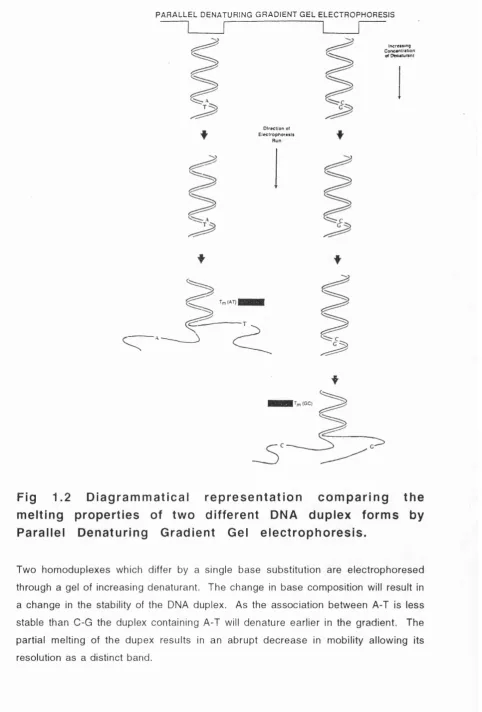
At the beginning of a denaturing gradient run, the double stranded (ds) DNA fragments run according to charge, size and shape into an increasing denaturing gradient until a point in the gradient is reached (corresponding to the Tm) at which the lowest melting domain unwinds and electrophoretic mobility is therefore reduced (Fig. 1.2). A single base pair substitution in this melting domain will alter the Tm such that the partially melted DNA focuses at a higher or lower point within the gradient.
PARALLEL DENATURING G R ADIEN T GEL ELECTROPHORESIS
Increisinq
D irection of Ele ctrophoresis
A
Fig 1.2 D ia g ra m m a tic a l re p re s e n ta tio n c o m p a rin g th e m e ltin g p ro p e rtie s of tw o d iffe re n t DNA d u p le x fo rm s by P a ra lle l D e n a tu rin g G ra d ie n t Gel e le c tro p h o re s is .
Two homoduplexes which differ by a single base substitution are electrophoresed
through a gel of increasing dénaturant. The change in base composition will result in
a change in the stability of the DNA duplex. As the association between A-T is less
stable than C-G the duplex containing A-T will denature earlier in the gradient. The
partial melting of the dupex results in an abrupt decrease in mobility allowing its
6-C
Tm G-C » Tn A > Tn B
Fig 1.3 Diagram m atical representation of the generation of a high melting domain through the incorporation of a
GC clamp at one end of the PCR product (Fodde et a/.,
1 9 9 4 ) .
Primers 1 and 2 generate a PCR product containing two separate melting domains, A
and B. The melting temperature, Tm, for domain A is greater than the TM of domain
B. Therefore mutations in the B domain cannot be resolved as the DNA duplex will
have completely dissociated. Through the addition of a GG clamp to either primer, in
this instance primer 1, an artificially high domain is generated, Tm G-C is much
greeted than the Tm of domain A or B. Therefore all potential mutations in either
Introduction Chapter 1
of all possible single base changes to close to 100% making it possible to resolve base changes anywhere within the amplified region.
DGGE resolves partially melted molecules. Strand dissociation leads to a loss of resolution. The irreversible transition due to strand separation can be shifted to higher tem peratures through the use of GC clamps. The sequence of highest sta b ility determ ines the tem perature at which the strand dissociate completely. This results in a loss in continuity of the transition curve of a perpendicular denaturing gradient gel (Fig 1.4).
C om puter analysis of the sequence of in te rest using two programs MELT87 and SQHTX (Lerman and Silverstein, 1987) enable the researcher to optim ise the conditions fo r DGGE analysis. MELT87 calculates the m elting map of a known sequence of DNA according to its nucleotide sequence. It plots the mid-melting point temperature at which each base pair is at 50% equilibrium between the helical and melted states as a fu n c tio n of its positio n along the DNA fra g m e n t. The electrophoretic conditions expected to provide optimal resolution and heteroduplex form ation are determ ined by displacem ent calculation using the SQHTX program.
Among the first genes analysed using DGGE were the mouse and human p globin genes (Sheffield et al., 1989). Since 1989, DGGE has found wide application in mutation detection including the genes for Cystic Fibrosis (Vidaud et a!., 1990; Fanen et a/., 1992; M ercier et al., 1993), hemophilia A (Kogan and Gitschier, 1990; Higuchi et al., 1991) and APC (Fodde, 1992). During the course of my studies, the DGGE technique has been applied as a mutation detection system, to a growing number of disease genes
Fig 1.4 Diagrammatical representation of perpendicular gradient gel electrophoresis.
The PCR product is electrophoresed perpendicular to a gradient of increasing
dénaturant. At the lower levels of dénaturant the product remains double stranded
until a concentration of dénaturant is reached where the fragment starts to melt.
This percentage dénaturant corresponds to the melting of the lowest melting domain.
In the example shown here the fragment contains a single melting domain. At higher
denaturing conditions the product completely dissociates into single strands and can
Introduction Chapter 1
of DGGE, initially described by Hovig et al. in 1991. It has mainly been applied to the p53 gene in human breast carcinoma and to the hypoxanthine phosphoribosyltransferase (HPRT) mutants. As with DGGE, the separation principle of CDGE is based on the melting behaviour of the DNA double helix of a given fragment. The gels used contain the same chemicals as for DGGE, but instead of a d é n a tu ra n t gra dien t, the gel is co n stitu te d of a uniform dénaturant concentration. The configuration of the molecule is constant through the gel and the resolution is a function of the distance travelled. A specific dénaturant concentration at which maximal separation between the wild-type and mutant fragments can be deduced from computer analysis using the algorithm s developed by Lerman and by direct experimentation using a set of constant dénaturant gels.
The dénaturation or melting of the double stranded DNA can also be achieved by tem perature and this m ethod is term ed temperature gradient gel electrophoresis (TGGE). As opposed to de na turin g chem ical gradients, TGGE induces no chem ical interactions with the macromolocules and creates a stable non diffusable gradient. The TGGE approach is based on the same theoretical background however as DGGE, (Riesner et al., 1989).
melting domain than the sequence containing the mutation). The m utation detection efficiency is therefore dram atically reduced to 20-60%. This might explain why this procedure has not been widely used so far. The use of different restriction enzymes in combination can overcome the poor efficiency to some extent and this modified system has been successfully used to screen for mutations (Gray ef a/.,1992; Burmeister et al., 1991, Krolewski a t a!., 1992).
1.3.2 Single strand conformation poiymorphism (SSCP).
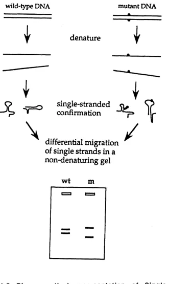
SSCP looks specifically at variation in single stranded molecules (Orita at a/., 1989). In non denaturing conditions, single-stranded DNA has a folded (tertiary) structure that is determ ined by intramolecular interactions and these in turn depend upon primary nucleotide sequence. A mutated sequence is therefore detected as a change in mobility caused by its altered folded structure (Fig 1 .5 ) . The high re s o lv in g po w e r of p o ly a c ry la m id e gel e le c tro p h o re s is m akes it p o s s ib le to d is tin g u is h m ost conform ational changes caused by subtle sequence differences such as a single base substitution in relatively sm all DNA fra g m e n t.
1.3.3 Heteroduplex analysis (HA).
wild-type DNA
mutant DNA
denature
I
I
\
single-stranded
confirmation
differential migration
of single strands in a
non-denaturing gel
I
^
I/
wt
m
Fig 1.5 Diagrammatical representation of Single Strand Comformation Poiymorphism (SSCP) (Grompe, 1993).
A single stranded molecule adopts a particular conformation as determined by its
intramolecular interactions. As the sequence of a product varies so will these
interactions result in different conformations and therefore altered mobility of the
strand. SSCP is based on the observation that these changes in conformation can have
the rhodopsin gene of autosomal dominant Retinitis Pigmentosa patients (Keen et a/.,1991) and has since been used to study a large number of additional diseases (Tassabehji at a/., 1 9 9 2 ; Artlich at a/.,1 9 9 2 ).
1.3.4 Chemical Cleavage of Nucleic Acid Heterodupiexes, also called Chemical Mismatch Cieavage (CMC).
The hydroxylam ine and osmium tetroxide chem ical cleavage method was first described by Cotton at al. in 1988. As in most other mutation detection systems the technique has been coupled with the PGR to generate target sequence (Fig 1.6).
A heteroduplex between a wildtype DNA molecule and mutant DNA (or RNA) is created by boiling and reannealing. M axam-Gilbert seq u e n cin g ch e m istry is then used to ch e m ica lly m od ify mismatched bases at the sites of mutations in the DNA-DNA or DNA-RNA heterodupiexes. Osmium tetroxide is used for the m od ifica tion of m ispaired thym idines and hydroxylam ine for mismatched cytosines. Adenosine and guanosine mismatches of the wild-type sense strand are detected by also labelling the anti-sense strand of wild-type DNA in the heteroduplex. The labelled DNA is cleaved by piperidine at the site of the m odification, and this is followed by denaturing polyacrylam ide gel electrophoresis (PAGE) and autoradiography.
heteroduplex between labeled wt and non labeled mutant strand
/
mismatch
electrophoresis detects cleaved
fragment
w ild-type mutant
full length probe
cleavage product
chemical modification o f mismatched bases by
hydroxylamine or osmium tetroxide
HA tj
m utation (mismatch)
piperidine cleaves modified mismatches
7 mismatch
Fig 1.6 D iag ram m atical re p re s e n ta tio n of chem ical
mismatch cleavage (CMC) (Grompe, 1993).
A heteroduplex between labelled (*) wild-type DNA and unlabelled mutant DNA is
shown. The mismatched base at the site of the mutation is chemically modified and
end-labeled only the fragm ent retaining the labeled end is observed.
1.3.5 RNase protection Assay: A m ism atch cle a v a g e
m e th o d
This method uses the ability of bovine pancreatic RNase to recognise and to cleave a significant percentage of single-base mismatches in RNA:RNA (Winter ef a/., 1985) or RNA:DNA (Myers et
a /.,1985) duplexes. A hom ogeneously labeled RNA probe, complementary to the gene being studied, is hybridised to cellular RNA or DNA, and the hybrids are digested with RNase A at the site of the mismatch. The resistant products are analysed by e le c tro p h o re s is in d e n a tu rin g p o ly a c ry la m id e g e ls and autoradiography. Point mutations in the gene or gene transcripts are detected by the presence of m ism atch-specific sub-bands. The size of the sub-bands is dependent on the position of the mismatches, therefore the mutations can be localised within the gene with an error of only a few nucleotides. This approach has been used in the identification of m utations in only a sm all number of genes which include the ras oncogene (W inter e t
a /.,1985), the HPRT gene (Gibbs and Caskey, 1987) and the retinoblastom a gene (Dunn et a/.,1989). However, since the introduction of PGR to generate target templates the technique in the main has been made obsolete.
1.3.6 Direct sequencing (DS)
Introduction Chapter 1
this. The asymmetric PCR method involves the reamplification of PCR product in a second reaction in w hich one of the oligonucleotide primers is in >100:1 excess of the other. This creates single-stranded PCR product which can be sequenced. The biotin method involves the biotinylation of one of the primers used fo r the PCR reaction. After the reaction, the double stranded PCR product is captured on an avidin-coated magnetic bead. The non-biotinylated strand is melted away with NaOH and the sequencing reactions are carried out on the im m obilized s in g le -s tra n d e d tem p late . The in tro d u c tio n of autom ated s e q u e n c in g using co m m e rc ia lly a v a ila b le m a ch in e s from Pharmacia and Applied Biosystems has increased the speed and efficiency of sequence analysis over conventional radioactive sequencing.
1.4 Model systems investigated.
The follow ing section briefly outlines the three model systems investigated in this study, the alpha 1 antitrypsin gene (AAT), a segment of the Y chromosome and the phenylalanine hydroxylase gene. These three model systems were used to evaluate the general applicability of DGGE for mutation detection analysis in the context of pathology and diagnosis of genetic disease and more generally in the detection of polymorphisms.
1.4.1 Alpha 1 antitrypsin (AAT).
AAT is a m a jo r p ro te in a s e in h ib ito r p re s e n t in high con cen tration s in the plasma. It is a glycoprotein w ith a m olecular w eight 52,000 which is synthesised m ainly in the liver. It inhibits a wide range of serine proteinases, although its principal target is thought to be neutrophil elastase (Heidtmann
It is associated with early onset pulmonary emphysema and a form of neonatal liver disease, both of which are associated with abnormally low levels of plasma AAT (Fagerhol et a/.,1981). The gene locus encoding AAT, called PI, is located on chromosome 14q31-32.3 (Darlington et a/.,1982) and displays high levels of g enetic polym orphism s at the protein level (W hitehouse e t
a /.,1989).
In this regard the existence of six common and well characterised point mutations (M l Ala, M l Val, M2, M3, S and Z), associated with variant protein phenotypes, made this an ideal system to evaluate the general applicability of DGGE for detection of single base pair polymorphism.
1.4.2 Y chromosome.
The Y chromosome covers 1.7% of the whole genome comprising about 5 X 1 0 ^ bp. The long arm (almost 70% of the Y chromosome) is composed of highly and moderately repeated sequences and constitutes the large heterochrom atic region. In contrast, the short arm contains the pseudoautosomal region (Y p l 1.2), which shows a high level of homology with the X chromosome. Y specific DNA probes from these regions are useful for various purposes including prenatal diagnosis of sex-linked disorders, follow ing up the host-versus graft cells in patients with bone marrow transplants from the opposite sex and analyses of sex chrom osom al disorders such as a Y /autosom e tra n slo ca tio n (Malaspina et al., 1990).
Introduction Chapter 1
4 6 ,5 1 5 n u c le o tid e s no p o ly m o rp h is m s w e re d e te c te d . Jakubicczka (1989) studied an additional 10 probes and only one polymorphism was detected. To date, similar RFLP studies have revealed polymorphisms at only 8 Y-specific loci (M alaspina e t al., 1990).
At the time of my study RFLP analysis had been the only approach used to screen for mutations on the Y chromosome. In this study, DGGE was used to look for variation in a 500 bp stretch of DNA from the Y p l 1.2-Ypter region, designated as pY-80 which had been completely characterised and had been demonstrated to be male specific (Tsukahara et a!., 1990).
Furthermore the MELT87 and SQHTX algorithms, devised by Lerman in 1987, to optimise the conditions for mutation detection using DGGE, were used prior to the practical investigations of this second model system. This also allowed me to evaluate the use of artificially mutated nucleotide primers as controls.
1.4.3 Phenylketonuria (PKU).
confirm ed an autosomal recessive transmission for the disease renam ing it PKU to reflect the high Phe level in affected individuals (P enrose,1935).
It was more than ten years later, in 1947, that the underlying biochemical pathway for PKU started to be deciphered by Jervis. He noticed that increased levels of tyrosine (Tyr) followed the ingestion of Phe in the blood of normal individuals but not in PKU patients. He confirmed these initial observations by comparing the transform ation of Phe into Tyr in post mortem liver tissue from normal and PKU affected individuals and found that normal individuals could transform Phe into Tyr, whereas this reaction was absent in PKU patients.
Udenfriend and Cooper later identified this enzymatic system as a hydroxylation step which converted Phe to Tyr (Udenfriend and Cooper, 1952). It soon became apparent, from later work by Mitoma et al. that two protein products were required to sustain Phe hydroxylation (Mitoma, 1956), a phenylalanine hydroxylase (PAH) and a pteridine reductase (BH4) (Kaufman, 1959, 1963). A diagram m atic representation of the Phe hydroxylation pathway in man, as described by Kaufman in 1976, is presented in Fig 1.7. Although a defect in either protein could be responsible for the PKU phenotype most patients lacked functioning PAH rather than pteridine reductase (Kaufman, 1976). It is now known that only about 2% of PKU cases result from the BH4 deficient form of PKU.
NM.
CH2 • CM *
L-Phenylalanine
COON
>2 HgO
Hydroxyiase
BH4
Tetrahydrobiopterin
BH2
Dihydrobiopterin
NM. I ' CM, ■ CM - COOH
L-Tyrosine
Quinonold
NADPH^-^
DIhydroblopte Reductase
NADPH + H^ rin
Fig 1.7 D ia g r a m m a t ic a l r e p r e s e n t a t i o n of the
of a neonatal screening program in 1959 in Great Britain (Gibbs and Woolf, 1959; Boyd, 1961). The first assay used was the urinary ferric chloride test originally developed by Foiling when he first described the disease. However, the discovery by Armstrong and Binkley in 1956 that excretion of phenylpyruvate is preceded by an elevation of Phe in serum led to the introduction of the Guthrie test (Guthrie, 1961; Guthrie and Susi, 1963), a bacterial inhibition assay for the detection of serum Phe levels. This gives an estimate of the frequency of PKU among Caucasians at approxim ately 1/10,000 live births corresponding to a carrier frequency of 1 in 50.
In addition to the neurological features of PKU, patients also exhibit hypopigm entation, other derm atological conditions and impaired postnatal growth among others (Scriver e fa /.,1 9 8 9 ) . Patients can be subdivided into 3 distinct groups; classical PKU, mild PKU and benign hyperphenylalaninem ia (G uttler, 1980). Classical PKU refers to the most severely affected class of hyperphenylalaninémie patients who would be profoundly retarded unless strict dietary therapy was implemented. Mild PKU refers to patients less severely affected than the previous group. They require dietery therapy but have a relaxed tolerance for Phe, r e la tiv e to th e c la s s ic a l g ro u p . F in a lly , b e n ig n h y p e rp h e n y la la n in e m ia d e s c rib e s h y p e rp h e n y la la n in é m ie individuals clinically normal without dietary therapy.
Introduction Chapter 1
exon 12 accounted for 34% of PKU alleles on a single haplotype background (Kalaydjieva et al., 1990).
Subsequently 8 RFLPs have been identified in or near the human PAH locus. Since these RFLPs are tightly linked to the PAH gene, they have been used to follow the transm ission of normal or m utant chrom osom es in PKU fam ilies (Lidsky et a!., 1985). Although over 70 haplotype patterns have been identified, most PKU chrom osom es are associated with haplotypes 1,2,3,4 in Caucasians populations. The application of haplotype analysis permitted the development of prenatal diagnosis of PKU (Riess e t
a /.,1987). This approach however has only limited application, for example prenatal diagnosis using RFLP analysis can only be provided to those fam ilies with a prior incidence of PKU. Unfortunately, over 95% of new cases of PKU are the result of random mating between previously undiagnosed carriers without a fam ily history and thus are undetectable by this kind of haplotype analysis.
To overcome these lim itations, the specific m olecular lesions responsible for PKU must be identified and characterised in each major population group, to allow more rapid screening of carrier individuals. The introduction of PGR coupled mutation detection systems (section 1.1.1.3 ) allowed more detailed analysis of the PAH gene using chemical cleavage mismatch (Cotton e fa /.,1 9 9 0 ) and sin g le strand co n fo rm a tio n p o lym o rp h ism (D o ckh o rn - Dworniczak et a!., 1991). By 1991, thirty one PAH mutations had been identified in PKU patients, spread over all 13 exons of the gene (Konecki et a/., 1991). The study also confirmed previous haplotype analyses which suggested that certain m utations predominate in a given population.
high degree of accuracy to screen an affected population. The objective of this study was to evaluate the DGGE technique as a rapid and reproducible method to identify and estim ate the frequency of PKU mutations in a South East of England affected population. It was anticipated that this would confirm previous observations that the phenotypic heterogeneity observed in PKU reflected an underlying heterogeneity of the PAH genotype and would help define the correlation between a pa rticu la r PKU mutation and its phenotype. In addition it was hoped that through a knowledge of the genotype of an individual the various levels of response of individuals to sim ilar diets could also be explained. Moreover screening services could be made available to fam ilies for prenatal diagnosis or carrier detection of the disease.
1.5 Aims and objectives.
Success in the isolation and characterisation of human genes responsible for genetic diseases coupled with the considerable m olecular he terogeneity at these loci, has placed grow ing emphasis on the im portance of mutation detection techniques both in coding and non-coding regions.
This thesis is concerned with the evaluation of one of these techniques : Denaturing Gradient Gel Electrophoresis (DGGE). This approach is tested in three model system s 1. the well characterised human alpha -1 - antitrypsin (AAT) gene, with a range of known mutations, 2. a segment of the Y chromosome w here there was no evidence fo r m uta tion s and 3. the ph en yla la nin e hydroxylase (PAH) locus w here co n sid e ra b le heterogeneity has been encountered in fam ilies affected with phenylketonuria (PKU).
Introduction Chapter 1
point mutations associated with variant protein phenotypes. The approach was largely experimental and involved a combination of perpendicular and parallel DGGE.
The computer algorithms designed by Lerman et al., 1987 were used to optimise the conditions for DGGE screening using the second model system, a segm ent from the Y chrom osom e designated as pY-80. The approach was used to screen for mutations across this interval.
Chapter 2 MATERIALS AND METHODS
2.1 Materials and Suppliers
Unless otherwise stated, solutions, plastic or glassware were sterilised by autoclaving at 15psi, 121^0 for 20-25 min.
G la s s w a r e
Standard laboratory glassware was washed in detergent and rinsed in double distilled water prior to autoclaving.
DGGE glass plates were purchased from Hoefer S cientific In stru m e n ts.
P l a s t i c w a r e
Disposable 50ml sterile screwtop tubes were purchased from Falcon (referred to as Falcon tubes). 1.5 ml microcentrifuge tubes (MCC) and 0.5 ml microcentrifuge tubes were purchased from Elkay and blue (1ml) and yellow (200|liI) pipette tips from Algen.
DGGE plastic clamps and cams were purchased from Hoefer S cie n tific Instrum ents.
General Reagents
General laboratory reagents were of analytical grade and were purchased from BDH unless specified below
Form am ide Sigma Chemical Co. Ltd
Materials and Methods Chapter 2
Xgal Agarose 3MM paper D etergent
(RBS 35 special glassware) Analytical Grade
Mixed Bed Resin
Bethesda Research Lab Sigma Chemical Co. Ltd Whatman
Pierce Biorad
R a d io n u c le o tid e s
[35s] dATPaS Amersham
Sequencing Reagents
A c ry la m id e
Ammonium persulphate Boric acid
N ,N ',N ',N '-te tra -
m e th yle th yle n e d ia m in e (TEMED)
Urea
Kochlight Ltd. Kochlight Ltd.
Sigma Chemical Co. Ltd
Sigma Chemical Co. Ltd Sigma Chemical Co. Ltd
Enzym es
Restriction enzymes
(EcoRI) BRL
K i t s
TA cloning kit
Sequencing version 2.0 kit
In v itro g e n United States
Biochemical Corporation
Taq polymerase
10 X buffer N ucleotides Mineral oil G lycero l
O lig on ucleo tides
Media Reagents
Bioexcellence, Anglian, Applied Biosystems, Promega
Applied Biosystems, Promega Pharm acia
BDH BDH
Oswells, or in house Applied Biosystems synthesiser
A g ar
A m p ic illin Kanamycin Trypton e Yeast Extract Glucose
D ifco
Sigma Chemical Co. Ltd Sigma Chemical Co. Ltd Oxoid Ltd.
Oxoid Ltd. BDH
General Solutions
TE buffer: lOmM Tris-HCI pH7.5, 0.1 mM sodium EDTA pH8.0
10 X loading buffer: 25% (w/v) ficoll (Mr 400kD), 0.25% (w/v) Orange G, 0.25M EDTA in 1 X TAE buffer
10 X TAE: 0.4M Tris-acetate, lOmM sodium EDTA, pH 8.0
10 X TBE: 90mM Tris borate, lOmM sodium EDTA, pH 8.3
DGGE Reagents
Acrylam ide/Bis 37.5: 1
Ammonium persulphate (APS) N ,N ',N ',N 'te tra
-Biorad
Materials and Methods Chapter 2
methylethylenediamine (TEMED) Sigma Chemical Co. Ltd
Urea Sigma Chemical Co. Ltd
Formamide Sigma Chemical Co. Ltd
Analytical grade mixed bed resin Biorad
Solutions used for DGGE and gel analysis
20 X TAE : SOOmM Tris-Hcl, 400mM Na Acetate, 20mM Na2 EDTA, pH 7,4 adjust with Acetic Acid
DGGE loading buffer: 20% ficoll, 10mM Tris/pH 7.8, 1 mM EDTA, 0.5% orange G, 0.5% xylene cyanol, 0.5% bromophenol blue
Media and solutions for microbiological manipulation
LB broth: (per 100ml) Bactotryptone 1g, Bacto-yeast extract 0.5g, NaCI 1g, Glucose 0.1 g
LB agar: (per 100ml) Bactotryptone 1g, Bacto-yeast extract 0.5g, NaCI 1g, 1.5g Bactor-agar
Cell resuspension solution : 50mM Tris-HCI pH 7.5, lOmM EDTA, lOOjLig/ml RNase A
Cell lysis solution 0.2M NaOH, 1% (w/v) SDS made fresh
Neutralisation solution : 2.55M, potassium acetate pH 4.8
Column Wash solution: 200mM NaCI, 20mM Tris-Hcl, pH7.5, 5mM EDTA (diluted 1:1 with 95% EtOH)
TE buffer: lOmM Tris-HCI, pH7, Im M EDTA