on osteoarthritic knee pain:
a randomised controlled trial
Henry Pollard, BSc, Grad Dip Chiro, Grad Dip AppSc, MSportSc, PhD
1*
Graham Ward, BSc, BE (Sc) MSc (hons) Mass, PhD
2Wayne Hoskins, B Chiro Sc
1Katie Hardy, BAppSci (Ex&SpSci)
1Background: Knee osteoarthritis is a highly prevalent
condition with a significant socioeconomic burden to society. It is known to effect sufferers through pain, loss of function and changes in health related quality of life. Management typically involves pharmacologic and/or exercise based therapy approaches to reduce pain. Previous studies have shown multimodal treatment approaches incorporating manual therapy to be efficacious. The aim of this study is to determine if a manual therapy technique knee protocol can alter the self reported pain experienced by a group of chronic knee osteoarthritis sufferers in a randomised controlled trial.
Methods: 43 participants with a chronic,
non-progressive history of osteoarthritic knee pain, aged between 47 and 70 years were randomly allocated following a screening procedure to an intervention group (n=26; 18 men and 8 women, mean age 56.5 years) or a control group (n=17; 11 men and 6 women, mean age 54.6 years). Participants were matched for present knee pain intensity measured on a visual analogue scale. The intervention consisted of the Macquarie Injury Management Group Knee Protocol whilst the control involved a non-forceful manual contact to the knee followed by interferential therapy set at zero.
Participants received three treatments per week for two consecutive weeks with a follow up immediately after the final treatment. Post-treatment Participants completed 11 questions including present knee pain intensity and feedback regarding their response to treatment utilizing
1 Macquarie Injury Management Group, Department of Health and Chiropractic, Macquarie University, NSW 2109, Australia. 2 Faculty of Health & Behavioural Sciences, Wollongong University, NSW 2522, Australia.
* Please address all correspondence to: Dr Henry Pollard, Macquarie Injury Management Group, c/o PO Box 448, Cronulla NSW, 2230 Australia. Email addresses: HP: [email protected] GW: [email protected] WH: [email protected]
KH: [email protected] © JCCA 2008.
Antécédents : Arthrose du genou ou gonarthrose est
une condition très présente, ce qui constitue un poids socio-économique important pour la société. On sait qu’elle affecte les personnes qui en souffrent, en leur infligeant des douleurs, des pertes de motricité et des atteintes à leur santé, en plus de s’attaquer à leur qualité de vie. La gestion du cas fait d’habitude appel à la pharmacologie et/ou à l’exercice, fondée sur des approches thérapeutiques pour atténuer la douleur. Des études antérieures ont démontré qu’une méthode de traitement combinée, y compris une thérapie manuelle, s’avérait efficace. L’objectif de la présente étude consiste à vérifier, dans un essai clinique comparatif randomisé, si une technique de thérapie manuelle, appliquée au protocole de traitement du genou, peut atténuer la douleur dont fait état un groupe de patients souffrant de gonarthrose chronique.
Méthode : 43 participants, âgés de 47 à 70 ans et
a visual analogue scale. Results were analysed using descriptive statistics.
Results: Prior to the intervention, there was no
significant differences in age or present knee pain intensity. Following treatment, the intervention group reported a significant decrease in the present pain severity (mean 1.9) when compared to the control group (mean 3.1). Response to treatment questions indicated that compared to the control group, the intervention group felt the intervention had helped them (intervention mean 7.0; control mean 3.4), felt it decreased their knee symptoms such as crepitus (intervention mean 6.0; control mean 3.4) and improved their knee mobility (intervention mean 6.4; control mean 3.4) and their ability to perform general activities (intervention mean 6.5; control mean 3.8). Importantly the MIMG Knee Protocol intervention group reported no adverse reactions during treatment.
Conclusions: A short-term manual therapy knee
protocol significantly reduced pain suffered by
participants with osteoarthritic knee pain and resulted in improvements in self-reported knee function immediately after the end of the 2 week treatment period.
(JCCA 2008; 52(4):229–242)
k e y wo r d s : chiropractic, musculoskeletal
manipulation, manual therapy, knee, pain, osteoarthritis, clinical trial
consistait en un contact manuel non énergique au genou, suivi par une thérapie interférentielle établie à zéro. Les participants ont reçu trois traitements par semaine pendant deux semaines consécutives, puis un suivi tout de suite après la fin du traitement. Après le traitement, les participants ont rempli un formulaire comptant 11 questions, dont une sur l’intensité de la douleur qu’ils ressentaient au moment de fournir leurs réponses et leurs commentaires sur leur réaction au traitement faisant appel à l’Échelle visuelle analogue. Les résultats ont été analysés en utilisant la grille de statistiques descriptives.
Résultats : Avant l’intervention, il n’y avait pas de
différence entre les âges et l’intensité de la douleur aux genoux. Après le traitement, le groupe d’intervention a rapporté une réduction importante de l’intensité de la douleur (moyenne de 1,9) par comparaison au groupe témoin (moyenne de 3,1). Les réponses aux questions sur le traitement indiquent que, par comparaison au groupe de contrôle, le groupe d’intervention a senti que le traitement avait fait du bien (moyenne du groupe d’intervention 7,0 ; groupe de contrôle, 3.4),a perçu une réduction des symptômes aux genoux, la crépitation articulaire, (moyenne du groupe d’intervention 6,0; moyenne du groupe de contrôle 3,4) et a amélioré la motricité de leurs genoux (moyenne d’intervention 6,4; groupe de contrôle 3,4) et leur capacité d’effectuer des activités générales (moyenne du groupe d’intervention 6,5; groupe de contrôle 3,8). Il est important de souligner que le Groupe d’intervention du protocole du genou MIMG a rapporté qu’aucune réaction indésirable ne s’était manifestée après le traitement.
Conclusions : Un protocole de thérapie manuelle du
genou a permis de réduire de manière importante la douleur pour les participants souffrant de gonarthrose et s’est traduit par l’amélioration de la motricité des genoux chez les participants, immédiatement à la fin des deux semaines de traitement.
(JACC 2008; 52(4):229–242)
Background
Osteoarthritis (OA) is one of the most prevalent articular disorders affecting humankind and a major cause of disa-bility and socioeconomic burden.1,2 The increasing impact
of such disorders on patients and healthcare systems has seen the designation of the Decade of Bone and Joint from 2000 to 2010.3 OA is a chronic degenerative disorder of
multifactorial aetiology, including acute and/or chronic insults from normal wear and tear, age, obesity, and joint injury.4,5 The true pathogenesis remains poorly
under-stood.1 OA is characterized by degradation of the articular
cartilage, resulting in an alteration of its biomechanical properties.6 This contributes to a focal loss of articular
cartilage, loss of joint space, osteophyte formation, focal areas of synovitis, periarticular bone remodelling and subchondral cysts.7 Evidence of knee osteoarthritic
change on radiographs increases with age8 and has been
found in 72.1% of symptomatic participants and 41.6% of asymptomatic participants aged 40 or older.9 However,
there is a low level of agreement between examiners in de-termining the degree of knee osteoarthritic change on radiographs10 and considerable variability in determining
the progression of OA radiographically.11 Furthermore,
evidence of radiological OA is not an accurate predictor of pain or disability.12,13 Radiological evaluation of knee
os-teoarthritis is of limited ability as a guide for management in most cases and it falls to more subjective measures of pain and disability to guide clinical practice.
At the knee joint, soft tissue changes can include de-creases in the strength of the quadriceps and sagittal range of motion, as well as increased soft tissue contrac-ture.14 Collectively these changes produce the typical
clinical picture of joint pain; worsening symptoms with activity and weight bearing, and stiffness developing at rest. These facilitate the decline in physical function and progression of disability.7 If advanced, OA may
ultimate-ly require total knee arthroplasty, a management option that is under scrutiny to evaluate its cost-effectiveness, particularly considering the revision rate15 and the
sub-stantial costs involved.16
The knee joint, along with other major weight bearing joints including joints of the spine and hip, are commonly subject to degenerative changes17. There is a higher
prev-alence of OA with advanced age18 and in females.18,19 In
fact, most knee pain in the elderly is due to OA.20 Knee
os-teoarthritis produces significant changes in health-related
quality of life, particularly physical, mental and social components of health.21,22 Determining accurate
preva-lence and incidence rates of knee osteoarthritis is difficult due to the lack of homogeneity in published studies.18
Fig-ures regarding prevalence of symptomatic knee osteoar-thritis in the general population vary, with estimates of 7.2% in those aged 40 or older,9 12.5% in those aged over
4523 and 14.8% in those aged 50 or older.24 OA in young
adults is most commonly a result of a specific injury to the knee, particularly intra-articular injury involving the ante-rior cruciate ligament (ACL).25 Ten years after ACL
inju-ry approximately half of all patients display clinical signs of knee osteoarthritis and extrapolating these results indi-cates that nearly all patients will have OA after 15–20 years.26 These figures appear regardless of whether
recon-structive surgery is performed.27 Former Finnish world
class athletes were found to have an increased prevalence of musculoskeletal disorders than the normal popula-tion.28 Swedish soccer and ice hockey players reported a
significant relationship with the presence of osteoarthritis, but only with previous knee injurues.29 However in
Aus-tralia, a significantly greater prevalence and severity of knee osteoarthritis, producing a twofold increased risk of knee replacement, was found in Australian Rules Football players.30 Occupational stresses including prolonged
kneeling and/or squatting and lifting may also increase the risk of knee osteoarthritis31.
The treatment of knee osteoarthritis is currently limit-ed to the management of symptoms rather than rlimit-educing disease progression.1 An evidence based approach to
management should include patient education about OA and its management, including pain management, options to improve function, decrease disability, and prevent or retard progression of the disease.32 Common current
treatment strategies involve pharmacological treatments, non-pharmacological treatments and surgical interven-tions. Analgesic and anti-inflammatory drugs are widely used in management,33 despite known serious adverse
ef-fects associated with long term NSAID use34 and doubts
about their efficacy.35 Paracetamol is the primary oral
an-algesic and, if successful, the preferred long term analge-sic.32 NSAIDs are considered in patients unresponsive to
paracetamol.32 Current best evidence suggests NSAIDs
may be beneficial in the reduction of pain in the short term, but there is no support for their long term use.36
inflam-mation and pain relief, however the short term pain re-duction provides relatively short lived benefits, and no difference in knee function is evident long-term.37
Intra-articular corticosteroids are indicated for flare up of knee pain, especially if accompanied with effusion.32 Recent
times has seen the advancement of alternative so-called ‘natural’ pharmaceutical options such as glucosamine and chondroitin.38 Supplementation use is supported by a
growing, but heterogeneous research base of mixed meth-odological quality than other pharmaceutical interven-tions.32 It has been demonstrated that these products have
a slower onset of action but their symptomatic effects tend to be more long lasting after the end of treatment.39
Invasive interventions may include arthroscopy and joint replacement surgery that are considered when other treat-ment modalities have failed and for patients who general-ly have more severe pain and disability with radiographic evidence of OA.32 In a randomised placebo-controlled
trial the outcomes after arthroscopic lavage or arthro-scopic debridement were no better than those after a pla-cebo procedure and at no point did either of the intervention groups report less pain or better function than the placebo group.40 Alternatively, replacement
sur-gery is considered an effective procedure in improving knee function, decreased pain, and may provide the op-portunity to resume a more active lifestyle.41
Whilst these forms of therapy help to deal with symp-toms, osteoarthritis is often viewed as a problem of biome-chanical function. In order to treat the large and growing number of sufferers, various treatment approaches outside the use of drugs are utilised. Thus, many sufferers visit practitioners who provide therapy intended to improve their function. To address the concerns of lost function, in-cluding the ability to ambulate, several forms of physical therapy have been advocated, with various strength-based and exercise programs the cornerstone of treatment. Pre-scription of an aerobic walking and quadriceps strength-ening exercise program had been used successfully, producing a reduction in both pain and disability.42 The
implementation of laterally wedged shoe orthotics has also been shown to provide symptomatic relief.43 Such
in-terventions are typically used in combination with phar-maceutical interventions.
A requirement also exists for simple and inexpensive treatment protocols to fill the void between medication, exercise and surgery. Multimodal approaches utilizing a
combination of exercises and individualized manual ther-apy (received twice weekly for 4 weeks) has resulted in significant improvements in knee pain and function when compared to a placebo therapy of sub-therapeutic ultra-sound in both the short term and long term follow up.44
Another trial compared clinic based treatment incorporat-ing supervised exercise, individualized manual therapy and a home exercise program over a four week period to a home exercise program.45 The results indicated that in
both group’s knee pain decreased and function improved in the short and long term. Another randomised control-led trial investigated high velocity thrust techniques (re-ceived 8 times over 3 weeks) to the knee compared with NSAIDs. They found no objective or subjective differ-ences between the groups; both were equally effective.46
Therefore, use of manual therapy should be offered as an alternative to pharmaceutical administrations.
Recently, there has been interest in research of the clinical efficacy of chiropractic manual therapy tech-niques for spinal structures.47 Whilst this interest is both
appropriate and desirable, much less attention has been focused upon chiropractic interventions directed towards peripheral joints. The application of chiropractic knee techniques has been previously documented in the litera-ture.46,48 Furthermore, little research has been directed
into chiropractic interventions for the aging population. The aim of this investigation was to determine if a/the Macquarie Injury Management Group (MIMG) knee pro-tocol can alter the self reported pain experienced by a group of chronic knee osteoarthritis sufferers compared to a control group in a randomised trial.
Methods
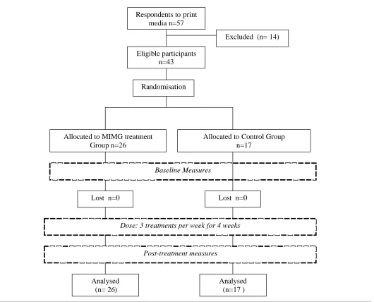
This study sought and received approval from the Mac-quarie University and the University of Wollongong Human Ethics Committees. Participants gave written in-formed consent prior to participation in the study. A CON-SORT diagram is provided for your reference (Figure 1).
Participants
Eligible participants n=43
Excluded (n= 14)
Randomisation
Allocated to MIMG treatment Group n=26
Allocated to Control Group n=17
Lost n=0
Analysed (n= 26)
Analysed (n=17 )
Baseline Measures
Lost n=0
Post-treatment measures
Respondents to print media n=57
Dose: 3 treatments per week for 4 weeks
were excluded as they experienced significant concurrent pain in the lower limb, 1 participant was excluded as they demonstrated significant varus deformity and one partici-pants was excluded as they suffered a concurrent golden staff infection in the lower limb. It was not investigated whether participants were currently undertaking concur-rent treatment or supplementation. Participants then com-pleted a knee pain questionnaire representing the present pain intensity on a graduated 10 centimetre rule, or visual analogue scale (VAS). The participants then drew a card
Table 1 Inclusion criteria Inclusion Criteria
Participants must be aged between 45 and 70 years and must suffer the following:
– A prior medical diagnosis of osteoarthritis in the knee(s) as per Forman et al (1983)
– Self reported mild to moderate knee pain of at least one year duration
– Self reported knee crepitus
– Self reported restricted range of motion and/or joint deformity of the knee
– No history of joint replacement therapy
– No recent history of meniscal or other knee surgery (less than 6 months)
Intervention Group
The intervention group received a MIMG chiropractic knee protocol, explained in Figures 2 and 3. It consists of a non-invasive myofascial mobilisation procedure and an impulse thrust procedure performed on the symptomatic knee of participants. It cases were OA was bilateral; mo-bilisation was perform on both knees. The momo-bilisation procedure directed a small, sustained load and specific force to the patellofemoral articulation in a pre-deter-mined direction of movement. This load was achieved through the active extension and flexion of the knee in the range starting from 90° of knee flexion to available full extension. During this movement, the patella is ac-tively mobilised in a supero-inferior direction in a plane directed tangentially to the patella. In this position, mini-mal compressive load is placed upon the patellofemoral articulation, as this movement is usually perceived as
painful in osteoarthritic patients. This allows the subject to actively articulate through knee flexion and not exces-sively tighten the quadriceps to cause a vector that com-presses the patella onto the femur. A positive orthopaedic test finding is pain reproduction upon compressing patel-lofemoral structures. The mobilization procedure stretch-es the joint capsule in the sagittal plane, gently mobilisstretch-es any restriction to normal movement within the limits of patient tolerance and likely loosens adhesions of the pa-tellofemoral articulation. In addition, it may be used on anterior thigh musculature to effectively mobilise tight myofascial thigh structures.
Control Group
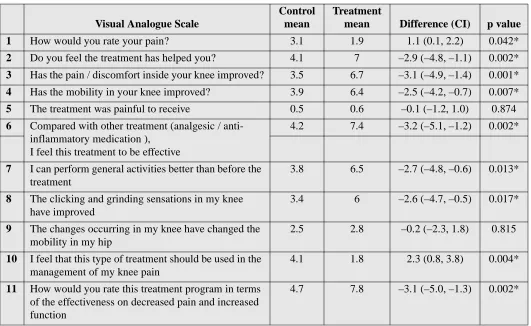
The control intervention consisted of a palmar contact to the knee without the application of force followed by in-terferential set at zero. The control group were told that Table 2 Change in 11 post study questions utilizing the visual analog scale
Visual Analogue Scale
Control mean
Treatment
mean Difference (CI) p value
1 How would you rate your pain? 3.1 1.9 1.1 (0.1, 2.2) 0.042*
2 Do you feel the treatment has helped you? 4.1 7 –2.9 (–4.8, –1.1) 0.002*
3 Has the pain / discomfort inside your knee improved? 3.5 6.7 –3.1 (–4.9, –1.4) 0.001*
4 Has the mobility in your knee improved? 3.9 6.4 –2.5 (–4.2, –0.7) 0.007*
5 The treatment was painful to receive 0.5 0.6 –0.1 (–1.2, 1.0) 0.874
6 Compared with other treatment (analgesic / anti-inflammatory medication ),
I feel this treatment to be effective
4.2 7.4 –3.2 (–5.1, –1.2) 0.002*
7 I can perform general activities better than before the treatment
3.8 6.5 –2.7 (–4.8, –0.6) 0.013*
8 The clicking and grinding sensations in my knee have improved
3.4 6 –2.6 (–4.7, –0.5) 0.017*
9 The changes occurring in my knee have changed the mobility in my hip
2.5 2.8 –0.2 (–2.3, 1.8) 0.815
10 I feel that this type of treatment should be used in the management of my knee pain
4.1 1.8 2.3 (0.8, 3.8) 0.004*
11 How would you rate this treatment program in terms of the effectiveness on decreased pain and increased function
the procedure was a micro current application that they should not be able to feel. The experimental protocol was performed so that participants were not aware to which group they were assigned. The participants were in-formed that one treatment might be more effective than another. The treatment regime consisted of 3 treatments per week for 2 consecutive weeks with a follow-up as-sessment after the final treatment.
Immediately following their involvement in the 2 week trial, participants completed 11 post treatment questions including present knee pain intensity and ques-tions regarding feedback on their response to treatment utilising a VAS. This scale was utilised as per previous researchers.49 The 11 short questions required a response
of between 0 and 10 on a 10 centimetre rule, and can be seen in Table 2. The minimum or zero point response on the VAS represented the response: none (Question 1), no effect (Questions 2, 10), no improvement (Questions 3, 4, 8), not painful (Question 5), not effective (Question 6, 11), and no change (Questions 7, 9). The 10 or maximum response on the VAS represented the following respons-es: unbearable (Questions 1,5), very effective (Questions 2, 6, 11), excellent improvement (Questions 3, 4, 8), much better (Questions 7, 9), and strongly disagree (Question 10). Gallagher reports a 13 mm difference on the VAS represents the smallest measurable change in pain severity that is clinically important.50
A post-intervention session was held after all the re-sults had been collected and the rere-sults tabulated. Partici-pants in the control group were offered the treatment program, of which all participants accepted but one.
Statistical Analysis
Statistical data was entered into power Macintosh com-puter, and utilised via a database soft ware package. Sta-tistical analysis utilised Minitab v8.2. Repeated ANOVA calculations were made to describe differences between the groups. The p value used for all analyses was p>0.05. Results were found to be statistically significant at the 5% level.
Results
Participants were randomly assigned to the intervention group (mean age 56.5 years) or a control group (mean age 54.6 years). Prior to the intervention no significant difference in present intensity knee pain between the in-tervention and control groups was evident (Table 3). It was a requirement that the participants had mild to mod-erate knee pain (as determined by a the McGill Pain Questionnaire). Following treatment the intervention group rated their pain less (1.9) while no change was not-ed in the control group (3.1) (Table 4). This change in pain in the intervention group was statistically significant when compared with the control (Table 3).
The results to the remaining 10 questions can be found in Table 2. When the participants were asked if the treat-ment helped them, the intervention group indicated a pos-itive response (7.0), which was significant when compared with the control group (4.1). Furthermore, when participants were asked if pain within the knee had improved, the intervention group (3.5) had significantly improved when compared with the control group (6.7). The participants were asked if a general improvement in Table 3 Changes in group pain scores between the control and treatment groups
Table 4 Changes between control group and treatment in pain scores
VAS n Pre-Test Mean (CI) Post-Test Mean (CI) p value
Control Group 17 3.5 (2.2, 4.7) 3.1 (2.1, 4.1) 0.602
Treatment Group 26 3.3 (2.6, 4.0) 1.9 (1.3, 2.6) 0.004*
VAS Difference (CI) p value
Pre-Test 0.2 (–1.1, 1.5) 0.771
knee mobility was noted since the treatment had begun (Question 4). The responses indicate a significant im-provement in the intervention group (6.4) greater than the control group (3.9). When asked if the clicking and grinding sensations (crepitus) in the knee had changed (Question 8), the intervention group (6.0) indicated a sig-nificant improvement when compared with the control group (3.4). The intervention group (6.5) also indicated a significantly improved ability to perform general activi-ties (Question 7) when compared to the control group (3.8). When asked to comment on whether their hip movement had been improved by the knee treatment (Question 4), the results indicated significantly improved mobility in the intervention group (6.4) when compared to the control group (3.9).
Following these questions several other questions were asked regarding the type of treatment that the participant received. When asked if the treatment was painful to re-ceive (Question 5) the participants’ responses indicated that little discomfort was experienced with the treatment; the results were similar for both the intervention group (0.6) and the control group (0.5). When asked to compare the short-term effect of their treatment to previous phar-macologic based prescriptions they had received (includ-ing analgesics and anti-inflammatory medication) (Question 6), the results demonstrated a significant sub-jective feeling of effectiveness for the intervention group (7.4) when compared to the control group (4.2). When asked if the treatment that they received should be includ-ed into the management protocol of their knee pain (Question 10), the results demonstrated a significant dif-ference between groups. Those in the intervention group (1.8) felt strongly that the management that they had received should be included in the management of “ar-thritis,” but the control group (4.1) were somewhat un-equivocal. Finally, the participants were asked to rate the treatment they received in terms of the effectiveness on decreased pain and increased function (Question 11). Again, the intervention group (7.8) rated the treatment as being more effective when compared to the equivocal re-sult of the control group (4.6).
Discussion
The results indicated that a MIMG knee protocol was successful in reducing self reported present intensity os-teoarthritic knee pain in the short-term and that this
change was statistically significant when compared with a control group. It is unlikely that the results for the inter-vention group can be explained in terms of a spontaneous remission or through natural resolution, as it was a re-quirement of the study for the knee pain to have been a chronic stable condition.
Research into arthritis and particularly OA has largely investigated medical interventions and physical therapy modalities including exercise. Much less emphasis has been placed on other manual therapy approaches. Several studies have investigated manual therapy for OA of the knee.44,45 employing protocols that included other forms
of therapy in a multi-modal approach. Our particular study employed one manual therapy discipline for effec-tive pain reduction in osteoarthritic knee patients.
An important consideration revealed in the post treat-ment questionnaire was the issue of pain and discomfort created by the treatment. Whilst concern may surround the use of manual therapy in the elderly,51 or in
degenera-tive cases, it is understood there are a range of chiroprac-tic methods suitable for certain patients and specific scenarios.52–55 Our results indicate that the treatment
caused little or no discomfort to the patients. Such find-ings are valuable as participant’s ages ranged from 47 to 70 years old. Whilst practitioner precaution is advised in dealing with patient conditions related to bone weakness, ligamentous laxity, deformity and tumour, much can be offered to the individual that has good bony and ligamen-tous integrity that also happens to suffer from osteoarthri-tis of the knee.
the procedure or at any time during the experimentation, meaning it is performed voluntary within their tolerance levels. This is an important first step in determining the limit to which force is used in the application of the man-ual therapy. It provides direct feedback to the practitioner about the degree of stiffness, limitation and pain present in the afflicted knee. The MIMG technique is a potentially useful addition to prehabilitation programs (rehabilitation aimed at improving range of motion, strength and reduc-ing swellreduc-ing prior to surgery). Of the conditions to which this procedure has been applied, only the leg with a marked degree of lateral instability (genu valgus or genu varus), or acute meniscal lesions seemed not to tolerate it. It has become a useful addition to many techniques often used to treat knee dysfunction.
The second part of the procedure utilises a manual therapy procedure that is not under the voluntary control of the patient. It involves the application of a longitudinal traction of the tibio-femoral joint in a manner designed to distract the knee and mobilise the joint in a near full ex-tension position. An impulse type thrust directed in the caudal direction is delivered to the knee of the patient. The leg of the patient is held in a position of light traction with the hands of the practitioner placed either side of the knee with the thumbs contacting on the tibial tuberosity and the fingers wrapping around the knee to the popliteal space. In addition to the above placement, the practi-tioner may optionally enhance the leverage available by placing the involved leg of the patient between the practi-tioner’s legs (at the level of the lower calf) in order to add further traction leverage. The object of this procedure is not to produce joint cavitation, more so to mobilise the joint. In cases of tibial rotational restriction, the pre-ma-nipulative set up could include a rotated tibia as a start point. The thrust component remains the same and is di-rected purely caudal in direction. Done correctly, this procedure is painless and has been used anecdotally to treat chronic meniscal injury. However, this procedure requires intact ligamentous and capsular structures to op-erate successfully. It also requires practice by the practi-tioner to acquire the motor skills necessary to perform the procedure.
Of interest to clinicians and patients alike, a significant treatment effect was found after only a short course of treatment. The study consisted of 3 treatments per week for 2 consecutive weeks, a total of 6 treatments that
pro-duced significant self-reported pain and dysfunction. Pre-vious studies have attempted to estimate the relationship between dosage and outcome parameters for low back pain56, headache57 and fibromyalgia.58 They found
be-tween 9–12 chiropractic treatments were feasible for pain relief and between 15 and 30 for quality of sleep and fa-tigue level. Further research should implement dosage characteristics of treatment modalities for improvements in valid and reliable measurement outcomes. This would hasten the transfer of information from researcher to the clinician.
The importance of the patellofemoral compartment in knee dysfunction and knee osteoarthritis is well estab-lished.59,60 Disease of the patellofemoral articulation can
cause pain, and be responsible for a great deal of difficul-ty in the everyday activities of squatting, using steps and stairs, kneeling, and rising up from chairs.61
Misalign-ment of the patella laterally has been proposed as a cause of the much of the pain associated with many patello-femoral conditions.62 These misalignment syndromes are
often referred to as “tracking” problems63 and are
classi-cally managed by physiotherapists through taping based protocols of the patella to correct the tracking problem.64
However, such protocols for knee osteoarthritis have shown it be no more effective than placebo in a ran-domised, double blind, placebo controlled trial.65
Preliminary findings of this study promote future re-search for chiropractic protocols in the management of OA and other similar degenerative disorders. Large Ran-domised clinical trials could investigate unimodal or multimodal chiropractic protocols. Further research should also attempt to address the dosage and duration of treatment required to resolve or manage a condition. Fu-ture investigations should study objective measurements of function and pain, with a medium to long term follow up to assess the duration of treatment effect or surgical intervention.
Limitations
dis-tance walked in 6 minutes would benefit future study. Once known, these data may be compared with the data gained from other approaches to the treatment of OA in the knee, and the pain and suffering that it causes in the older population.
Another limitation was the absence of strict exclusion criteria based around the use of concurrent therapies or additional supplementation. Investigation of these varia-bles in future study can provide stronger evidence on the effectiveness of a manual therapy intervention for OA of the knee.
Finally, the outcomes of this study were assessed immediately following a 2 week intervention period. It outlines the short-term effects of this protocol on osteo-arthritis, however further research is necessary to investi-gate long-term results of such an intervention for osteoarthritis. The clinical relevance of a short-term treat-ment program for osteoarthritis, which is chronic in na-ture, is uncertain.
Conclusions
The MIMG manual therapy knee protocol outlined in this research demonstrated significant short-term relief of self-reported pain and dysfunction in participants with knee osteoarthritis. In addition, no participants in either group reported adverse effects/discomfort with interven-tion. In light of these findings, it is recommended that further research be conducted to determine the utility of this protocol in patients not achieving satisfactory pain management with traditional approaches of exercises and medication for knee osteoarthritis. Further research should also focus on the duration of the clinical effects as measured by the reduction of symptoms in medium and long-term objective measures of pain and disability.
Competing interests
No funding was received in the preparation of this manu-script. The authors have no conflict of interest directly re-lated to the content of the manuscript. The investigators do not stand to benefit from the commercial use of the protocol or the teaching of this protocol.
Authors’ contributions
HP conceived of the study, participated in its design, con-structed the literature review, provided treatment to the Participants, and helped to draft and edit the manuscript.
GW participated in the design and helped edit and draft the manuscript.
WH assisted with the literature review and helped edit and draft the manuscript.
KH assisted with the literature review and helped edit and draft the manuscript.
All authors read and approved the manuscript.
Acknowledgements
No Source of funding was used in the preparation of this manuscript. The authors have no conflict of interest that is directly relevant to the content of this manuscript.
References
1 Jackson BD, Wluka AE, Teichtahl AJ et al. Reviewing knee osteoarthritis – a biomechanical perspective. J Sci Med Sport 2004; 7(3):347–357.
2 Wu CW, Kalunian KC. New developments in osteoarthritis. Clin Geriatr Med 2005; 21(3):589–601. 3 Bjorklund L. The Bone and Joint Decade 2000–2010.
Inaugural meeting 17 and 18 April 1998, Lund, Sweden. Acta Orthop Scand Suppl. 1998; 281:67–80.
4 Mandelbaum B, Waddell D. Etiology and pathophysiology of osteoarthritis. Orthopedics 2005; 28(2 Suppl):s207–214. 5 Cicuttini FM, Spector TD. Genetics of osteoarthritis. Ann
Rheum Dis 1996; 55(9):65–67.
6 Pearle AD, Warren RF, Rodeo SA. Basic science of articular cartilage and osteoarthritis. Clin Sports Med 2005; 4(1):1–12.
7 Mahajan A, Verma S, Tandon V. osteoarthritis. J Assoc Physicians India 2005; 53:634–641.
8 Felson DT, Naimark A, Anderson J et al. The prevalence of knee osteoarthritis in the elderly. The Framingham osteoarthritis Study. Arthritis Rheum 1987; 30(8):914–918. 9 Du H, Chen SL, Bao CD, et al. Prevalence and risk factors
of knee osteoarthritis in Huang-Pu District, Shanghai, China. Rheumatol Int 2005; 25(8):585–590.
10 Vilalta C, Nunez M, Segur JM, et al. Knee osteoarthritis: interpretation variability of radiological signs. Clin Rheumatol 2004; 23(6):501–504.
11 Ravaud P, Giraudeau B, Auleley GR, et al. Variability in knee radiographing: implication for definition of
radiological progression in medial knee osteoarthritis. Ann Rheum Dis 1998; 57(10):624–629.
12 Odding E, Valkenburg HA, Algra D, et al. Associations of radiological osteoarthritis of the hip and knee with locomotor disability in the Rotterdam Study. Ann Rheum Dis 1998; 57(4):203–208.
14 Scott WN, Insall JN, Kelly MA. Arthroscopy and Meniscectomy: Surgical Approaches, Anatomy, and techniques. In: Surgery of the knee. Edited by Insall JN. New York, Churchill Livingstone 1993; 165–216. 15 Best JT: Revision total hip and total knee arthroplasty.
Orthop Nurs 2005; 24(3):174–179.
16 Bourne RB, Maloney WJ, Wright JG. An AOA critical issue. The outcome of the outcomes movement. J Bone Joint Surg Am 2004; 86-A(3):633–640.
17 Altman R, Asch E, Bloch D, et al. Development of criteria for the classification and reporting of osteoarthritis: classification of osteoarthritis of the knee. Arthritis Rheum 1986; 29:1039–1049.
18 D’Ambrosia RD: Epidemiology of osteoarthritis. Orthopedics 2005; 28(2 Suppl):s201–205.
19 Zhang Y, Xu L, Nevitt MC, et al. Comparison of the prevalence of knee osteoarthritis between the elderly Chinese population in Beijing and whites in the United States: The Beijing OA Study. Arthritis Rheum 2001; 44(9):2065–2071.
20 Felson DT: Epidemiology of hip and knee OA. Epidemiologic Reviews 1988; 10:1–24.
21 Salaffi F, Carotti M, Stancati A, et al. Health-related quality of life in older adults with symptomatic hip and knee OA: a comparison with matched healthy controls. Aging Clin Exp Res 2005; 17(4):255–263.
22 van der Waal JM, Terwee CB, van der Windt DA, et al. The impact of non-traumatic hip and knee disorders on health-related quality of life as measured with the SF-36 or SF-12. A systematic review. Qual Life Res 2005;
14(4):1141–1155.
23 Bedson J, Jordan K, Croft P. The prevalence and history of knee osteoarthritis in general practice: a case-control study. Fam Pract 2005; 22(1):103–108.
24 Kacar C, Gilgil E, Urhan S, et al. The prevalence of symptomatic knee and distal interphalangeal joint osteoarthritis in the urban population of Antalya, Turkey. Rheumatol Int 2005; 25(3):201–204.
25 Roos EM: Joint injury causes knee osteoarthritis in young adults. Curr Opin Rheumatol 2005; 7(2):195–200.
26 Myklebust G, Bahr R. Return to play guidelines after anterior cruciate ligament surgery. Br J Sports Med. 2005; 39(3):127–131.
27 Von Porat A, Roos EM, Roos H. High prevalence of osteoarthritis 14 years after an anterior cruciate ligament tear in male soccer players: a study of radiographic and patient relevant outcomes. Br J Sports Med 2004; 38:263. 28 Kujala UM, Sarna S, Kaprio J, et al. Hospital care in later
life among former world-class Finnish athletes. JAMA 1996; 276(3):216–220.
29 Thelin N, Holmberg S, Thelin A. Knee injuries account for the sports-related increased risk of knee osteoarthritis. Scand J Med Sci Sports. 2006 Oct; 16(5):329–333.
30 Deacon A, Bennell K, Kiss ZS, et al. osteoarthritis of the knee in retired, elite Australian Rules footballers. Med J Aust 1997; 166(4):187–190.
31 McMillan G, Nichols L. osteoarthritis and meniscus disorders of the knee as occupational diseases of miners. Occup Environ Med 2005; 62(8):567–575.
32 Jordan KM, Arden NK, Doherty M, et al. Standing Committee for International Clinical Studies Including Therapeutic Trials ESCISIT: EULAR Recommendations 2003: an evidence based approach to the management of knee osteoarthritis: Report of a Task Force of the Standing Committee for International Clinical Studies Including Therapeutic Trials (ESCISIT). Ann Rheum Dis 2003; 62(12):1145–1155.
33 McColl GJ. Pharmacological therapies for the treatment of osteoarthritis. Med J Aust 2001; 19; 175 Suppl:S108–111. 34 Pham K, Hirschberg R. Global safety of coxibs and
NSAIDs. Curr Top Med Chem 2005; 5(5):465–473. 35 Dieppe PA, Frankel SJ, Toth B. Is research into the
treatment of osteoarthritis with NSAID’s misdirected? Lancet 1992; 341:353–354.
36 Bjordal JM, Ljunggren AE, Klovning A, et al. Non-steroidal anti-inflammatory drugs, including cyclo-oxygenase-2 inhibitors, in osteoarthritic knee pain: meta-analysis of randomised placebo controlled trials. BMJ 2004, 329(7478):1317.
37 Bellamy N, Campbell J, Robinson V, et al. Intraarticular corticosteroid for treatment of osteoarthritis of the knee. Cochrane Database Syst Rev 2005; (2):CD005328. 38 Owens S, Wagner P, Vangsness CT Jr. Recent advances in
glucosamine and chondroitin supplementation. J Knee Surg 2004; 17(4):185–193.
39 Morreale P, Manopulo R, Galati M, et al. Comparison of the antiinflammatory efficacy of chondroitin sulphate and diclofenac sodium in patients with knee osteoarthritis. J Rheumatol 1996; 23:1385–1391.
40 Moseley JB, O’Malley K, Petersen NJ, et al. A controlled trial of arthroscopic surgery for osteoarthritis of the knee. N Engl J Med 2002; 347(2):81–88.
41 Kane RL, Saleh KJ, Wilt TJ, et al. The functional outcomes of total knee arthroplasty. J Bone Joint Surg Am 2005; 87(8):1719–1724.
42 Roddy E, Zhang W, Doherty M. Aerobic walking or strengthening exercise for osteoarthritis of the knee? A systematic review. Ann Rheum Dis 2005; 64(4):544–548. 43 Marks R, Penton L. Are foot orthotics efficacious for
treating painful medial compartment knee osteoarthritis? A review of the literature. Int J Clin Pract 2004; 58(1): 49–57.
45 Deyle GD, Allison SC, Matekel RL, et al. Physical therapy treatment effectiveness for osteoarthritis of the knee: A randomized comparison of supervised clinical exercise and manual therapy procedures versus a home exercise program. Phys Ther 2005; 85(12):1301–1317. 46 Tucker M, Brantingham JW, Myburg C. Relative
effectiveness of a non-steroidal anti-inflammatory medication (Meloxicam) versus manipulation in the treatment of osteo-arthritis of the knee. Eur J Chiro 2003; 50:163–183.
47 Nelson CF, Lawrence DJ, Triano JJ, et al. Chiropractic as spine care: a model for the profession Chiropr Osteopat 2005; 13:9.
48 Hoskins WT, Pollard HP. Successful management of hamstring injuries in Australian Rules footballers: two case reports. Chiropr Osteopat 2005; 13(1):4.
49 Ogon M, Krismer M, Sollner W, et al. Chronic low back pain measurement with visual analogue scales in different settings. Pain 1996; 64(3):425–428.
50 Gallagher EJ, Liebman M, Bijur PE: Prospective validation of clinically important changes in pain severity measured on a visual analog scale. Ann Emerg Med 2001,
38:633–638
51 Blackman J, Prip K. Mobilisation techniques. Edinburgh, Churchill Livingstone 1988:45–54.
52 Cooperstein R, Killinger LZ. Chiropractic Techniques in Care of the Older Patient. In: Brian Gleberzon Editor. Chiropractic care of the older patient. Butterworth Heineman. Oxford, UK. 2001; 359–383.
53 Killinger LZ. Chiropractic Adjusting Techniques in the Care of Aging Patients. In: Redwood D, and Cleveland CC III, editors. Fundamentals of Chiropractic Text. Mosby Publishers. 2003
54 Bergman TF, Larson L. Manipulative therapy and older persons. Top Clin Chir0.1996; 3:56–65.
55 Killinger LZ. Chiropractic adjusting and the “aging” patient. J Amer Chiropr Assoc. Nov. 2003; 40:26–28.
56 Haas M, Groupp E, Aickin M, et al. Dose response for chiropractic care of chronic cervicogenic headache and associated neck pain: a randomized pilot study. J Manipulative Physiol Ther 2004; 27:547–553. 57 Haas M, Groupp E, Kraemer DF. Dose-response for
chiropractic care of chronic low back pain. Spine J 2004; 4:574–583.
58 Hains G, Hains F. A combined ischemic compression and spinal manipulation in the treatment of fibromyalgia: a preliminary estimate of dose and efficacy. J Manipulative Physiol Ther 2000; 23:225–230.
59 McAlindon TE, Snow S, Cooper C, et al. Radiographic patterns of knee osteoarthritis in the community: the importance of the patellofemoral joint. Ann Rheum Dis 1993; 51:844–849.
60 Ledingham J, Regan M, Jones A, et al. Radiographic patterns and associations of osteoarthritis of the knee in patients referred to hospital. Ann Rheum Dis 1993; 52:520–526.
61 Iwano T, Kurosawa H, Tokayama H, et al.
Roentgenographic and clinical findings of patellofemoral osteoarthrosis. With special reference to its relationship to femorotibial osteoarthrosis and etiologic factors. Clin Orthop Rel Res 1990; 252;190–197.
62 Morrissey M. Reflex inhibition of thigh muscles in knee injury. Causes and treatment. Sports Med 1989; 7:263. 63 McConnell J. The management of chondromalacia
patellae: a long term solution. Aust J Phys Ther 1986; 32:215–219.
64 McConnell J. The physical therapist’s approach to patellofemoral disorders. Clin Sports Med 2002; 21(3):363–387.
65 Bennell KL, Hinman RS, Metcalf BR, et al. Efficacy of physiotherapy management of knee joint osteoarthritis: a randomised, double blind, placebo controlled trial. Ann Rheum Dis 2005; 64(6):906–912.