Lit c hfi el d , D a m i e n a n d Do n o v a n , Ti m ( 2 0 1 6 ) Wo r t h a q u i c k loo k ? I ni ti al s c e n e p r e vi e w s c a n g u i d e e y e m o v e m e n t s a s a f u n c ti o n of d o m a i n-s p e cific e x p e r ti s e b u t c a n a l s o h a v e u n f o r e s e e n c o s t s . Jo u r n al of E x p e r i m e n t al P sy c h olo g y: H u m a n P e r c e p ti o n a n d P e rf o r m a n c e , 4 2 ( 7). p p . 9 8 2-9 9 4 .
Do w n l o a d e d fr o m : h t t p ://i n si g h t . c u m b r i a . a c . u k /i d/ e p ri n t/ 1 9 4 1 /
U s a g e o f a n y i t e m s f r o m t h e U n i v e r s i t y o f C u m b r i a’ s i n s t i t u t i o n a l r e p o s i t o r y ‘I n s i g h t ’ m u s t c o n f o r m t o t h e f o l l o w i n g f a i r u s a g e g u i d e l i n e s .
Any it e m a n d it s a s s o ci a t e d m e t a d a t a h el d i n t h e U niv e r si ty of C u m b r i a ’s in s ti t u ti o n al r e p o si t o r y I n si g h t ( u nl e s s s t a t e d o t h e r wi s e o n t h e m e t a d a t a r e c o r d ) m a y b e c o pi e d , di s pl ay e d o r p e rf o r m e d , a n d s t o r e d i n li n e wi t h t h e JIS C f ai r d e a li n g g ui d eli n e s ( av ail a bl e
h e r e) fo r e d u c a t i o n al a n d n o t-fo r-p r ofi t a c tiviti e s p r o v i d e d t h a t
• t h e a u t h o r s , ti tl e a n d full bi blio g r a p h i c d e t ail s of t h e it e m a r e ci t e d cl e a rly w h e n a n y p a r t
of t h e w o r k is r ef e r r e d t o v e r b a lly o r i n t h e w ri t t e n fo r m
• a h y p e rli n k/ U RL t o t h e o ri gi n al I n si g h t r e c o r d of t h a t it e m is i n cl u d e d i n a n y ci t a ti o n s of t h e w o r k
• t h e c o n t e n t is n o t c h a n g e d i n a n y w a y
• all fil e s r e q ui r e d fo r u s a g e of t h e it e m a r e k e p t t o g e t h e r wi t h t h e m a i n it e m fil e. Yo u m a y n o t
• s ell a n y p a r t of a n it e m
• r e f e r t o a n y p a r t of a n it e m wi t h o u t ci t a ti o n
• a m e n d a n y it e m o r c o n t e x t u ali s e it i n a w a y t h a t will i m p u g n t h e c r e a t o r ’s r e p u t a t i o n
• r e m ov e o r a l t e r t h e c o py ri g h t s t a t e m e n t o n a n it e m . T h e full p oli cy c a n b e fo u n d h e r e.
Alt e r n a t iv ely c o n t a c t t h e U niv e r si t y of C u m b ri a R e p o si t o ry E di t o r b y e m a ili n g
Running Head: Domain-specific expertise and initial scene processing
Worth a quick look? Initial scene previews can guide eye movements as a function of
domain-specific expertise but can also have unforeseen costs
Damien Litchfield1 *
Tim Donovan2
1Department of Psychology
Edge Hill University, UK
2Medical & Sport Sciences,
University of Cumbria, UK
____________________________
*Corresponding author:
Department of Psychology, Edge Hill University, Ormskirk, L39 4QP, UK
Email: [email protected]
Abstract
Rapid scene recognition is a global visual process we can all exploit to guide search. This
ability is thought to underpin expertise in medical image perception yet there is no direct
evidence that isolates the expertise-specific contribution of processing scene previews on
subsequent eye movement performance. We used the flash-preview moving window
paradigm (Castelhano & Henderson, 2007) to investigate this issue. Expert radiologists and
novice observers underwent 2 experiments whereby participants either saw a 250ms scene
preview or a mask before searching for a target. Observers looked for everyday objects from
real-world scenes (Experiment 1), and searched for lung nodules from medical images
(Experiment 2). Both expertise groups exploited the brief preview of the upcoming scene to
more efficiently guide windowed search in Experiment 1, but there was only a weak effect of
domain-specific expertise in Experiment 2, with experts showing small improvements in
search metrics with scene previews. Expert diagnostic performance was better than novices in
all conditions but was not contingent on seeing the scene preview, and scene preview actually
impaired novice diagnostic performance.Experiment 3 required novice and experienced
observers to search for a variety abnormalities from different medical images. Rather than
maximising the expertise-specific advantage of processing scene previews, both novices and
experienced radiographers were worse at detecting abnormalities with scene previews. We
discuss how restricting access to the initial glimpse can be compensated for by subsequent
search and discovery processing, but there can still be costs in integrating a fleeting glimpse
of a medical scene.
Keywords: medical image perception, scene perception, eye movements, flash-preview
Introduction
Detecting significant clinical findings from medical images is a key component of how
expert practitioners make life-saving decisions (Beam, Krupinski, Kundel, Sickles, &
Wagner, 2006; Field, 2014). Since medical image perception is a difficult task, even for
expert radiologists, research over the last few decades has sought to understand what
influences performance and what are the perceptual and cognitive reasons for why errors of
up to 30% still persist (Krupinski, 2010). This body of research was largely based on the
search for cancer from static chest radiographs and mammograms, but to address 21st century
developments is now exploring a range of digital (Jaarsma, Jarodzka, Nap, Merrienboer, &
Boshuizen, 2014; Krupinski et al., 2006) and volumetric imaging modalities (Bertram, Helle,
Kaakinen, & Svedström, 2013; Drew et al., 2013b; Phillips et al., 2013). Nevertheless, at the
heart of prevailing models of medical image perception (e.g., Nodine & Kundel, 1987;
Kundel, Nodine, Conant, & Weinstein, 2007) is that within the first glimpse, the expert
observer holistically processes the medical image and subsequently makes efficient
search-related eye movements to potentially abnormal areas to support diagnostic decision-making.
The importance of the initial glimpse in relation to diagnostic accuracy was realized
early on by Kundel and Nodine’s (1975) tachistoscopic experiments, in which they found that
experts could correctly detect 70% of abnormal images, even though such images were only
presented for 200ms (Carmody, Nodine & Kundel, 1981; Evans, Georgian-Smith,
Tambouret, Birdwell, & Wolfe, 2013; Mugglestone, Gale, Cowley & Wilson, 1995). The
idea that holistic processing was integral to expert performance was also established by
experiments that disrupted holistic processing, by requiring search through segmented
(Carmody, Nodine, & Kundel, 1980) or rotated images (Oestmann, Greene, Bourgouin,
Linetsky, & Llewellyn, 1993). Similarly, the efficiency in which expert observers search
observers, experts are more likely to find abnormalities and do so faster and with fewer eye
movements (Donovan & Litchfield, 2013; Kundel, Nodine, & Carmody, 1978; Kundel & La
Follette, 1972; Kundel, Nodine, Krupinski, & Mello-Thoms, 2008; Manning, Ethell,
Donovan, & Crawford, 2006). These enhancements are domain-specific in nature, as
although expert radiologists may have better sensitivity in medical image discrimination tasks
compared to novices (Sowden, Davies, & Roling, 2000), experts do not perform any better at
general visual search tasks (Nodine & Krupinski, 1998).
One of the key principles of the holistic model (Kundel et al., 2007) is that the rapid
initial holistic processing helps constrain search to suspicious areas in the image, and that
with increasing expertise in medical image perception it is more likely that guidance towards
abnormalities will be based on initial holistic processing. However, whilst there is supporting
evidence for two distinct streams of information processing, 1) rapid initial holistic
processing, and 2) slower processing relating to search and discovery (Kundel et al. 2008),
how these two processes interact so as guide subsequent eye movements is not well
understood. Thankfully, alongside this account of medical image perception, numerous
psychological and computational models (e.g., Torralba, Oliva, Castelhano, & Henderson,
2006; Wolfe Evans, Võ, & Greene, 2011) have also been investigating global and local
processing to address how observers are able to rapidly recognise the scene category, or
‘scene-gist’, and infer what objects would be in such scenes, where they are likely to be
located, and how the initial glimpse of a scene guides real-world search (Biederman,
Mezzanotte, & Rabinowitz, 1982; Greene & Oliva, 2009). Indeed, there is substantial overlap
in the literature on scene perception and medical image perception (for a recent review see
Drew, Evans Võ, Jacobson, & Wolfe, 2013a).
One of the main problems with the holistic model is that there is no direct evidence that
performance as a function of expertise. Instead, inferences are made based on how observers
perform under tachistoscopic conditions where eye movements are prevented, or by analysing
time-to-first fixation data. For example, data from several mammography studies showed that
57% of all cancers were fixated within 1 second, whereas the remaining cancers fixated in
subsequent search (Kundel et al. 2008). However, we recently showed that with chest x-rays,
only 33% of cancers were fixated within 1 second, whereas 56% of cancers were fixated
within 2 seconds (Donovan & Litchfield, 2013). As a result, we cannot always equate the
visual processing across imaging modalities. A more problematic issue with time-to-first
fixation data is that it is obtained from eye tracking experiments under free viewing
conditions, whereby the observer has constant access to the whole scene via peripheral
vision, making it difficult to isolate the specific contribution of the initial scene preview on
subsequent eye movement behaviour (Donovan & Litchfield, 2013). In the present study we
make use of the recently developed gaze contingent ‘flash-preview moving window’
(FPMW) paradigm (Castelhano & Henderson, 2007) as it dissociates the initial scene
representation from the ongoing scene representation obtained during search.
In the FPMW paradigm observers are shown a brief preview of a scene (or control) and
then asked to search for a target object within the same scene whilst their peripheral vision is
restricted to a small gaze-contingent window. Typically, target objects are detected faster
with scene previews as eye movement metrics reflect greater efficiencies in initiating and
executing windowed search, and this suggests that the initial representation of the scene can
be retained in memory and used to plan subsequent eye movements (see also Hollingworth,
2009). These improvements in search are thought to be the product of the initially generated
scene representation interacting with the target knowledge activated from viewing the
presented target word (Hillstrom, Schloley, Liversedge, Benson, 2012), or picture
target object is not visible during the preview but only found through windowed search,
thereby confirming the benefit of scene-context processing, irrespective of any additional
local target processing that could occur when targets are actually present during previews
(Castelhano & Henderson, 2007; Võ & Henderson, 2010).
So far the FPMW paradigm has helped reveal the time-course of the initial
representation (Hillstrom, et al., 2012; Võ & Henderson, 2010), the individual differences
associated with initial scene processing (Võ & Schneider, 2010), and the extent to which
semantically consistent objects are processed within scenes (Castelhano & Heaven 2011; Võ
& Henderson, 2011). Crucially, however, it is our experience with specific scenes and objects
that allows initial scene processing to be exploited, for subsequent eye movements to be
guided more effectively towards task relevant areas, and for decisions to be made faster as a
function of expertise (Gegenfurtner, Lehtinen, & Säljö, 2011; Reingold & Sheridan, 2011).
To our knowledge, no actual study has been conducted using the FPMW paradigm using
experts and novices, and thereby directly confirming whether domain-specific knowledge
contributes to the effective processing of the initial representation.
Using the FPMW paradigm we compare the performance and eye movement behaviour
of expert radiologists and novice observers (psychology students) as they search for everyday
objects from real-world scenes (Experiment 1), or lung nodules from chest x-ray images
(Experiment 2). We also compare novice observers and experienced radiographers as they
search through a variety of medical image types looking for different pathologies
(Experiment 3). Across all experiments, the guiding rationale is that only when viewing
familiar scenes would prior knowledge facilitate key processing decisions. Assuming that
initial scene processing would be exploited based on domain-specific expertise (Donovan &
Litchfield, 2013; Drew et al., 2013a; Kundel et al., 2007; Wolfe et al., 2011), it is expected
targets and show more efficient eye movement behaviour when presented with scene
previews before search (for both real-world scenes and medical images). However, it is
expected that novices will only benefit from scene previews when viewing scenes that they
are highly familiar with (real-world scenes). Note that in all experiments the target will be
visible during the preview. Whilst this means that scene guidance and local target processing
could facilitate search within these scenes, these effects should be additive, and therefore lead
to a stronger (if not purer) scene preview benefit compared to mask preview. It is our
intention that if stronger scene preview effects can first be established then subsequent
studies can further isolate the contribution of the respective processing mechanisms.
Experiment 1
Method
Participants
There were 28 participants consisting of 14 experts (13 male; mean age = 46.8 years)
and 14 novices (9 male; mean age = 21.2.years). Experts were all board-certified consultant
radiologists for the NHS with a minimum of 10yrs medical image perception experience,
whereas the novices were all psychology undergraduates with no experience of medical
images or the nodule detection task. All participants had normal or corrected-to-normal
vision, and all participants completed Experiment 1 followed immediately by Experiment 2.
Stimuli and apparatus
For Experiment 1, the stimuli were 40 full-colour photographs of real-world scenes
taken from the LabelMe database (Russell, Torralba, Murphy, & Freeman, 2008), a
repository of copyright-free images. Half were indoor scenes (e.g., kitchens, offices, living
presented on a 19-in. CRT monitor (1024 x 768 pixels, 120 Hz). The scenes subtended 24.24°
x 18.18° of visual angle when viewed from 57 cm. Each scene contained a single unique
target object with an average size of 3.58° x 3.55°. Across all scenes there was an equal
probability of the target occurring on either the left or right side of the image. The mask
preview was created in Adobe Photoshop and consisted of a random array of coloured pixels
(Experiment 1) or greyscale pixels (Experiments 2 & 3).
Eye movements were recorded using an EyeLink 1000 desktop eye tracker (SR
Research Ltd, Mississauga, Canada) and stimuli were presented via Experimental Builder
software. Calibration points of eye position were only accepted if they had an average
resolution less than 0.5° visual angle. Rectangular interest areas were created that best fit
each target object. The key performance metrics were Response Time (RT), defined as the
time from the onset of the windowed search screen until button press, and Accuracy (%
targets correctly identified). To assess the efficiency of search as a function of preview and
expertise, we examined the time until first target fixation (search latency) and number of
fixations until first target fixation. We also examined the initial saccadic latency and initial
saccadic amplitude of the first eye movement as these measures represent the first response
relating to the rapid processing of the scene preview and the readiness to initiate search
(Hillstrom, et al., 2012; Võ & Henderson, 2010; for a recent medical imaging equivalent, see
Pietrzyk, McEntee, Evanoff, Brennan, & Mello-Thoms, 2014).
Procedure
Eye movements were calibrated using a 9 point calibration and validation. Participants
were instructed that they would have up to 15 seconds to search for a target from a real-world
scene under windowed viewing conditions, and that on some trials they may be shown a brief
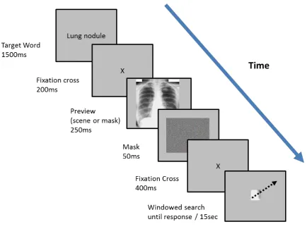
sequence for each condition using the FPMW paradigm. Participants were presented with a
black target word indicating the identity of the target object for 1500ms. Then a fixation cross
for 200ms, and then either a scene preview or mask preview for 250ms, followed by a mask
for 50ms. Following a second fixation cross for 400ms, windowed search of the scene began.
A 2.5° radius window was used to restrict the field-of view during search and to detect a
target participants pressed a gamepad button whilst directly fixating the target. By presenting
the target word before the scene preview, we tried to control for the fact that in Experiment 2
the target object (lung nodule) was always known before scenes were presented. Four
separate practice trials were presented beforehand to familiarise participants with the
procedure followed by 2 blocks of 20 trials. Targets were only considered correctly identified
if a fixation was within the target AOI during button press. Participants only saw each scene
once and so scene/condition combinations were counterbalanced across participants. Trials
were presented in a randomized order for each participant and the whole experiment took
approximately 20min.
<< Insert Figure 1 about here >>
Results
All data were subjected to a 2 x 2 mixed measures ANOVA with preview (scene, mask)
as a within-participant factor, and expertise (novice, expert) as a between participant factor.
For all measures only trials with correct responses were analysed. With the exception of
accuracy, we expected all response metrics would be more efficient with scene preview than
mask preview, but that there would be no difference between the expertise groups on any of
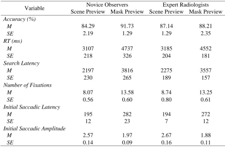
<< Insert Table 1 about here >>
Performance
Overall search accuracy averaged 88% (ranging from 60% to 100%) but did not differ
as a function preview, F(1, 26) = 2.62, p = .12, η2= .09, or expertise, F(1, 26) = .03, p = .87,
η2< .01, and there was no interaction, F(1, 26) = 1.48, p = .24, η2= .05. RTs averaged
3895ms across conditions and there was a main effect of preview, F(1, 26) = 77.46, p < .001,
η2= .74. However, there was no main effect of expertise, F(1, 26) = .34, p = .86, η2< .01, and
there was no interaction, F(1, 26) = 0.60, p = .48, η2 = .01. For both groups of observers RTs
were faster for scene preview than mask preview.
Search-related eye movements
Search latency averaged 2961ms across conditions and there was a main effect of
preview, F(1, 26) = 115.94, p < .001, η2= .81. However, like the RT measures, there was no
main effect of expertise, F(1, 26) = .11, p = .74, η2< .01, and no significant interaction, F(1,
26) = 1.58, p = .22, η2= .01. The number of fixations averaged 10.91 across conditions and
there was a main effect of preview, F(1, 26) = 126.95, p < .001, η2= .82. However, there was
no main effect of expertise, F(1, 26) = .11, p = .74, η2< .01, and no significant interaction,
F(1, 26) = 1.28, p = .72, η2= .01. All these search metrics indicate that targets were identified
faster and in fewer eye movements for scene preview than mask preview, but were not
modulated in any way by expertise group.
First eye movement of search
Theinitial saccadic latency averaged 236ms across conditions and there was a main
effect of preview, F(1, 26) = 28.99, p < .001, η2= .53, but no effect of expertise, F(1, 26) =
amplitude averaged 2.03° visual angle across conditions and there was a main effect of
preview, F(1, 26) = 141.36, p < .001, η2= .84. Once again however there no main effect of
expertise, F(1, 26) = 80, p = .38, η2= .03, and no interaction, F(1, 26) = .12, p = .73, η2< .01.
The first eye movement of search following a scene preview was both faster and of larger
amplitude than following a mask preview, however, once again there was no difference
across expertise groups.
Discussion
In line with previous research (e.g., Castelhano & Henderson, 2007; Võ & Henderson,
2010), all metrics indicated search for everyday objects from real-world scenes was more
efficient when participants were presented with a scene preview than a mask preview.
Following a 250ms glimpse of the upcoming scene, participants were quicker to initiate
search and faster to fixate and identify the target. Importantly, there was no difference
between the two expertise groups on any of the measures in Experiment 1. This is consistent
with previous comparisons studies between experts and lay observers (Nodine & Krupinski,
1998), which show that experts in medical image perception do not demonstrate superior
visual processing in search tasks outside their domain-specific expertise. Moreover, our
results suggest novice observers were exploiting the initial glimpse of the scene in the exact
same manner as expert radiologists. Experiment 2 attempts to isolate the expertise-dependent
contribution of initial scene processing and eye guidance by requiring the same participants
to this time search for lung nodules from chest-x-rays using the FPMW paradigm.
Experiment 2
Method
Stimuli and apparatus
A testbank of 60 chest x-ray images were used in Experiment 2, 36 images were
abnormal and contained a single nodule located within the lung fields, and 24 images did not
contain a nodule. Nodules were defined as discrete opacities in the lung field or mediastinum
measuring between 5–30mm in diameter, and all nodules were histopathologically proven.
The chest x-ray images subtended 22.48° x 20.44°, and lung nodules had an average size of
3.30° x 3.30°. We have successfully used this testbank in previous studies to establish
expertise-related differences in search (Donovan & Litchfield, 2013), and how search-related
eye movement behaviour can be used as learning cues for other observers (Litchfield, Ball,
Donovan, Manning, & Crawford, 2010). For methodological reasons, medical image
perception research typically adopts a 50% prevalence rate as this helps characterise observer
performance, as it is important to not only detect targets, but to refrain from making false
positive decisions on normal images. Since the primary interest of the present study is how
quickly observers identified abnormalities to maximise the number of valid samples in the
final analysis we adopted a prevalence rate of 60% in Experiment 2, and to be consistent with
Experiment 1, we adopted a 100% prevalence rate in Experiment 3. It should be noted,
however, that the prevalence rate in clinical settings can be substantially lower than this, and
that low prevalence is a contributing factor as to why such targets are missed in medical
image perception (Nakashima, Kobayashi, Maeda, Yoshikawa, & Yokosawa, 2013; see also
Wolfe, Brunelli, Rubinstein, & Horowitz, 2013).
Procedure
Although the timings in which the medical images were shown were identical to
Experiment 1, adapting the FPMW paradigm to a medical imaging task meant that there were
included normal images that did not contain a target. Participants were told that if they did
not identify a target within the 15 second maximum limit, then that trial would be coded as
normal. As such, if observers finished searching the image within this time and believed the
image was normal then they should allow the timer run out. This timeout feature was a key
logistical constraint that has been used previously (e.g., Carmody et al., 1981), and it ensured
that observers only had one response button to press if a nodule was detected and so were not
making additional response compatibility judgements during this already demanding task.
The second key difference was that the search targets (lung nodules) were much more
difficult to find in Experiment 2. Consistent with FPMW studies, the search targets in
Experiment 1 were everyday objects that once fixated are easily recognised. In contrast, lung
nodules are notoriously difficult to correctly identify, even in free viewing tasks. It is
therefore standard practice in medical imaging tasks to obtain a confidence rating for each
decision so that such information can be used in receiver operating characteristic (ROC)
analysis to more accurately characterise performance (Chakraborty, & Berbaum, 2004; Green
& Swets, 1966). Accordingly, once a participant had identified what they thought to be a
nodule by pressing the gamepad button whilst looking at the suspected target, before
proceeding to the next image participants were required to provide a 1-4 confidence rating (4
being highly confident) regarding their decision.
As the novice observers had no experience with medical images or lung nodules,
before beginning the experiment two practice chest-rays containing a nodule were first shown
to participants so that they understood what targets they were looking for. A further two
separate practice trials (1 abnormal, 1 normal) were presented using the FPMW paradigm
beforehand to help participants familiarise themselves with the modified procedure.
participants. Trials were presented in 2 blocks of 30 trials in a randomised order and the
whole experiment took approximately 30min.
Results
All data were subjected to a 2 x 2 mixed measures ANOVA with preview (scene, mask)
as a within-participant factor, and expertise (novice, expert) as a between-participant factor.
For all measures, analysis was restricted to only trials where targets were correctly detected.
Since Experiment 2 involves a medical imaging task, we provide 2 analyses of diagnostic
performance. To provide a clear comparison between Experiment 1 and previous FPMW
studies, we first report accuracy levels based on the % of target nodules correctly identified
(i.e., ignoring true/false negative decisions on normal images). We then report observer
performance based on jackknife free-response ROC (JAFROC) analysis, which has been
validated as a more sensitive measure of diagnostic decision-making than ROC (Chakraborty,
& Berbaum, 2004). JAFROC was calculated using the freely available RJafroc software
(http://www.devchakraborty.com) which uses the number of true positives and false positives
observers report and their respective confidence ratings to produce a single figure of merit for
each observer. This figure of merit represents the likelihood that a true positive will be given
a higher confidence rating than a false positive. This single measure is superior to ROC
because it simultaneously takes into account decision confidence and location information.
Traditional ROC does not take into account location information but simply requires the
observer to state whether the image contains an abnormality or not, without actually having
to localize it. This can lead to problematic situations where an observer views an abnormal
image, reports the image is abnormal, and so according to ROC the observer is making a
correct decision (i.e., true positive). However, that observer could be stating the image is
and so ROC can overestimate observer performance. With JAFROC, a decision is only
counted as a true positive if the observer correctly identified the location of the abnormality.
Consistent with Experiment 1 and other FPMW studies, a correct response is determined by
the participant directly fixating the suspected target and pressing the response button.
JAFROC uses a chance level of .50 and we have previously shown that expert performance is
usually represented by a figure-of-merit of .75 or above (Donovan & Litchfield, 2013;
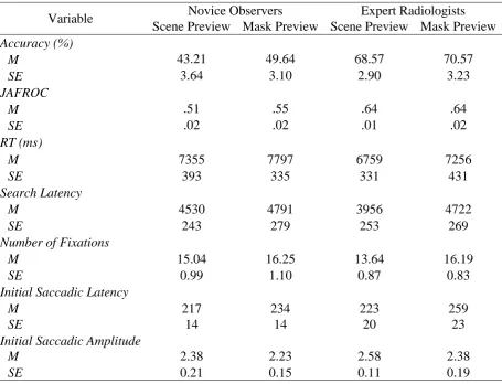
Litchfield et al., 2010). A summary of means can be seen in Table 2.
<< Insert Table 2 about here >>
Performance
Overall accuracy averaged just 58% (novices ranging from 22% to 82%; experts
ranging from 44% to 88%). There was no main effect of preview, F(1, 26) = 3.29, p = .08, η2
= .11, and there was no interaction, F(1, 26) = .91, p = .35, η2= .03. There was however, a
main effect of expertise, F(1, 26) = 21.95, p < .001, η2= .46, with experts detecting more
nodules (70%) than novice observers (46%). Moreover, performance assessed by JAFROC
revealed a number of significant effects. There was a main effect of preview, F(1, 26) = 6.40,
p = .018, η2= .15, a main effect of expertise, F(1, 26) = .20.25, p < .001, η2= .44, and a
significant preview x expertise interaction, F(1, 26) = 7.22, p = .012, η2= .19, with experts
better at detecting nodules than novice observers.
In unpacking the interaction, the simple main effect analyses revealed some rather
surprising findings. Experts performed better than novices in both the scene preview
condition, F(1, 26) = 26.42, p < .001, η2= .50 , and mask preview condition, F(1, 26) =
12.74, p < .001, η2= .34. However, denying experts the initial glimpse of the image did not
preview conditions, F(1, 26) = .01, p = .91, η2< .01. In contrast, novice observers actually
performed better in the mask preview condition than the scene preview condition, F(1, 26) =
13.61, p = .001, η2= .52. As mentioned above, the JAFROC analysis is a much more
sensitive measure of diagnostic performance than simple accuracy levels. We return to
potential explanations of these findings in the discussion. Finally, RTs averaged 7291ms
across conditions and there was no main effect of preview, F(1, 26) = 2.85, p = .10, η2= .10,
no main effect of expertise, F(1, 26) = .34, p = .86, η2= .06, and there was no interaction,
F(1, 26) = 0.01, p = .92, η2 < .01.
Search-related eye movements
The overallsearch latency averaged 4499ms across conditions and there was a
borderline main effect of preview, F(1, 26) = 4.19, p = .051, η2= .13. However, there was no
main effect of expertise, F(1, 26) = 1.40, p = .25, η2= .05, and no significant interaction, F(1,
26) = 1.02, p = .32, η2= .03. The overall number of fixations averaged 15.28 across
conditions and there was a main effect of preview, F(1, 26) = 4.96, p = .035, η2= .16, but no
main effect of expertise, F(1, 26) = .48, p = .50, η2= .02, and no interaction, F(1, 26) = .68, p
= .44, η2= .02. Contrary to our predictions, for novices as well as experts, the scene preview
led to faster search latencies and fewer fixations compared to mask preview conditions.
First eye movement
Initial saccadic latency averaged 233ms across conditions but there was no main effect
of preview, F(1, 26) = 2.52, p = .12, η2= .09, expertise, F(1, 26) = .63, p = .44. η2 = .02, and
no interaction, F(1, 26) = .32, p = .57, η2= .01. Likewise, the initial saccadic amplitude
averaged 2.03° across conditions but there was no main effect of preview, F(1, 26) = 1.56, p
p = .85, η2< .01. These measures indicated there was no difference in the speed or amplitude
of the first eye movement of search following a scene preview compared to mask preview, or
any influence of domain-specific expertise.
Discussion
One of the first issues to note is that for both groups of observers, the accuracy in
detecting targets in this task was much lower than Experiment 1. Nevertheless, as one would
expect, novices performed much worse than experts in detecting nodules from chest x-rays
images. In addition, our JAFROC analysis confirmed that across all conditions, experts (M =
.64) outperformed novices (M = .53) but also that novice observers actually performed worse
in the scene preview condition (M = .51) compared to the mask preview condition (M = .55).
We have shown in previous research (e.g., Donovan & Litchfield, 2013) that novices
searching for lung nodules are more likely to fixate regions in the image that contain
nodule-like distractors, but which are in fact normal anatomy (e.g., the hilar and mediastinum). Since
rapidly distinguishing normal anatomy from pathology is a hallmark of expertise, one
potential explanation for why novices in Experiment 2 performed worse in the scene preview
condition is that whilst encoding the initial scene representation, novices may have been
biased towards these distractors regions, which experts with years of experience would have
learned to attenuate. Indeed, novices made significantly more false positives in scene preview
(M = 9.57) than mask preview (M = 7.43), t(13) = 5.30, p < .001, Cohen’s d = .38) which
would have contributed to the lower JAFROC figure-of-merit. In contrast, expert observers
showed no such difference in false positives between scene preview (M = 4.07) and mask
preview (M = 4.21), t(13) = -.20, p = .85 Cohen’s d = .04). This difference in false positive
novices was not because they thought abnormal images were normal and therefore gave up
search, but rather that they mistook normal features for pathology.
One of the key aims of this study was to establish the expertise-based contribution of
initial scene processing on diagnostic and search performance. In applying the FPMW
paradigm to medical imaging, we were surprised to find that experts showed no advantage in
diagnostic performance (either in accuracy or JAFROC) in the scene preview condition.
Providing an initial glimpse of the scene did not appear to contribute to expert performance,
and as discussed above, actually reduced novice performance. Moreover, in the mask preview
condition performance can only be attributed to slower processing relating to search and
discovery, and not rapid initial holistic processing (Kundel et al. 2008). The fact that experts
were better than novices in the mask preview condition but showed no greater advantage with
scene previews indicates that the importance of search and discovery should not be
underestimated as a marker of expert diagnostic performance.
Previous research has used eye-tracking measures such as time-to-first-fixation as an
indirect measure of rapid holistic processing (Kundel et al. 2008). The FPMW paradigm
provides a more rigorous manipulation of the initial scene preview on subsequent eye
movement performance. As shown in Table 2, we found that both novices and experts fixated
nodules faster (M = 4530ms, M = 3956ms) and in fewer eye movements (M = 15.04, M =
13.64) when provided with an initial glimpse of the upcoming medical image. This suggests
that whilst there was not a diagnostic advantage of scene previews, there was a search
advantage for both observer groups. However, this facilitation of search-related eye
movements was a much smaller effect than that observed in Experiment 1. For example, the
search latency effect sizes for the scene preview benefit in Experiment 1 was η2= .81, but
only η2= .13 in Experiment 2. Indeed, only search latency and number of fixations showed
first eye movement of search that typically accompanies such scene preview benefits
(Castelhano & Henderson, 2007; Hillstrom, et al., 2012; Pietrzyk et al., 2014).
Given the weak nature of this effect in the medical image perception task, we also
examined expert and novice search performance in isolation. A scene preview did not enable
novices to fixate nodules quicker, (Mdiff = -260ms), t(13) = -.77, p = .457, Cohen’s d = .27),
or in fewer eye movements(Mdiff = -1.21), t(13) = -1.15, p = .272, Cohen’s d = .31) than the
mask preview condition. Whereas for experts, the effect of scene preview was approaching
significance for search latency (Mdiff = -765ms), t(13) = -2.08, p = .058, Cohen’s d = .78) and
in the number of eye movements made (Mdiff = -2.55), t(13) = -1.93, p = 075, Cohen’s d =
.80). According to Cohen (1988) these effects of scene preview for experts could be
considered as medium to large. However, to put these effect sizes in the context of
Experiment 1 using the same individual analysis, this showed that the scene preview effects
for real-world scenes were at least twice as large and in this case did enable novices to fixate
real-world targets quicker (Mdiff = -1620ms), t(13) = -8.57, p < .001, Cohen’s d = .1.74) and
with fewer eye movements(Mdiff = -5.52), t(13) = -9.04, p < .001, Cohen’s d = 2.55), and
there were similarly strong effects for experts regarding search latencies (Mdiff = -1282ms),
t(13) = -6.68, p < .001, Cohen’s d = 1.97) and number of fixations (Mdiff = -4.51), t(13) =
-6.96, p < .001, Cohen’s d = 1.70) .Taken together, this suggests that if domain-specific
expertise in medical image perception is modulating how the initial scene is processed, its
effect above and beyond our shared expertise in initial scene processing is weak at best.
A possible reason for these weak effects could be because the same image type and
target type was searched repeatedly throughout Experiment 2. The scene preview benefit has
so far been demonstrated as a robust effect that diminishes after the first few fixations
(Hillstrom et al., 2012). However, like Experiment 1, in all FPMW studies different scenes
contrast, observers in Experiment 2 were repeatedly accessing broadly the same scene
guidance and target knowledge and so this could have minimized the scene preview
advantage. Even with a mask preview, observers still knew they would always be searching
through chest x-ray images for lung nodules and could have exploited that single scene-gist
and target template information to help guide search. Because novices could also take
advantage of these repeated search conditions, experts may have been prevented from
showing their faster processing of these domain-specific scenes and outmatch novice search.
With enough trials, repeated search for the same target within the same scene will decrease
search times, but change the task, even within the same scene, and that search benefit is lost
(Võ & Wolfe, 2012). If we were to use a range of medical image types and different
abnormalities as search targets this should once again maximise the advantage of the scene
preview, but specifically for the experts, as it is they that should be faster at recognising the
scene-gist and accessing the appropriate target knowledge of where to look. Experiment 3
was designed to directly address this issue by varying the medical image type and pathology
type across trials. In addition, to be more consistent with Experiment 1 where robust scene
preview effects were found, we adopted a 100% prevalence rate so participants only needed
to detect the target on each image, and no longer had to make any normal decisions or
confidence ratings.
Experiment 3
Method
Participants
We recruited 22 novices (9 male; mean age = 20.9 years) and 19 experienced
radiographers (8 male; mean age = 32.7 years). Radiographers were all trained in detection of
reporting radiographers for the NHS. We have previously shown that on nodule detection
tasks (e.g., Donovan & Litchfield, 2013; Manning et al., 2006) and skeletal fracture tasks
(Donovan, Manning, Phillips, Higham, & Crawford, 2005) experienced radiographers
demonstrate comparable detection performance to radiologists, with both groups typically
finding skeletal images easier than chest x-rays.
Stimuli and apparatus
The testbank consisted of 100 abnormal images from 3 different imaging modalities: 30
chest x-ray images, 20 single axial slice CT or MRI brain images (half each), and 50 skeletal
digital x-ray images. All images contained a single discrete pathology and were clinical cases
that had previously been reported by a consultant radiologist. The 30 chest images were
randomly selected from the 36 abnormal chest images used in Experiment 2, and as such, the
pathology was still a single nodule located within the lung fields. The pathology for the 20
brain images were all brain haemorrhages or tumours, whereas the pathology for the 50
skeletal images were all bone fractures. All images were presented on the same monitor as
the previous experiments and subtended 22.44° x 23.27° for chest images, 18.07° x 19.09°
for brain images, and 27.46° x 31.44° for skeletal images, and the abnormalities were all of
comparable size (lung nodules: 3.31° x 3.33°, brain haemorrhages/tumours: 3.86° x 4.17°,
fractures: 3.89° x 3.40°). Since we only analyse the eye movement data of correct (i.e., true
positive) decisions our 100 % prevalence rate should maximise the number of valid samples
in the final analysis and ensure we have sufficient power to detect scene preview effects
without being confounded by fatigue effects (Krupinski, Berbaum, Caldwell, Schartz, & Kim,
2010).
Participants were given practice examples of the 3 image types and the pathology they
would be searching for and were told that there would be a single pathology on every image.
All timings of how images were shown were identical to Experiment 2. A critical difference
was that rather than searching for the same target (lung nodule) across the same image type
(chest), participants searched for pathology on the given image. The target word (pathology)
was presented before the scene preview and this generic word was chosen as it did not
indicate the upcoming image type. Presentation of image type (chest, brain, skeletal) and
preview (scene, mask) conditions were randomised across 4 blocks of 25 trials. Participants
saw each image once and preview was counterbalanced. Trials were terminated either by
button press or timed out after 15s. Note that as there were no normal (i.e., target-absent)
images in the present study, a timeout response could in no way be considered a positive
aspect of performance. Likewise, since the focus was on accuracy rather than JAFROC, no
confidence data was collected after each decision was made. There were 2 practice trials at
the start to familiarise participants with the procedure and the whole experiment took
approximately 40min.
Results
All data were subjected to a 2 x 2 mixed measures ANOVA with preview (scene, mask)
as a within participants factor and expertise (novice, experienced) as between participant
factors. For all measures only trials with correct responses were analysed.
Performance
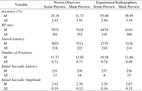
Overall accuracy across all images and conditions averaged just 44% (novices ranging
from 2% to 46%; experienced radiographers ranging from 32% to 80%). There was a main
= 60.88, p < .001, η2= .61, However, there was no interaction, F(1, 39) = 0.01, p = .92, η2 <
.01. As expected, experienced radiographers were much better at detecting a range of
pathologies (57%) than novice observers (30%). However, as shown in Table 3, rather than
finding a scene preview benefit, both groups of observers were significantly worse at
detecting pathologies with a scene preview (novice = 28%, experienced = 56%) compared to
a mask preview (novice = 32%, experienced = 59%).
RTs across all images and conditions averaged 6977ms and there was no main effect of
preview, F(1, 39) = 2.55, p = .12, η2= .06, and no interaction, F(1, 39) = 0.06, p = .81, η2 <
.01. However, there was a main effect of expertise, F(1, 39) = 9.92, p < .01, η2= .20, with
experienced radiographers faster at detecting a range of pathologies (M = 6308ms) than
novice observers (M = 7645ms).
<< Insert Table 3 about here >>
Search-related eye movements
The overallsearch latency averaged 3060ms across all images and conditions.
However, unlike the RT findings, there was no main effect of preview, F(1, 39) = 2.23, p =
.14, η2= .06, no main effect of expertise, F(1, 39) = 0.91, p = .35, η2= .02, and no
interaction, F(1, 39) = 0.02, p = .88, η2 < .01. The overall number of fixations before finding
pathology averaged 11.46 across conditions and mirrored the search latency non-significant
findings. There was no main effect of preview, F(1, 39) = 1.77, p = .19, η2= .04, no main
effect of expertise, F(1, 39) = 0.43, p = .52, η2= .01, and no interaction, F(1, 39) = 0.19, p =
.66, η2 < .01.
Initial saccadic latency averaged 248ms across conditions, however, there was no main
effect of preview, F(1, 39) = 0.30, p = .59, η2< .01, no main effect of expertise, F(1, 39) =
2.54, p = .12, η2= .06, and no interaction, F(1, 39) = 0.44, p = .51, η2 = .01. Similarly, the
initial saccadic amplitude averaged 2.32°, yet there was no main effect of preview, F(1, 39) =
1.91, p = .18, η2= .05, no interaction, F(1, 39) = 0.13, p = .72, η2 < .01, whereas the main
effect of expertise was approaching significance, F(1, 39) = 3.93, p = .054, η2= .09. Taken
together, all eye movement metrics failed to demonstrate a scene preview benefit for novice
or experienced observers examining a range of medical images using the FPMW paradigm.
Discussion
Experiment 3 focused on clarifying the weak scene preview benefit observed in
Experiment 2 by systematically increasing the range of medical image types and pathologies
and thereby maximise the benefit of the scene preview. Consistent with previous research
(e.g., Donovan et al., 2005; Donovan & Litchfield, 2013; Manning et al., 2004), Experiment
3 found that experienced radiographers could detect more pathologies and do so faster than
novices. However, rather than maximising the scene preview benefit compared to mask
preview, Experiment 3 found that both novices and experienced radiographers were worse at
detecting pathologies with a scene preview than a mask preview, and all eye movement
metrics confirmed there was no search related advantage of the scene preview.
Results from Experiment 2 hinted that scene previews could have unforeseen costs in
terms of diagnostic performance, but this was only found for novice observers, not experts.
By randomising medical image and pathology type, Experiment 3 replicated the finding that
scene previews impaired novice performance, but also that scene previews impaired
experienced radiographers that are currently practicing in hospitals. This scene preview
supposed to be exploited with experience in order to enhance performance (e.g., Donovan &
Litchfield, 2013; Drew et al., 2013a; Krupinski, 2010; Kundel et al., 2007; Wolfe et al.,
2011). Whilst we adopted a 100% prevalence rate in Experiment 3 so as to be more
consistent with Experiment 1 and to ensure we had adequate power to detect scene preview
effects, knowing that there was always pathology could have led to a change in decision
thresholds and led to an increase in false positives. However, this changing of decision
thresholds purely based on prevalence would still not account for our pattern of results, and
specifically why the scene preview impaired detection of targets relative to mask preview.
Examining the accuracy effects in more detail, it is evident from Tables 2 and 3 that
there was a clear drop in overall accuracy (approx 15%) between Experiment 2 and
Experiment 3. Experienced radiographers frequently demonstrate comparable detection
performance to experts in nodule detection tasks (Donovan & Litchfield, 2013; Litchfield et
al., 2010; Manning, Barker-Mill, Donovan, & Crawford, 2005; Manning et al., 2006) and
fracture detection tasks (Donovan et al., 2005). As such, we do not believe this drop in
accuracy was due to a lack of expertise, but instead due to increasing task demands. This is
supported by the fact there was also a comparable drop in novice accuracy (from 45% in
Experiment 2, to just 30% in Experiment 3). The key question though is how this more
demanding task contributed to the impairment in detection for scene preview compared to
mask preview.
In all previous research where accuracy has been reported using the FPMW paradigm
(e.g., Castelhano & Henderson, 2007; Võ & Henderson, 2010, 2011; Võ & Schneider, 2010)
observers have always had to switch between different images and target knowledge across
trials and never before has an accuracy impairment been found for scene preview. As such, it
is not as if switching creates a generic cognitive-load issue that could give rise to accuracy
images could have led to the specific scene preview impairment is how observers were able
to filter out the inherent distractors in the image under the different preview conditions. In the
scene perception tasks, there is always only one search target in the scene and efforts are
made to select images carefully so as to minimise target-like distractors. Likewise in medical
imaging tasks, there is also often only one search target – a genuine pathology that has been
verified by consultant radiologists beforehand. However, what is inevitable with medical
images is that normal anatomy can provide many potential target-like distractors that are
inherent to the image (Wester et al., 1997). Some abnormalities have poor visual conspicuity
(Krupinski, Berger, Dallas, & Roehrig, 2003) and together with the co-presence of target-like
distractors this means that even when fixating directly at a target for several seconds it can
often be declared as not being an abnormality (Kundel et al., 1978; Manning, Ethell, &
Donovan, 2004) or instead that a normal feature is identified as the abnormality (Wester et
al., 1997). Indeed, when a suspected abnormality is difficult to disambiguate from normality
its spatial location may have to be relied upon to make correct decisions (Carmody, Kundel &
Toto, 1984; Donovan & Litchfield, 2013). When a medical image is flashed, the observer
processes the gist of the image, and potentially detects pathology, even if the abnormality
cannot later be localized (Evans et al., 2013). It may be that flashing the same type of images
(chest) in Experiment 2 allowed expert observers (but not novices) to better discriminate
between targets and distractors. In contrast, switching between image types in Experiment 3
may have meant that both novice observers and experienced observers were more susceptible
to the distractors inherent in the images following the scene preview. Conversely, the mask
preview prevented observers from immediately processing the gist as well as potential targets
and distractors, and this may have mitigated any further susceptibility to distractors. The
implication is that the costs of perceiving this initial glimpse must have outweighed any
Taken together, these substantial issues in detection accuracy may fundamentally
explain why we did not find robust search benefits of scene preview when attempting to
apply the FPMW paradigm to this domain-specific task. The unforeseen costs of scene
previews and the implications this has are further explored in the general discussion.
General Discussion
The aim of the present study was to establish how the initial scene representation
guides search as a function of domain-specific expertise. Using the FPMW paradigm
(Castelhano & Henderson, 2007) two experience groups (expert radiologists, psychology
students) searched for everyday objects from real-world scenes (Experiment 1), and lung
nodules from chest x-ray images (Experiment 2), whereas a second sample of observers
(experienced radiographers, psychology students) searched for a variety of pathologies from
different medical image types (Experiment 3). Consistent with previous research (e.g.,
Castelhano & Henderson, 2007; Võ & Henderson, 2010) we found strong scene preview
effects for the observers in Experiment 1, as both expert radiologists and psychology students
were able to exploit a brief glimpse of the upcoming scene to guide search. However, in this
first application of the FPMW to a specific expertise domain, we found only weak effects of
scene preview in Experiment 2 using these same participants. This suggests that experts were
not substantially better than novices at exploiting the scene preview of medical images.
Overall, both groups of observers were able to find abnormalities in medical images faster
and with fewer eye movements following a brief glimpse of the scene. However, it was only
when we examined the preview effects of each group in isolation that search metrics of
experts (but not novices) seemed to improve with scene preview. Moreover, these
improvements in search did not translate into benefits in diagnostic performance. Experts
medical scene did not lead to additional improvements in decision-making, and in fact,
further impaired novice performance. Experiment 3 was designed to tease out these weak
scene preview effects by requiring novice observers and experienced radiographers to
examine a greater range of medical image and target types, thereby maximising the expertise
advantage of receiving the scene preview to guide search. Instead, we discovered unexpected
findings that corroborate the results of Experiment 2; scene previews of medical images led
to impaired accuracy compared to mask preview for both novice and experienced observers
and there was still no search benefit for scene preview.
At a descriptive level, the holistic model of medical image perception (Kundel et al.,
2007; Nodine & Kundel, 1987) helps account for the well documented expertise differences
in search and diagnostic performance (Nodine & Mello Thoms, 2010; Reingold & Sheridan,
2011). The ability to exploit the initial glimpse of the scene is at the core of the holistic
model (Kundel et al., 2007) but is also a key component of scene perception research
(Castelhano & Henderson, 2007; Torralba et al., 2006; Wolfe et al., 2011). By using the
FPMW paradigm to control the contribution of the initial glimpse on subsequent search as a
function of expertise, the present study extends previous eye-tracking research that has until
now only been able to indirectly investigate these issues, either via tachistoscopic studies
(Carmody et al., 1981; Kundel et al., 1975; Evans et al., 2013) or free viewing studies
(Donovan & Litchfield, 2013; Kundel et al., 1978; Kundel et al., 2008; Manning et al., 2004).
We first discuss why we only observed a weak expertise advantage of processing the scene
preview in Experiment 2, and then elaborate on the explanation we put forward in
Experiment 3, as to how an initial scene preview of a medical image could impair the
detection of targets as found in Experiment 2 and Experiment 3.
First, a major component of how the scene-context guides search is that observers learn
2007; Castelhano & Heaven, 2010). As a result, the scene-context can guide search towards
likely target locations, even when targets were not present in previews. Critically, however,
unlike scene perception and the search for everyday objects, the scene-context of the chest
x-ray is not particularly predictive as to the location of lung nodules, as these targets can appear
anywhere within the lung fields (Båth et al., 2005). Contextual guidance of the scenes (e.g.,
Torralba et al., 2006) would therefore promote the outer lung fields as highly probable search
areas but would not be able to narrow down search guidance much further on a given image,
and instead would rely on subsequent search-related eye movements to discount non-target
areas.
Second, it would seem there is a better target template in the visual search for
real-world scenes than in medical images (Malcolm & Henderson, 2009; Vickery, King, & Jiang,
2009). As mentioned previously, lung nodules can be difficult to identify and the medical
images often contains numerous distractors from normal anatomy that closely resemble the
features of nodules (Krupinski et al., 2003; Wester et al., 1997). A better target template
would allow for greater sensitivity to target signals and attenuation of distractors that do not
share target similar features, and can help guide search.Taking both these issues into
consideration, one reason why only a weak expertise advantage in search was found for scene
preview in Experiment 2 was because the targets were difficult to identify and were not in a
predictable location.
Notwithstanding these issues, an alternative explanation for the weak scene preview
could have simply been because the same type of image and target type was searched
repeatedly throughout Experiment 2. To rule out this alternative explanation, Experiment 3
increased the variety of image types and pathology across trials. However, rather than
enhancing the scene preview benefit, there was no scene preview benefit for novice or
compared to mask preview. In the discussion of Experiment 3 we highlighted that
experienced radiographers have comparable performance to radiologists in these specific
medical imaging tasks (e.g., Donovan et al., 2005; Donovan & Litchfield, 2013; Manning, et
al., 2005, 2006) and so the reason why experienced radiographers were impaired along
novices with scene preview is unlikely to be because the radiographers lacked sufficient
expertise. Moreover, we put forward that one of the reasons why experts in Experiment 2
were not likewise impaired with scene preview, could have been because repeatedly flashing
the same type of images (chest) in Experiment 2 may have allowed expert observers to better
discriminate between targets and distractors. Here we elaborate on this explanation by
drawing on the mechanisms that may have allowed experts to do this.
Apart from the global-local processing already discussed, medical image perception
must be reliant on additional processes such as the way the visual system adapts to images
and consistent patterns of stimulation (Webster, 2011). For example, Webster and colleagues
(Kompaniez-Dunigan, Abbey, Boone, & Webster, 2015) recently demonstrated that when
pathology was easy to discriminate, encouraging adaptation of visual processes by repeated
exposure did not increase detection performance. However, when pathology was more
difficult to distinguish from the background, adaptation via repeated exposure to images did
enhance performance. In some respects, this weighting of signals is similar to what in
medical image perception is known as the application of a pre-whitening filter (Eckstein,
Pham, Abbey, & Zhang, 2006), which enables the observer to discount the normal anatomic
background noise (De Vries, Hooge, Wertheim, & Verstraten, 2013).
Tying all these aspects together, experts have better visual sensitivity to abnormalities
than novices (cf. Sowden et al., 2000) and generally outperform them (Donovan & Litchfield,
2013; Nodine & Mello Thoms, 2010; Reingold & Sheridan, 2011), but also, by repeating the
of processing the distractors visible in the preview, and therefore, maintained the same
detection performance for both scene preview and mask preview (cf., Kompaniez-Dunigan et
al. 2015). In contrast, by manipulating the image type in Experiment 3 we may have
disrupted the observer’s ability to become sensitive to pathology and attenuate distractors
within the experiment. Just like not appropriately applying a pre-whitening filter (Eckstein et
al., 2006), these processes could have a significant impact on decision-making performance
in medical imaging, yet are not so crucial in target detection for real-world scenes. For
example, the detection of targets in real-world scenes can make use of other channels of
information, such as colour, which are irrelevant to grey-scale medical images, but can
nonetheless affect how the gist is exploited in the first place (Nijboer, Kanai, de Haan, & van
der Smagt, 2008).
One FPMW study that is worth pointing out here in relation to the costs of processing
the initial glimpse is Võ and Schneider (2010). Although they did not find the same accuracy
impairments we observed in the present study, they made some very relevant discussion
points as to the relationship between local and global processing, which overlap in many
ways with our own points. Võ and Schneider (2010) examined the individual differences in
the ability to process scene previews by comparing those participants that could later describe
the differences between the previews conditions (the ‘conscious-report’ group), with those
that could not (the ‘no report group’). Again, showing that the FPMW paradigm can lead to
surprising results, the conscious-report group did not gain a search benefit from viewing an
identical preview containing the background scene context and the inherent objects within,
but instead this group could exploit a preview that only consisted of the background scene
context. In contrast, the no report group benefited mostly from identical previews. Võ and
Schneider (2010) argued that this meant the additional objects in the identical preview may
scene priors from the local and global pathway. In other words, there was such a thing as too
much information in the preview. In this context, our mask preview could be considered a
convenient way of blocking out the interference of competing global and local signals and
allowed the observers to just focus search and decision-making to what can be seen through
the moving window. This suggests that aside from the distinct visual processes used to
optimise medical imaging performance, individual differences of how previews are encoded
and exploited needs further research, as our current findings also force us to question the real
benefits of scene previews, particularly when targets are not so easy to detect.
Overall, we have found with the FPMW paradigm that the scene preview can disrupt
observer performance in unexpected ways. Although the processing that takes place based on
the initial glimpse of the scene is thought to be integral to expert medical image perception
performance (Kundel et al., 2007), research has already highlighted that there are limits as to
the types of decisions that can be made solely based on this initial glimpse (Carmody et al.,
1981; Evans et al. 2013). Our present study goes further than this by demonstrating that when
the initial glimpse is controlled, but search is still allowed, decision-making can sometimes
be better when no prior glimpse is available. Given that the ability to exploit the initial
glimpse of the scene is at the core of the holistic model (Kundel et al., 2007) and a key
component of scene perception research (Castelhano & Henderson, 2007; Torralba et al.,
2006; Wolfe et al., 2011) our findings add to the existing FPMW research (e.g., Castelhano
& Henderson, 2007; Hillstrom, et al., 2012; Võ & Henderson, 2011; Võ & Schneider, 2010),
but highlight that further studies are needed to systematically investigate the target-distractor
relationship within scenes.
In addition, this study represents just one domain of visual expertise. As we have found,
we cannot easily generalize findings from scene perception research using FPMW to our
to other visual expertise domains. Instead, how an initial scene is processed and exploited for
search as function of expertise may depend on the specific parameters of the search task, and
as such, we encourage research using the FPMW paradigm in other domains.
Using a medical image perception task, we found weak expertise benefits in search
from scene previews, but such search benefits were later overshadowed by scene preview
impairments in detection. Medical image perception is a difficult task, but experienced
observers find ways of dealing with the consistent patterns of stimulation to help reach better
decisions. Clearly, there is a real impetus to both further understand and improve
performance on such tasks (Beam et al., 2006; Field, 2014; Krupinski, 2010), and in doing so,
this also provides new perspectives on scene perception and expertise. What is refreshing is
that with the FPMW paradigm (Castelhano & Henderson, 2007), we may now have the better
tools in which to test out potential explanations, and identify ways that enhance or impair
Acknowledgements
This work was supported by the Edge Hill University Research Investment Fund and a
Phillips Medical Systems grant. We would also like to sincerely thank the action editor,
James Enns, as well as Jeremy Wolfe and the two anonymous reviewers for their constructive
References
Båth, M., Håkansson, M., Börjesson, S., Kheddache, S., Grahn, A., Ruschin, M., ... &
Månsson, L. G. (2005). Nodule detection in digital chest radiography: introduction to
the RADIUS chest trial. Radiation Protection Dosimetry, 114, 85-91.
Beam, C. A., Krupinski, E. A., Kundel, H. L., Sickles, E. A., & Wagner, R. F. (2006).
The place of medical image perception in 21st-century health care. Journal of the
American College of Radiology, 3, 409-412.
Bertram, R., Helle, L., Kaakinen, J. K., & Svedström, E. (2013). The effect of expertise
on eye movement behaviour in medical image perception. PloS one, 8, e66169.
Biederman, I., Mezzanotte, R. J., & Rabinowitz, J. C. (1982). Scene perception:
Detecting and judging objects undergoing relational violations. Cognitive Psychology,
14, 143–177.
Carmody, D. P., Kundel, H. L., & Toto, L. C. (1984). Comparison scans while reading
chest images: Taught but not practiced. Investigative Radiology, 119, 462–466.
Carmody, D. P., Nodine, C. F., & Kundel, H. L. (1980). Global and segmented search for
lung nodules of different edge gradients. Investigative Radiology, 15, 224–233.
Carmody, D. P., Nodine, C. F., & Kundel, H. L. (1981). Finding lung nodules with and
without comparative visual search. Perception & Psychophysics, 29, 594–598.
Castelhano, M. S., & Heaven, C. (2010). The relative contribution of scene context and
target features to visual search in scenes. Attention, Perception, & Psychophysics, 72,
1283–1297.
Castelhano, M. S., & Heaven, C. (2011). Scene context influences without scene gist: Eye
movements guided by spatial associations in visual search. Psychonomic Bulletin &
Castelhano, M. S., & Henderson, J. M. (2007). Initial scene representations facilitate eye
movement guidance in visual search. Journal of Experimental Psychology: Human
Perception and Performance, 33, 753-763.
Chakraborty, D. P., & Berbaum, K. S. (2004). Observer studies involving detection and
localization: Modeling, analysis and validation. Medical Physics, 31, 2313–2330.
Cohen, J. (1988). Statistical power analysis for the behavioral sciences (2nd ed.). New
Jersey: Lawrence Erlbaum.
De Vries, J. P., Hooge, I. T., Wertheim, A. H., & Verstraten, F. A. (2013). Background,
an important factor in visual search. Vision research, 86, 128-138.
Donovan, T., & Lit