GAP JUNCTIONS IN THE ADULT AND DEVELOPING
CEREBRAL CORTEX OF THE RAT: REGIONAL DIFFERENCES
IN THEIR DISTRIBUTION AND CELLULAR LOCALIZATION OF
CONNEXINS
By
BAGIRATHY NADARAJAH
Submitted to the University of London for the Degree of
Doctor of Philosophy
Department of Anatomy and Developmental Biology University College London
ProQ uest Number: 1 0 0 5 5 4 3 6
All rights reserved
INFORMATION TO ALL U SE R S
The quality of this reproduction is d ep en d en t upon the quality of the copy subm itted. In the unlikely even t that the author did not sen d a com plete manuscript
and there are m issing p a g e s, th e se will be noted. Also, if material had to be rem oved, a note will indicate the deletion.
uest.
ProQ uest 1 0 0 5 5 4 3 6
Published by ProQ uest LLC(2016). Copyright of the Dissertation is held by the Author. All rights reserved.
This work is protected against unauthorized copying under Title 17, United S ta tes C ode. Microform Edition © ProQ uest LLC.
ProQ uest LLC
789 East E isenhow er Parkway P.O. Box 1346
ACKNOW LEDGEMENTS
I would like to thank Professor John Pam avelas for his supervision and encouragem ent throughout this project.
M y thanks to D r. M arina M ione for her helpful advise and suggestions on several scientific aspects, Brett H arris for his technical assistance, D r. Dim itra Thom aidou for her help with W estern blots. D r. loannis Pappas and Audrey Dooley for their help in preparing dissociated cell cultures, John Cavanagh for his helpful suggestions and assistance in preparing graphical illustrations, Alex Lavdas for his help and suggestions, M ark Turmaine for his contribution to freeze-fracture electron m icroscopy and Kate W hitley for her help in photography and confocal m icroscopy.
I would like to take this opportunity to thank Professor How ard Evans,
U niversity o f W ales College o f M edicine, for providing me with connexin antibodies and for inviting me to carry out some experiments of N orthern blot analysis in his laboratory, Anna-M arie Jones, for carrying out the N orthern blot analysis and Drs. E lvira M ambetisaeva, Patricia Nicholas, David Llewlyn and Juan D ier for their help during my stay in Cardiff.
I would like to acknowledge the M ahapola Trust Committee o f Sri Lanka for awarding me a postgraduate scholarship to the UK, the British Council for their adm inistrative services, the CVCP for the ORS award. University o f London M edical School for the A stor fellowship, and the department o f Anatomy and Developmental Biology, U CL for providing me with some financial support.
P U B L IC A T IO N S A R IS IN G F R O M T H I S W O R K
N a d a ra ja h B, Thomaidu D, Evans W H and Pamavelas JG (1996) Gap Junctions in the adult cerebral cortex: regional differences in their distribution and cellular expression of connexins. J. Comp. Neurol. 376: 326-342.
TABLE OF CONTENTS
page number
Acknowledgements
Publications arising from this work Table of Contents
Abstract Abbreviations
Chapter One - introduction
Chapter Two - expression of connexins in the adult cortex Chapter Three- expression of connexins in the developing cortex Chapter Four - biochemical analyses of connexins in the cortex Chapter Five - gap junctions in the cortex: electron microscopical
analysis
ABSTRACT
Neurons and glia of the cerebral cortex arise from the ventricular zone lining the telencephalic ventricles. Recent physiological evidence has demonstrated the presence of low resistance pathways between cortical neurons during corticogenesis and neuronal circuit formation, and proposed a role for gap junctions in the formation o f neuronal domains which eventually may lead to the establishment of cortical columns. Gap junctions, the morphological correlates of electrical coupling and low resistance pathways, are plasma membrane channels composed o f connexin proteins that connect the cytoplasm o f contiguous cells. To date, thirteen mammalian connexins (Cxs) have been described in rodents o f which Cxs 26, 32 and 43 are the m ajor isoforms expressed in the adult and developing brain.
Using immunocytochemistry, W estern blotting. Northern blotting and electron microscopy (thin-section and freeze-ffacture), the present study examines the frequency of gap junctions, the cell types involved in the formation o f these junctions, gene expression of gap junction proteins, the connexins, and their distribution in the adult and developing cerebral cortex of the rat.
neurons and glia, of which the junctions between neurons and astrocytes are of interest as they may implicate the presence of a non-synaptic signalling pathways between these two cell types.
LIST OF ABBREVIATIONS ABC- avidin biotin complex
ANOVA - analysis of variance CP - cortical plate
Cx - connexin
DAB - 3,3 diaminobenzidine DIG - digoxigenin
DMEM - Dulbecco’s modified Eagle’s medium E - embryonic day
E face - exoplasmic face
FITC - fluorescene isothiocyanate GFAP - glial fibrillary acidic protein HBSS - Hank’s balanced salt solution IZ - intermediate zone
LV - lateral ventricle
MAP2 - microtubule associated protein 2 MZ - marginal zone
p - probability P - postnatal day
P face - protoplasmic face PBS - phosphate buffered saline SDS- sodium dodecyl sulfate SEM - standard error of the mean SVZ - sub ventricular zone
VZ - ventricular zone
CHAPTER ONE
List Of Contents
1.1. Cytology of the rat cerebral cortex - 9
1.2. Neocortical Development in the rat brain - 10 1.2.1. Laminar organization of the developing telencephalic vesicle- 10 1.2.2. Heterogeneity of the ventricular zone - 16
1.2.3. Radial migration of neurons - 21
1.2.4. Tangential migration of neurons - 23
1.2.5. Development of cortical areas - 23
1.3. Gap junctions: structure, expression and functions - 25
1.3.1. Structure of plasma membrane - 25
1.3.2. Structure of gap junctions: historical perspective - 25 1.3.3. Structure of gap junctions: current understanding - 30 1.3.4. Biochemical characterization of gap junctions - 30 1.3.5. Structure and expression of connexins - 32
1.3.6. Formation of gap junctions - 36
1.3.7. Regulation of gap junctions - 38
1.3.8. Degradation of gap junctions - 40
1.3.9. Gap junctions as morphological correlates of
electrical coupling - 40
1.3.10. Evidence for metabolic coupling through gap junctions - 41 1.3.11. Evidence for intercellular calcium signalling
through gap junctions - 43
1.3.12. Gap junctions in pathological conditions - 44
1.4. Gap junction mediated communication in the nervous system - 46
1.5. Gap junctions in the neocortex - 49
1 .1 .
C ytology o f the Rat C erebral C ortex
The cerebral cortex o f the rat is a laminated structure measuring 1.2-1.5 mm in thickness. It is organized into functionally distinct areas that show distinctive cytoarchitectural features. Sections through the thickness of the cortex indicate that neurons are organized into six layers. Neurons in each layer show a characteristic size, shape and packing density, and share physiological properties and axonal connections (Gilbert, 1983).
Layer I, which lies beneath the pial surface, is sparsely populated with neurons. Layers II and III, which do not show a distinct boundary between them, contain small and medium sized neurons displaying ovoid or triangular shaped cell bodies. Layer IV, the granular layer, appears distinct with its small and closely packed neurons. Layer V is characterized by loosely packed neurons with triangular or polygonally shaped cell bodies and layer VI contains small and closely packed neurons some of which are oriented horizontally.
Every layer contains each o f the two m ain classes of neurons recognized in the cortex, the pyramidal cells (projection neurons) and nonpyramidal cells (intemeurons). Pyram idal neurons utilise the excitatory amino acids L-glutamate or L-aspartate as neurotransmitters, and exert potent excitatory action in the cortex and in their target areas (Fagg and Foster, 1983; Dori et al., 1992). Nonpyramidal cells utilise GABA and wield a powerful inhibitory effect on neurons of the cortex (Fagg and Foster, 1983). O f the macroglia, astrocytes are present in all layers o f the cortex, whereas oligodendrocytes are mostly dispersed in the lower layers (Pamavelas et al., 1983).
ipsilateral or contralateral hemispheres. Pyramidal neurons layer V send their axons to subcortical structures such as the superior colliculus and the pons, whereas neurons of layer VI project to the thalamus (Pamavelas et al., 1989). The afferent fibres that project into the cortex can be grouped into subcortical and cortico-cortical connections. Subcortical afférents originate in the thalamus, basal forebrain, brainstem and midbrain, while cortico-cortical afferents consist callosal and ipsilateral connections.
Two morphological types of synapses have been described in the cerebral cortex: asymmetrical and symmetrical synapses (Colonnier, 1968) which correspond to Gray’s type I and type II, respectively (Gray, 1959). Asymmetrical synapses are characterized by a prominent postsynaptic density, and symmetrical synapses by a thin postsynaptic density (De Felipe and Farinas, 1992). The major source of asymmetrical synapses are pyramidal neurons and extrinsic cortical afferents, whereas the vast majority of symmetrical synapses are of intrinsic origin and are formed by the
nonpyramidal neurons (Peters and Jones, 1984). Since pyramidal neurons and most cortical afferents are excitatory in function, it is generally assumed that axon terminals forming asymmetrical synapses are excitatory. Likewise, axon terminals of the GABA- containing nonpyramidal cells forming symmetrical synapses are generally assumed to be inhibitory (Colonnier, 1981; De Felipe and Farinas, 1992).
1.2.
Neocortical Development in the Rat Brain
1.2.1. Laminar Organization of the Developing Telencephalic VesicleVentricular Zone
ventricular zone (VZ). Due to active interkinetic nuclear migration (see 1.1.2) o f the cells, the V Z has been described as ‘pseudo stratified’ germinal m atrix by Sauer (1935); it changes into a stratified matrix as cortical neurogenesis commences. The structures bordering the ventricles include rounded mitotic cells and end-feet of processes belonging to nonmitotic cells. Specialized junctional complexes exist between all mitotic cells and processes bordering the ventricles (Shoukimas and Hinds, 1978).
M arginal Zone
The marginal zone (MZ) appears at the pial surface of the developing brain forming the outermost layer. This subpial zone, initially comprising the cytoplasmic processes o f VZ cells (Boulder Committee, 1970), eventually becomes populated by horizontally oriented neurons, the Cajal-Retzius cells, which are the earliest neurons to differentiate in the embryonic cerebral cortex (Rickmann et al., 1977; Raedler and Raedler, 1978).
Intermediate Zone
The next step in corticogenesis is the establishment of an intermediate zone (IZ) between the MZ and the VZ as postmitotic neurons migrate away from the ventricular surface. The IZ contains a mixture of cells of variable orientations and a rich matrix of fibres; the latter comprise the early cortical afferents and efferents. The upper portion of this zone is made up of many horizontally oriented cells and the lower portion contains mostly radially aligned cells. It is generally assumed that a substantial proportion of cells in the IZ are neurons migrating to the CP (Gadisseux et al., 1990). The neuronal population of this zone is to some extent compartmentalized into small clusters by vertical and horizontal axon bundles (Gadisseux et al., 1990).
Subventricular Zone
Cortical Plate
Fig. 1.1. Cortical VZ of a rat brain at gestation day 16.
Note the radial alignment of the columnar cells that form this zone and the rounded mitotic cells at the ventricular surface. Scale bar: 200/xm.
V
Adherence to the inside-out rule appear to be more strict in cat and monkey than in rodents (McConnell, 1995).
Subplate
The subplate is a cell-rich layer beneath the cortical plate where polymorphic cells predominate. Autoradiographic studies have shown that subplate cells and Cajal- Retzius cells o f layer 1 are the oldest cortical neurons (Luskin and Shatz, 1985b; Chun et al., 1987). Recent studies on the development o f the thalamocortical connections in primates and cats suggest that thalamocortical axons accumulate in the subplate and ‘w ait’ for the layer IV neurons to arrive before growing into the cortex (Shatz, 1992). Few subplate cells persists into adult life scattered sparsely in the white matter whüe the m ajority o f these cells are eliminated by programmed cell death (Chun and Shatz, 1989; Valverde et al., 1995).
1 .2 .2 . H eterogeneity o f the Ventricular Zone
undergo interkinetic movement as they traverse the separate phases of the mitotic cycle. The S-phase is initiated when the nuclei are at the outer margin of the epithelium, and then they descend towards the ventricular surface in the course of DNA synthesis and G2-phase. After M-phase, ascent of the nuclei takes place in G1-phase (Takahashi et al., 1993).
Other investigators who used pulse and/or cumulative tritiated thymidine labelling and electron microscopy have supported the notion that the VZ contains a homogeneous population of cells at asynchronous mitotic cycles undergoing interkinetic nuclear migration (Sidman et al., 1959; Sauer and Walker, 1961; Hinds and Ruffet, 1971; Seymour and Berry, 1975). According to Fujita (1964), the VZ comprises a single precursor cell population giving rise first to neuroblasts during early development and then to glial and ependymal cells. Pulse labelling studies and electron microscopic examination, which failed to disclose any ultrastructural variations that might indicate differences in the developmental potential between progenitor cells, also suggested that the VZ comprises a homogeneous cell population sequentially giving rise to neuronal and then glial cells.
results were obtained using antibodies to a neurofdament protein (NF), a neuron specific marker, in the VZ of the chick brain and spinal cord, suggesting that morphologically undifferentiated neuroepithelial cells express a neuron-associated beta tubulin isotype very early during the neuroblastic differentiation, and this expression is sustained even in the mature neuronal populations (Caccamo et al., 1989). The interpretation of these immunocytochemical studies is that the mitotic cells that are positive for GFAP or NF are committed to glial or neuronal lineages indicating the presence of two separate cell populations in the VZ. Further, the glial phenotype is expressed prior to the cell's final division (Levitt et al., 1981). However, these studies do not show whether there might be a stage at which VZ cells are indifferent.
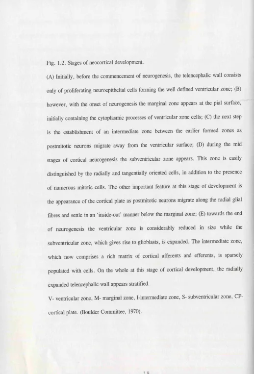
Fig. 1.2. Stages of neocortical development.
(A) Initially, before the commencement of neurogenesis, the telencephalic wall consists only of proliferating neuroepithelial cells forming the well defined ventricular zone; (B) however, with the onset of neurogenesis the marginal zone appears at the pial surface, initially containing the cytoplasmic processes of ventricular zone cells; (C) the next step is the establishment of an intermediate zone between the earlier formed zones as postmitotic neurons migrate away from the ventricular surface; (D) during the mid stages of cortical neurogenesis the subventricular zone appears. This zone is easily distinguished by the radially and tangentially oriented cells, in addition to the presence of numerous mitotic cells. The other important feature at this stage of development is the appearance of the cortical plate as postmitotic neurons migrate along the radial glial fibres and settle in an ‘inside-ouf manner below the marginal zone; (E) towards the end of neurogenesis the ventricular zone is considerably reduced in size while the subventricular zone, which gives rise to glioblasts, is expanded. The intermediate zone, which now comprises a rich matrix of cortical afferents and efferents, is sparsely populated with cells. On the whole at this stage of cortical development, the radially expanded telencephalic wall appears stratified.
Fig. 1.3. Laminar organization of the developing telencephalic wall of a rat brain at gestation day 17.
LV- lateral ventricle, VZ- ventricular zone, SVZ- subventricular zone, IZ- intermediate zone, SP- subplate, CP- cortical plate, MZ- marginal zone, PS- pial surface.
. PS
g
r%-\ i i
m
m
p
fm
f
m
iÆûm
m
Although the neuroepithelial cells do not show morphological features to suggest an association with particular areas of the adult neocortex, molecular markers have now been identified that define heterogeneous regions in the VZ of the forebrain. Genes such as Emx, Otx, murine homologous of zebrafish Pax and a number of Pou-domain genes are developmentally regulated and show characteristic patterns of distribution within the developing mammalian forebrain (Boncinelli et al., 1993; Mione and Pamavelas, 1994).Therefore, it is now evident that the VZ comprises a heterogeneous population of precursor cells.
1.2.3. Radial Migration of Neurons
One of the proposed models for the generation of the cortex was put forth by Rakic in 1988. According to his scheme (protomap model), future cytoarchitectonie areas of the cortex are specified in the neuroepithelium and recapitulated in the developing cortical plate by a point to point migration of the cortical neurons along a radial glial scaffolding. Thus, the neuroepithelium is programmed to generate area- specific cortical neurons that would migrate out radially and the relative positions and sizes of areas of the cerebral cortex are prespecified (O’Leary et al., 1994).
fetal monkey neocortex has revealed that the entire length of a migrating neuron maintains an intimate contact with the radial fibre (Rakic, 1972). Computer aided reconstructions of serial sections show that the leading processes of the migrating neurons often extend pseudopodia onto more than one radial fibre, thus giving the impression that cell migration is an active process involving extension and retraction of the leading process (Rakic et ah, 1974).
interactions, may initiate detachment and differentiation of neurons (Rakic, 1985). More recent studies have demonstrated the transient presence of a novel polypeptide in radial glial cells comprising a plasmalemal microdomain between the migrating neuron and supporting glia (Cameron and Rakic, 1994; Anton et al., 1996).
1.2.4. Tangential Migration of Neurons
The presence of a substantial proportion of horizontally oriented cells in the SVZ and IZ cannot be satisfactorily accounted by the theory of neuronal migration along radially oriented glial fibres. It has been observed that some cells in the IZ undergo rotation and are dispersed tangentially (O’Rourke et al., 1992, 1995). Similarly, lateral dispersion of progenitors within the VZ has also been demonstrated (Fishell et al., 1993). The use of genetically modified retrovirus vectors in lineage- tracing experiments have demonstrated that daughters of a single cortical progenitor cell can spread to widely separated areas of the cortex (Walsh and Cepko, 1992). Further, recent evidence has shown that as many as 30% of cortical neurons may migrate tangentially to reach their destination (O’Rourke et al., 1995; Tan et al., 1995). These results strongly implicate non-radial mode of cell migration and are inconsistent with the notion that neocortical neurons are precommitted to areal identities as proposed by the protomap model (McConnell, 1992).
1.2.5. Development of Cortical Areas
transplanted into the somatosensory cortex of the host brain (Stanfield and O'Leary, 1985; Schlagge and O'Leary, 1991). These results imply that boundaries between cortical areas do not appear to be determined at early stages of development, suggesting that the fetal neocortex is functionally equipotential and may be viewed as one large developmental compartment within the forebrain as hypothesized by the protocortex model (O’Leary, 1989). The protocortex model, which emphasizes the role of epigenetic influences on the development of cortical areas, proposes that the VZ cells are not programmed to generate cortical neurons committed to a particular areal fate. Rather, the VZ is specified to generate neurons to form a laminated cortex, a variety of cortical cell types and a common local circuitry (Schlaggar and O’Leary, 1991). However, the generated neurons may have the potential to develop the range of features associated with different neocortical areas and would require instructions from thalamic afferents to develop area-specific connections and architecture (O’Leary et al., 1994).
1.3. Gap Junctions: Structure, Expression and Functions
1.3.1. Structure of the Plasma MembraneIn thin sections, plasma membranes appear as trilaminar structures with two 25 Â electron opaque lines separated by 35 Â electron transparent space. The electron opaque lines correspond to the hydrophilic surface of the membranes and the electron transparent centre to the hydrophobic region of the lipid bilayer. According to the fluid mosaic membrane’ model of Singer and Nicolson (1972), the lipid bilayer forms the membrane matrix. The proteins of the membrane are divided into peripheral and integral based on their positions (Alberts et al., 1994). Peripheral proteins are associated with the membrane surface, while the integral proteins are partly embedded and partly protruding from the membrane and, thus, may traverse the entire thickness.
Membranes can be prepared for electron microscopic examination by thin- sectioning, freeze-etching and negative staining. In freeze fracturing, the frozen membranes are cleaved along the central hydrophobic plane of the bilayer region giving rise to two complementary fracture faces exhibiting randomly distributed particles (40- 160Â) on a smooth background. The smooth area probably corresponds to the lipid bilayer region, whereas the particles are the integral proteins embedded within the lipid matrix (Staehelin, 1974).
1.3.2. Structure of Gap Junctions: Historical Perspective
membrane and, thereby, differentiated these from the tight junctions. However, it was the work of Revel and Kamovsky in 1967 that clarified the current understanding of gap junctional structures. These investigators, using their newly developed colloidal lanthanum negative staining method, were able to relate the hexagonal lattice structures visualized in tangential sections to a junction in thin sections of tissue stained en bloc
Fig. 1.4. A three dimensional model of gap junction channels between two apposing plasma membranes (from Makowski et al., 1977; courtesy Dr. David Becker).
gap of 2-4 nm
interacting plasma m em branes
channel 1.5 nm in diam eter
tw o connexons in register form ing an open channel between
adjacent cells
connexon composed of
Fig. 1.5. A gap junction as defined in thin section electron micrographs.
Note the heptalaminar appearance and the central gap of 2-3 nm (arrow) between the apposing plasma membranes. Scale bar: 40 nm.
Fig. 1.6. A gap junctional plaque as observed in freeze-fracture replicas.
Note the connexons appearing as 8-9 nm intramembranous particles and complimentary pits on the exoplasmic (E) and protoplasmic (P) faces, respectively. Scale bar: lOOnm.
1.3.3. Structure of Gap Junctions: Current Understanding
In thin section electron micrographs, gap junctions are defined as heptalaminar structures between intimately apposed plasma membranes separated by a gap of 2-3 nm. In freeze fracture preparations, gap junction plaques appear as intramembranous particles and complementary pits of 8-9 nm on protoplasmic and exoplasmic faces respectively. It is now known that gap junctions are clusters of plasma membrane channels which directly link the cytoplasmic compartments of neighbouring cells. A gap junction channel consists a pair of hemichannels or connexons one located in each of the two junctional membranes. In vertebrates, a connexon has - 2 .5 nm central channel and projects across the 2-3 nm extracellular gap to meet the apposing channel (Bennett et al., 1991). These connexons span the full length of the membrane and are packed closely together. X-ray diffraction studies have shown that each connexon is made up of six subunits and from more recent studies it is known that each subunit corresponds to a single integral protein molecule termed “connexin” (Severs, 1995).
1.3.4. Biochemical Characterisation of Gap Junctions
Fig. 1.7. The proposed molecular models for connexin topology
A. Model describing the six connexin subunits arranged to form the central channel (adopted from Kumar and Gilula, 1996).
B. Topological model for connexins: each connexin molecule contains four membrane spanning regions (M1-M4) with cytoplasmic amino (NHj) and carboxy (COOH) termini (adopted from Becker et al., 1995).
A
S i x P rotein S ubunits (C onnexins)
E nclosing C hannel
\ Lipid B ilayer
B
NH2
membrane
cytoplasm
cDNA was first synthesised and cloned from rat and human liver (Paul, 1986; Kumar and Gilula, 1986) while, Cx 43 cDNA was made from rat heart (Beyer et al., 1987).
1.3.5. Structure and Expression of Connexins
Gap junction proteins were first named according to their tissue of origin and apparent molecular weight determined by SDS-poly acrylamide gel electrophoresis (PAGE). However, different gap junction gene products are currently distinguished, principally based on predicted molecular mass (KDa) of the proteins (Beyer et al., 1987). Low stringency hybridization and polymerase chain reaction cloning from genomic libraries have increased the putative rodent connexins to 13, comprising a multigene family of two distinct lineages, class I or B (eg. Cxs 26, 31 and 32) and class II or a (eg. Cxs 37, 40, 43 and 45). All family members possess a similar gene structure exhibiting about 50% sequence homology (Dermietzel and Spray, 1993).
apposing connexin. The four membrane spanning regions (M1-M4) of each connexin molecule displays the a-helical structure and twists around each other (Unwin, 1989). The extracellular domains of all connexins contain three cystein residues located at identical positions (Rahman and Evans, 1991). This high conservation suggests an important structural function in docking of the hemichannel and/or in channel opening (Dahl et al., 1992). The presence of six subunits allows for a larger and more permeable channel than could be obtained with pentameric or tetrameric structure as in the super families of ligand gated or voltage gated channels (Bennett et al., 1991).
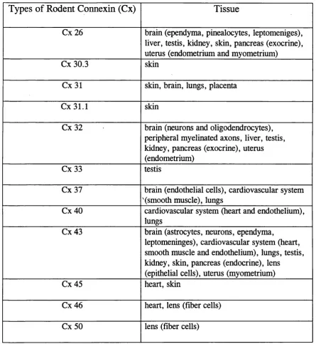
Table 1.1. List of known rodent connexins and their tissue expression (adapted from Dermietzel and Spray, 1993, and Kumar and Gilula, 1996).
Types of Rodent Connexin (Cx)
Tissue
Cx 26 brain (ependyma, pinealocytes, leptomeniges), liver, testis, kidney, skin, pancreas (exocrine), uterus (endometrium and myometrium)
Cx 30.3 skin
Cx31 skin, brain, lungs, placenta
Cx31.1 skin
Cx32 brain (neurons and oligodendrocytes), peripheral myelinated axons, liver, testis, kidney, pancreas (exocrine), uterus (endometrium)
Cx 33 testis
Cx 37 brain (endothelial cells), cardiovascular system '(smooth muscle), lungs
Cx40 cardiovascular system (heart and endothelium), lungs
Cx 43 brain (astrocytes, neurons, ependyma,
leptomeninges), cardiovascular system (heart, smooth muscle and endothelium), lungs, testis, kidney, skin, pancreas (endocrine), lens
(epithelial cells), uterus (myometrium)
Cx 45 heart, skin
Later it was reported that rat Cx 32 and Cx 26 (Barrio et aL, 1991) as well as murine Cx 40 and Cx 37 (Henneman et al., 1992) can form functional heterotypic gap junction channels. In contrast to the reports of Werner et al. (1989) and Swenson et al. (1989), White et al. (1995), using a similar xenopus model system, demonstrated that oocytes injected with Cx 32 and Cx 43 mRNA do not show any electrical coupling when the endogenous xenopus connexin (Cx 38) was blocked with antisense oligonucleotides. These results suggest that the permeability of gap junctions is a connexin specific parameter determined by the protein subunits constituting the channel. Further, the homotypic and heterotypic channels formed by the same connexins differ in their permeability (White et al., 1994).
As an alternative to the oocyte expression system, cultured mammalian cell lines that show low levels of gap junctional communication were transfected with connexin cDNAs. Examples of such cell line systems are Hela human cervix carcinoma cells (Henneman et al., 1992; Traub et al., 1994), C6 rat glioma cells (Zhu et al., 1991) and N2A murine neuroblastoma cells (Beyer et al., 1992). Although both oocyte and mammalian cell expression system yielded comparable results, in a recent study using the Hela cell model system Elfgang et al. (1995) has demonstrated that dye transfer is different between several connexin transfectants even though the junctional conductance is similar, suggesting that the permeability of different connexins to cellular molecules may vary. Further, they have shown by transfer of Lucifer yellow that B-type connexins (eg. Cx 32 and Cx 26) can form functional channels only with each other and not with a-type connexins (eg. Cx 43 and Cx 40). However, one cannot exclude the possibility of electrical conductance between the a and B type connexin transfectants.
hemichannels has not been illustrated. This suggests that hemichannels or connexons are homo-oligomeric in nature, composed of a single constituent connexin (Risek et ah,
1994). Epstein and Gilula in 1977 showed that gap junctional coupling can occur across species and even across vertebrate classes in vitro. However, the question remains whether the cells that coupled made the same connexin or atleast a connexin of the same type.
Recent advances in the field of molecular biology has made specific gene ablation possible. Thus, mice in which Cx 43 genes were ablated gave birth to homozycote embryos that died shortly after birth due to cardiac malformations. Histological examination of these homozycote embryos revealed no overt abnormalities in organs expressing Cx 43, except in the heart which was deformed (Paul, 1995; Reaume et al., 1995). Cultures of astrocytes from these knock-out animals expressed higher levels of Cx 40, compared to controls (Spray et al., 1995). These results suggest that most cells in the absence of a particular connexin gene can upregulate the expression of a complementary connexin, which may not be present normally at detectable levels and, thereby, maintain intercellular communication.
1.3.6. Formation of Gap Junctions
into gap junctional plaques. In general, oligomerization of most membrane proteins takes place in the endoplasmic reticulum (ER) (Hurtley and Helenius, 1989). Thus, assembled oligomers of connexins have been shown in the membranes of ER of BHK cells transfected with Cx 32 cDNA (Kumar et al., 1995). Similarly, analysis of subcellular fractions of rat liver also showed that connexins oligomerize in the ER (Rahman et al., 1993). In contrast, another study showed that, unlike other integral proteins, connexins undergo post-ER oligomerization into connexons within the trans- Golgi complex (Musil and Goodenough, 1993). Such post-ER oligomerization has been implicated in preventing the possible intracellular pairing of connexons and subsequent formation of gap junction plaques within the ER.
molecules such as N-CAM or L-CAM (Mege et al., 1988; Musil et al., 1990; Musil and Goodenough, 1991: Meyer et al., 1992). However, it has been shown that although cell adhesion molecules may contribute to the formation of gap junctions through a feedback mechanism involving a signal transduction pathway, they are apparently not essential for gap junction formation (Kumar and Gilula, 1996).
Studies, using Novikoff hepatoma cells and eye lens fibre (Decker et al., 1974; Johnson et al., 1974), have shown that in the first 30 minutes of cell reassociation the two adjacent membranes, relatively poor in intramembranous particles (IMP) on the P fracture face, flatten to make the ‘formation plaque’. Large 10 nm IMP appear on the P face and almost simultaneously small particles also appear intermingled with the large ones. At this stage, the intercellular space is rapidly reduced between the membranes, from 10 nm to 2-3 nm. After 60 minutes of cell reassociation, the large 10 nm particles apparently disappear leaving behind the completed gap junctions, suggesting that the large 10 nm particles may be the precursor connexons (Peracchia, 1980).
1.3.7. Regulation of Gap Junctions
cytoplasmic pH (Turin and Warner, 1977), free Ca (Rose and Loewenstein, 1975; Loewenstein, 1981), voltage (Spray et aL, 1979) and cyclic nucleotides (Saez et aL, 1986).
Modulation of gap junctions by neurotransmitters was first demonstrated in pancreatic acinar cells treated with acetylcholine (Findlay and Petersen, 1982). Recently, dopamine has been shown to modulate gap Junction permeability in the rat neostriatum (Cepada et aL, 1989) and in amacrine cells of the rabbit retina (Hampson et aL, 1992). Noradrenaline is another neurotransmitter that modulates the permeability of gap junctions (Saez et aL, 1991).
Peptides and steroid hormones, including estrogen and progesterone, may also regulate gap junction formation and degradation. Estrogen stimulates the gap junction formation while progesterone inhibits the same. De novo protein synthesis has been shown to be necessary for the formation of gap junction precursors in the rat myometrium. Inhibition of protein synthesis with cyclohexamide (inhibits translation) and actinomycin D (inhibits transcription) has been shown to reduce gap junction size and number (Garfield et aL, 1988). Other studies, however, indicate that de novo
protein synthesis may not be required for gap junction formation in all cells (Kannan and Daniel, 1978).
Conductance of gap junctions may be gated by voltage (Spray et aL, 1985). In
1.3.8. Degradation of Gap Junctions
The apparent life span of connexins in tissue culture conditions is several hours (Traub et al., 1989). Gap junctions are removed from the plasma membrane by internalization of the entire junction (Larsen, 1983). Initially, one of the coupled cells interdigitates into the other followed by endocytosis of the interdigitation. The resultant vesicle with a double membrane structure is known as an ‘annular gap junction’ and is ultimately degraded within lysosomes.
1.3.9. Gap Junctions as Morphological Correlates of Electrical Coupling
Knowledge of intercellular exchange of small ions comes from measurements of electrical resistance between neighbouring cells. Low intercellular resistance indicates a free exchange of ions between coupled cells, but does not allow one to discriminate between positively and negatively charged ions. Low resistance junctions were first recognised and studied in excitable tissues by Furshpan and Potter (1957), but such junctions were also detected in non-excitable tissues (Loewenstein and Kanno, 1964). Loewenstein (1966) reported that electrotonic junctions that are responsible for low resistance pathways must contain continuous passages for ions to flow between two connected cytoplasms. Such passages, he suggested, must be limited by high resistance seals or diffusion barriers at the level of the intercellular space to prevent the leakage of ions to the exterior.
of gap junctions became separated, while the intercellular resistance increased to complete cell uncoupling. A year later, Dreifuss et al., (1966) clearly showed that gap junctions are responsible for the electrical coupling in cardiac muscle tissue. In this study, cardiac muscle which contains both gap junctions and desmosomes, was incubated in , Ca^"*" free medium that separated the desmosomes leaving the gap junctions intact. Following this treatment, adjacent cells in the tissue were still found to be ionically coupled.
Another direct demonstration for the involvement of gap junctions in cell coupling came from the study of induced synchrony in spontaneously beating mouse and chick myocardial cells in culture (Goshima 1969, 1970). In these studies, cells that beat at different frequencies became synchronised as soon as they came in contact with one another. Synchronous beating must have been produced by the spread of current through newly formed gap junctions, since myocardial cells that were in contact with mouse-L-cells, incapable of forming gap junctions, did not acquire synchrony.
1.3.10. Evidence for Metabolic Coupling Through Gap Junctions
These authors concluded that the metabolism of IPP negative cells was altered by the exchange of substances through intercellular channels.
The most convincing study in this area, performed by Gilula et al. (1972), was a three-way correlation between metabolic and ionic coupling and ultrastructural localization of gap junctions. In this study, Don hamster (IPP positive cells), a variant DA hamster (IPP negative cells) and A9 mouse fibroblast (IPP negative cells) cell lines were used. When Don cells, marked with carnitine, were mixed with unlabelled DA or A9 cells, DA cells that were in contact with Don cells incorporated tritiated hypoxanthine at a reduced level, whereas the A9 cells never incorporated label above background. Similarly, ionic coupling was found between Don-DA cells, DA-DA cells and Don-Don cells but not between Don-A9 or A9-A9 cells. Morphological analysis revealed the presence of gap junctions at the electron microscopical level between pairs of cells that showed metabolic and ionic coupling but not between cells where physiological coupling was absent. Since gap junctions were the only type of junctions that were detected in these cultures, it was concluded that the simultaneous occurrences of metabolic and ionic coupling depends on the presence of gap junctions.
respond by an increase in spontaneous beat frequency, only when cell-cell contact was present. The interpretation was that the cAMP synthesized in the grannulosa cells had diffused to the myocardial cells through the gap junctional coupling.
To determine the range of sizes of particles that pass through gap junctions, many investigators have used molecules of different sizes tagged with fluorescence or colour. Lucifer yellow and neurobiotin are some of the widely used tracers to investigate the presence of gap junction coupling as well as to study their selective permeability.
1.3.11. Evidence for Intercellular Calcium Signalling Through Gap Junctions
wave that was communicated to multiple surrounding cells. These results provide direct evidence that intercellular Ca^"^ signalling occurs through gap junctions.
A plausible model suggests that rise of cytoplasmic Ca^"^ stimulates phospholipase C to break down inositol lipid. Inositol 1,3,5-trisphosphate (IP3), a lipid metabolite, is capable of releasing Ca^'*’ from an internal store and, thereby, perpetuating the wave of phospholipase C activity (Harootunian et al., 1991; Meyer and Stryer, 1991). The role of IP3 in Ca^"^ was shown by Saez et al (1989). These authors demonstrated that gap junctions are permeated by Ca^'*’ and IP3 by testing for Ca^"^ response to micro injections of IP3, a second messenger. It was shown that the injected
IP3 diffused through gap junctions and released intracellular Ca^"^. This has been later confirmed by the prevention of Ca^^ waves with IP3 receptor antagonists (Boitano et al., 1992; Sneydetal., 1994).
1.3.12 Gap Junctions in Pathological Conditions
32 coding regions have been amplified, from genomic DNA of the CMTX patients using PGR and have been expressed in the Xenopus oocytes (Bruzzone et al., 1994). None of these mutants formed functional gap junctions between paired oocytes, supporting the notion that such mutations of Cx 32 resulting in CMTX involves loss of junctional communication. However, it would appear that some of the Cx 32 mutations would be less disruptive to channel function than others, a possible explanation for the varying degrees of severity seen among CMTX patients (Spray and Dermietzel, 1995).
In the VAH inherited disorder, which manifests from mild to lethal forms of severity, appears to arise by fundamental perturbations in the left-right patterning during cardiac development (Britz-Cunningham et al., 1995). It has been shown that in patients with VAH, point mutations are present in phosphorylation sites of Cx 43 expressed in heart (Britz-Cunningham et al., 1995). Further analysis of transfected cells with one of the mutant Cx 43 genes showed abnormal regulation of protein kinases.
1995). Similar response has been noted in the expression of astroglial Cx 43 in ipsilateral facial nucleus following facial nerve lesions (Rohlmann et al., 1994).
It was suggested more than a decade ago that electrical coupling is strengthened between hyper-excitable neurons with electrotonic connections synchronizing neuronal activity in regions that were proned to epileptogenesis (Dudek et al., 1983, 1986). This has been supported by the recent findings of synchronized spontaneous neuronal activity (Baimbridge et al., 1991). Further evidence that coupling strength might increase in hyper-excitable brain regions comes from the study in which epileptic foci exhibited marked increase in the expression of Cx 43 mRNA in epileptic cortices compared with nonepileptic controls (Naus et al., 1991). Furthermore, several reports have provided evidence that anti-epileptic drugs block gap junction channels (Spray and Dermietzel, 1995).
The use of cellular and molecular approaches has made it possible to investigate where particular connexins are expressed in the CNS. It is now known that Cxs 26, 32 and 43 are the major gap junctional proteins that are expressed in the CNS (Dermietzel and Spray, 1993). Connexin 43 is expressed in undifferentiated ectodermal cells and subsequently in both neural and lateral ectodermal cells. As the brain vesicles become established and with the closure of neural tube, both Cxs 26 and 43 are expressed in abundance (Dermietzel et al., 1989; Ruangvoravat and Lo, 1992; Yancy et al., 1992).
In situ immunolabelling and primary cultures of neuronal and non-neuronal cells have shown that in the adult brain, Cx 43 is expressed in astrocytes, leptomeninges, ependyma and endothelial cells (Dermietzel et al., 1989, 1991; Micevych and Abelson, 1991; Yamamoto et al., 1989, 1990). The expression of Cx 32 has been shown in oligodendrocytes, and among neuronal subpopulations in the brainstem, mesencephalon, cerebral cortex, basal ganglia (Nagy et al., 1988; Dermietzel et al., 1989 ; Micevych and Abelson 1991). Connexin 26, expressed during development of the brain is later confined to non-neuronal tissues such as the ependyma and leptomeninges (Dermietzel et al., 1989). Dermietzel et al. (1989) also showed that the amount and pattern of connexin expression changes during development. These findings strongly suggest that the developing brain requires a particular complement of connexins and the functional properties may be important for the epigenetic events that determine the fate of the neuroepithelial derivatives (Dermietzel and Spray, 1993).
and details of junctional morphology. Gap junctions have been described between neurons of the mouse / trigeminal nucleus (Hinnrichsen and Larramendi, 1968), basket cells of the rat cerebellar cortex (Sotelo and Llinas, 1972), dendritic appendages/ of cerebellar glomeruli (Sotelo, 1975) and between axon terminals and neuronal j perikarya in the retina (Dowling and Boycott, 1966). Ultrastructural analysis has shown the occurrence of gap junctions between glial cells involving astrocytes and oligodendrocytes (Massa and Mugnaini, 1982). However, no experimental evidence is yet available to illustrate heterologousfoupling between these two cell types. In vitro
studies have shown the presence of weak electrical coupling in the absence of dye passage between oligodendrocytes and astrocytes (Ransom and Kettenman, 1990). There is also increasing physiological evidence that suggests glial cells are capable of extensive cell-cell communication including glial to glial and bi-directional glial to neuronal signalling (Charles, 1994). Neuro-glial electrical coupling has been previously demonstrated in vitro by Walker et al (1969), and recently direct Ca^^ signalling from astrocytes to neurons has been suggested to occur through gap junctions ( Nedergaard 1994). More recently, Nedergaard et al., (1995) have shown that gap junctions are required for the propagation of spreading depression (SD) and suggested that Ca^'*’ waves in vitro share the same features of SD in vivo.
In the adult retina, gap junctions are present between amacrine cells and ganglion cells in a highly ordered manner. They have been identified with tracer studies, and electron microscopical approaches (Fujisawa et al., 1976; Penn et al., 1994) in the vertical pathway from photoreceptors to ganglion cells, and linking specific cell types in mosaic arrays (Mobbs et al., 1988; Vaney et al., 1991; Vaney, 1994; Cook and Becker, 1995).
1.5.
Gap Junctions in the Neocortex
During early corticogenesis, the neuroepithelial cells of the ventricular zone are coupled into clusters by Lucifer yellow permeable gap junctions, which may be analogous to ‘proliferative units’ observed in primate neocortex. The number of cells within the coupled clusters decrease with differentiation of precursor cells and neuronal migration (Lo Turco and Kreigstein, 1991). As gap junction channels have a unitary conductance of 100 ps, it follows that approximately 13 such channels connect one cell to another in a cluster. Finally these investigators have concluded that many, if not all the cells of a cluster, are neuroblasts. Although, the gap junction proteins within these clusters have not yet been identified, the most probable candidates are Cxs 26 and 43 (Dermietzel et al., 1989, 1993).
decreased to 40% by 18 days and to 20% in the adult cortex. In a previous study by Gutnick and Prince (1981), 44% of the adult guinea pig cortical neurons coupled by Lucifer yellow, were confined to superficial layers. Similarly, injection of Lucifer yellow into physiologically identified glial cells resulted in coupling of surrounding glial cells (Gutnick et al., 1981).
Peinado et al. (1993b) using Neurobiotin, a low molecular weight intracellular tracer, have demonstrated that dye coupling occurs primarily through dendrosomatic and dendrodendritic contacts. These investigators, with the aid of gap junction blockers, demonstrated that Neurobiotmcoupling to surrounding neurons is mediated through gap junctions. Further, their study also showed that cortical cells exhibits a cell-type selectivity in coupling. Thus, injections of neurons failed to label cells that were morphologically identified as glia and vice-versa. Among neurons too, injection of smooth stellate cells resulted in clusters containing only nonpyramidal cells (Peinado et al., 1993b).
through gap junctions the rest of the cells either electrically or biochemically, (Kandler and Katz, 1995).
Gap junctional coupling in the adult neocortex has been speculated to be involved in the mediation of subthreshold oscillations responsible for synchronizing neuronal firing. Weak coupling between adult cortical neurons may be necessary to orchestrate collective behaviour in neuronal assemblies to ensure that the influence of no single neuron predominates (Peinado et al., 1993a). Gap junctions, the morphological correlates of dye transfer and electrical coupling, have been sighted in the motor and sensory areas of the adult primate neocortex (Sloper and Powell, 1972) and rat frontal cortex (Peters, 1981). However, neuronal gap junctions have not yet been described with electron microscopy in immature neocortex. It is possible that functional coupling can sometimes persist without organized gap junctions due to low spatial density (Connors et al, 1983).
1.6.
Aims of the this Study
(Hatten and Mason, 1990; Fishell and Hatten, 1991; Roberts et al., 1993), no evidence has yet been documented relating gap junction mediated interaction between the neuroepithelial progenitors to corticogenesis. In this context, the Cx 43 gene knocked- out mice which showed high levels of Cx 40 labelling in the brain, suggests that the role of gap junction mediated intercellular communication is essential for the normal development of the brain.
As a first step in a long term investigation of gap junction mediated interaction and their modulatory effects in the developing and mature cerebral cortex, the present study was designed to examine the major connexins (Cxs 26, 32 and 43) expressed in the cerebral cortex, with respect to their time of expression, distribution and abundance. Although the existence of low resistance pathways mediated by gap junctions have been demonstrated in the developing cortex, gap junctions or their constituent connexins mediating such pathways have not been characterized. Thus, using quantitative immunocytochemistry in frozen sections, the expression, pattern and density distribution of Cxs 26, 32 and 43 were examined in the brains of embryonic (embryonic days 12, 14, 16 and 19) and postnatal (postnatal days 0, 3, 7, 14, 21, 28 and 60) rats. In the adult rats, these analyses were performed in cytoarchitectonically and functionally distinct cortical areas to ascertain possible differences in the expression of connexins which may reflect the intrinsic variation of the respective cortical areas.
To characterize the cortical cells expressing the constituent connexins and to investigate multiple connexin expression, double immunolabelling of sections and acute preparations of dissociated cortices were performed using antibodies directed against cell specific markers and connexins. In addition, to characterize and assess the potential of the developing neuroepithelial cells expressing connexins, in vitro studies were conducted using a dissociated cell culture system.
CHAPTER TWO
List of Contents
2.1. Introduction - 55
2.2. Materials - 56
2.3. Methods - 57
2.3.1. Methods of fixation and Processing of Tissue for Connexin
immunocytochemistry - 57
2.3.2. Characterization of anti-connexins 32 and 43 antibodies - 62 2.3.3. Connexin immunocytochemistry using
immunofluorescence - 62
2.3.4. Control experiments - 63
2.3.5. Quantitative analyses - 64
2.3.6. Statistical analyses - 65
2.3.7. Characterization of cells expressing connexins - 65
2.3.8. Double-immunofluorescence - 67
2.4. Results - 68
2.4.1. Specificity of connexin antibodies - 68 2.4.2. Distribution of connexin immunoreactivity in the
cerebral cortex - 68
2.4.3. Cell specificity of connexins - 69
REGIONAL DIFFERENCES IN THE DISTRIBUTION AND CELLULAR
EXPRESSION OF CONNEXINS IN THE ADULT CEREBRAL CORTEX
2.1. Introduction
It is widely believed that intercellular communication between cortical neurons takes place through chemical synapses. Although recent studies have provided evidence of developing cortical neurons being extensively tracer coupled, similar studies have shown that adult neurons are coupled to a lesser degree (see 1.4). However, the demonstration of connexins in different regions of the adult brain, including neocortex, is suggestive of a non-synaptic mode of communication mediated by gap junctions in the mature brain (Yamamoto et al., 1990; Micevych and Abelson,
1991).
The mammalian cerebral cortex is subdivided into areas characterized by unique cytoarchitectonie features and functions. The visual cortex of the rat, consisting of 3 distinct cytoarchitectonie areas, is located in the occipital region of the cerebral hemispheres. Area 17, the primary visual cortex, bordered by area 18 and 18a is approximately 1.5 mm thickness (Krieg, 1946). The primary somatosensory cortex, area 3, is the largest sensory area occupying most of the lateral and dorsolateral cortical walls above the gustatory area (Bayer and Altman, 1991). This region has received considerable attention in rodents due to discrete cortical representation in layer IV of individual facial vibrissae, the ‘barrels’ (Woolsey and | Van der Loos 1970). The motor cortex, area 6, located near the frontal pole, is the primary source
the
of corticospinal tract (Bayer and Altman, 1991). The present study examines the A'
2.2. Materials
Animals'. 20 adult Sprague Dawley albino rats weighing between 250 - 300 g were used in this study.
Chemicals’. 0.1 M phosphate buffered saline (PBS) - 1 tablet/100 ml of distilled water. 4% paraformaldehyde in 0.1 M PBS - a stock solution of 8% paraformaldehyde was made by dissolving 8 g in 100 ml of distilled water at 65° C to which 5-6 drops of 0.1 M NaOH was added until the solution became clear. This solution was then diluted to 4% with 0.2 M PBS.
liquid nitrogen, anaesthetic ether, isopentane, sucrose, OCT, L-lysine, methanol, citiflor, poly-L-lysine, laminin.
Cell culture media’. Hanks balanced salt solution (HBSS-Sigma), trypsin, EDTA, DNAse (Boehringer), Dulbecco’s modified Eagle’s medium (Sigma), foetal calf serum (Gibco).
Immunochemicals and reagents’, normal goat serum (Sigma), pre-immune rabbit serum (Sigma), bovine serum albumin, anti-connexin antibodies (donated by Prof. W.H.Evans, University Wales College of Medicine, Cardiff), anti-MAP-2 monoclonal antibody (Boheringer), anti-GFAP monoclonal antibody (Sigma), biotinylated- goat anti-mouse and goat anti-rabbit secondary antibodies (Vector), goat anti-rabbit-conjugated to FITC (Sigma), streptavidin Texas red and streptavidin fluorescene (Amersham).
2. 3. Methods
2.3.1. Methods of Fixation and Processing of Tissue for Connexin
Immunocytochemistry
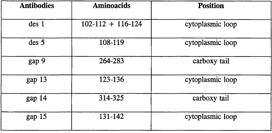
Table 2.1. Amino acid sequences of oligopeptides synthesized to raise different Cx 32 (des 1, des 5, gap 9) and Cx 43 (gap 13, gap 14 and gap 15) antibodies and their
positions in the parent proteins
Antibodies Aminoacids Position
des 1 102-112 + 116-124 cytoplasmic loop
des 5 108-119 cytoplasmic loop
gap 9 264-283 carboxy tail
gap 13 123-136 cytoplasmic loop
gap 14 314-325 carboxy tail
Fig. 2.1. Localization of the different Cx 43 (gap 13, 14, and 15) oligopeptides in the parent connexin molecule. Note that these epitopes are located in the cytoplasmic domain and the carboxy tail, the least conserved regions, of the connexin molecule.
/
membrane
cytoplasm
NH2 GAP 15
GAP 13
Fig. 2.2. Localization of the different Cx 32 (gap 9, des 1 and des 5) oligopeptides in the parent connexin molecule. Note that these epitopes are located in the cytoplasmic domain and the carboxy tail, the least conserved regions, of the connexin molecule.
membrane
cytoplasm
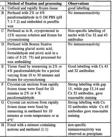
Table 2.2. A study conducted to test the efficiency of different anti Cxs 32 and 43 antibodies and to optimize tissue fixation for quantitative immunocytochemistry
Method of fixation and processing Observations A Unfixed and rapidly frozen tissue good labelling B Perfused with 2% or 4%
paraformaldehyde in 0.1 M PBS (pH 7.1-7.2) and embedded in paraffin wax
No immunoreactivity
C Perfused as in B, cryoprotected in 12% sucrose solution and frozen for cryosectioning
Non-specific labelling of nuclei with Cxs 32 and 43 antibodies
D Perfused with Bouins fixative (containing glacial acetic acid, formaldehyde and picric acid in a ratio of 5:25; 75) and processed for wax embedding
No immunoreactivity
E Tissue fixed by immersing in 2% or 4% paraformaldehyde for a period varying from 10 to 30 minutes and frozen for cryosectioning
Good labelling with Cx 43 and 32 antibodies
F Cryostat-cut sections from rapidly frozen tissue were fixed for 10 minutes in 2% or 4 %
paraformaldehyde
Strong labelling with gap 15, while gap 13,14 and Cx 32 antibodies, gave reasonable staining G Cryostat cut sections from rapidly
frozen tissue were fixed by immersing in methanol for 10 minutes at room temperature or at 4°C
Strong labelling with Cx 32 antibodies while Cx 43 antibodies gave reasonable staining
H Fixed with a mixture containing acetone and methanol (1:1)
non-specific
2.3.2. Characterization of Anti Connexins 32 and 43 Antibodies
Oligopeptides consisting of short sequences of amino acids coupled to hemocyanin were synthesized by FMOC polyamide solid phase chemistry and used to generate polyclonal antibodies in rabbits. These antibodies were characterized by immunoblotting and immunocytochemistry in tissues where the expression of connexins is well documented. For example, the immunoreactivity of Cx 32 was tested in rat liver, as gap junctions between hepatocytes are known to be composed predominantly (90%) of Cx 32 (Paul, 1986). Similarly, contractile cardiac tissue was chosen to demonstrate Cx 43 immunoreactivity as gap junctions between intercalated discs are known to be composed of Cx 43 (Gourdie et al., 1991). The gap 15 antibody, raised against the rodent Cx 43, has been used to study the expression of this protein by immunocytochemistry in mouse breast and heart (Monaghan et al.,
1994, 1996; Becker et al., 1995), and by Western blotting in rodent heart (Becker et al., 1995). In addition, it has been shown to recognize Cx 43 in porcine endothelial cells, heart and lung tissues of pig, rat and guinea pig, using immunocytochemistry and Western blotting (Carter et al., 1996). The des 5 antibody has been similarly studied by Western blotting in rodent liver (Rahman and Evans, 1991; Rahman et al.,
1993).
2.3.3. Connexin Immunocytochemistry Using Immunofluorescence
thickness, collected on poly-L-lysine coated glass slides* were fixed with 4% paraformaldehyde for Cx 43 immunoreactivity, while, those used for Cx 32 labelling were treated with methanol. After fixation, all sections were rinsed in 0.1 M PBS for 5 minutes and blocked for non-specific labelling with a solution containing O.IM lysine and 0.1% bovine serum albumin followed by treatment with 5% normal goat serum in O.IM PBS, each for 20 minutes. All sections were incubated with one of the connexin antibodies overnight at 4°C in a humid chamber. Preliminary studies carried out with varying dilutions (1:50, 1:100, 1:150, 1:200, 1:250 and 1:500) of antibody solutions showed optimal connexin labelling at 1:100 dilution. Incubations with primary antibodies were followed by second and third layers of biotinylated goat anti-rabbit (diluted 1:100) and streptavidin fluorescein (diluted to 1:100) each for 2 hour at room temperature, with two 10 minutes washes in between using 0.1 M PBS. All sections were mounted with citiflour, a water based antifade medium, and analysed using a laser-scanning confocal microscope.
* Preparation o f poly-L-Lysine coated glass slides: glass microscopic slides were immersed in concentrated chromic acid overnight, followed by thorough washing in running tap water (3-4 hour) and distilled water. Cleaned slides were then immersed in poly-L-lysine solution, diluted to 1:10, fo r at least 10 minutes and dried at 37°C.
2. 3. 4. Control Experiments
Peptide competition tests: oligopeptides of gap 15 and des 5 were added to aliquots of corresponding antisera (diluted to 1:100) to make a series of dilutions with final concentrations as 20, 40, 60, 80 and 100 pg/ml (w/v). The solutions were well mixed using a vortex and allowed to stand for 30 minutes at room temperature before applying to sections.
Negative controls were performed on sections of brain, liver and heart tissue with pre-immune serum (10% in 0.1 M PBS) or by processing for immunocytochemistry in the absence of primary antibodies.
2. 3. 5. Quantitative Analyses
A total of 20 serial sections, 10 pm thick, cut saggitally from the midline of each of 5 animals were used for quantitative analysis of Cxs 32 and 43 immunoreactivity. Alternate sections were used to analyze each connexin expression and for every section analyzed, from three cytoarchitectonically distinct areas of the cortex (visual, somatosensory, and motor cortex) (Fig. 2.3), a sequential series of non-overlapping images together spanning the entire thickness of the cortex were collected. Analysis of density distribution and size of immunoreactive particles was carried out according to the method described by Gourdie et al. (1991), using the PC- Image image analysis package. Briefly, a 3 x 3 median filter was passed over the whole image to remove background noise and single pixels. The pixel intensity threshold was then adjusted to a range from 70-140 on the 0-255 level grey scale,
ranging from 141-255 in order to measure the brighter particles. This was carried out in all images collected from 10 sections from each of 5 animals per connexin.
Images collected through the cortical thickness of the three areas were photographed using TMX-100 film. Light micrographs, printed at x 2.5 enlargement,
I the
were assembled into cortical strips extending from the pial surface to^white matter in the same sequence of order as the images were collected. The punctate immunolabelling was then copied on to tracing paper overlaid on the cortical strips.
*
A preliminary study was carried out with 30 images selected from sections labelled with varying levels o f Cx 32 and Cx 43 immunoreactivity. Analyses o f this trial study showed that at a minimal threshold of 70 in the 0-256 grey scale level, almost all the labelled particles were demarcated in the binary image.
2. 3. 6. Statistical Analyses
Average levels of connexin immunoreactivity for each cortical area was determined by summing up the labelled puncta in all the images spanning the cortical thickness. Differences in the expression of a connexin in the three cortical areas were examined using one way - analysis of variance (ANOVA) test on the data collected from 5 animals (n=5). To examine for size difference between the immunoreactive particles of the two connexins unpaired, two tailed. Students t-test was used.
2.3.7. Characterization of Cells Expressing Connexins