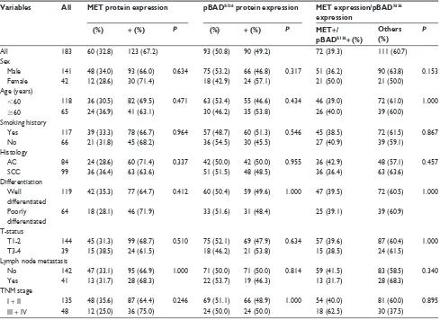
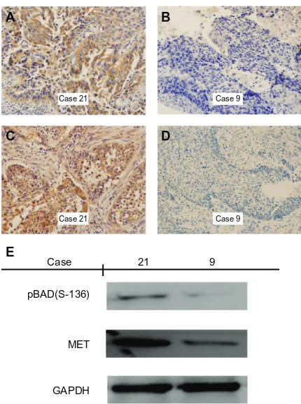
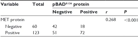
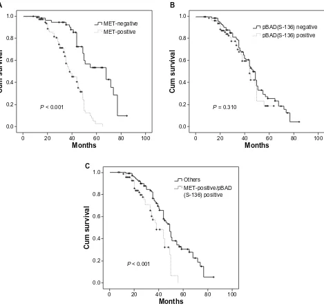
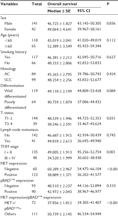
Expression and prognostic relevance of MET and phospho-BAD in non-small cell lung cancer
Full text
Figure
Related documents
The PowerOnt ontology model presented in this paper is specifically designed to model nominal, typical and real power consumption of each device in a home and its modular design
Para ilustrar la natu- raleza de la orientación normativa dominante y el riesgo de centrarse en ella en la investigación y el análisis, traigo al texto extractos de tres proyectos
This study aimed to assess if the participant with lesser degree of glucose intolerance increases the incidence of false positive diagnosis of gestational diabetes mellitus with
TQM relies on soft concepts like leadership, respect, integrity, trust, honesty, commitment, customer satisfaction, openness and high ethics, clarity of vision, problem solving
RESULTS AND DISCUSSION: A total of 40 ethnomedicinal plant species including herb, shrub, tree, and vine distributed across 29 families were documented in the study
http://ijessr.com Page 103 This study aims to explore and study how aesthetic education able to gain the student’s critical consciousness as the impact of
The recommendation made by Bain and Company Consultants that most drastically impacted the operations of De Beers was the vertical integration to include retailing into the De
On the other hand, a path planning algorithm, based on Fast Marching, has been used to generate a path that takes into account both locomotion modes and the best one depending on
