Stem cell horizons in intervertebral disc
degeneration
Joseph Ciacci1
Allen Ho1,2
Christopher P Ames3
Rahul Jandial4
1Division of Neurosurgery, University
of California, San Diego, La Jolla, California, USA; 2Del E Webb
Neurosciences, Aging and Stem Cell Research Center, The Burnham Institute for Medical Research, La Jolla, California, USA; 3Department
of Neurological Surgery, University of California, San Francisco, San Francisco, CA, USA; 4Division
of Neurosurgery, Department of Surgery, City of Hope Cancer Center, Duarte, CA, USA
Correspondence: Rahul Jandial Division of Neurosurgery, Department of Surgery, City of Hope Cancer Center, Duarte, CA, USA
Tel +1 626 471 700 Fax +1 626 471 7344
Email rahuljandial@hotmail.com
Abstract: Intervertebral disc degeneration remains a pervasive and intractable disease arising from a combination of aging and stress on the back and spine. The growing fi eld of regenera-tive medicine brings the promise of stem cells in the treatment of disc disease. Scientists and physicians hope to employ stem cells not only to stop, but also reverse degeneration. However, there are many important outstanding issues, including the hostile avascular, apoptotic physi-ological environment of the intervertebral disc, and the diffi culty of obtaining mesenchymal stem cells, and directing them towards chondrocytic differentiation and integration within the nucleus pulposus of the disc. Given the recent advances in minimally invasive spine surgery, and developing body of work on stem cell manipulation and transplantation, stem cells are uniquely poised to bring about large-scale improvements in treatment and outcomes for degenerative disc disease. In this review we will fi rst discuss the cellular and molecular factors infl uencing degeneration, and then examine the effi cacy and diffi culties of stem cell transplantation.
Keywords:intervertebral disc degeneration, stem cells, disc disease, mesenchymal stem cells, stem cell transplantation
Introduction
Two anatomically distinct regions comprise the cartilaginous intervertebral discs (IVD) of the human spine. Concurrently, the nucleus pulposus and annulus fi brosus provide both fl uid and viscoelastic support within the IVD. First, the central nucleus pulposus (NP) occupies the internal structure of disc. It is fi lled with collagen type II extracellular matrix (ECM) and hydrophilic proteoglycans. The unique extracellular matrix of the NP provides “shock absorbing” capacity to the IVD derived from the water content of its components. The NP is enveloped by the second component of the IVD, the annulus fi brosus (AF). The AF is mainly collagen type I, and forms a fi brotic circumferential boundary to the more liquid NP (Paesold et al 2007). Consequently, the AF functions to gird the viscoelastic NP and provide structural integrity and resistance to its extrusion when compressive forces are applied to the NP.
The cellular composition of the IVD forms three separate sections. The concentric lamellae of collagen I fi bers of the AF surround the NP. The NP contains two unique types of cells: a population of primitive notochordal cells, and a population of chondrocytic cells. The former are most likely the vestige of an embryonic notochord cell that directed the development of the IVD and spine. They disappear in humans after approximately 10 years of age. This may be due to differentiation into chrondrocytic cells or apoptosis. Both regions of the IVD are bound above and below by endplates of cartilage. Some experts believe that these cells play a role in the IVD niche that provides for successful mesen-chymal stem cell (MSC) differentiation into the cells of the NP (Hunter et al 2003).
Mechanisms of disc degeneration
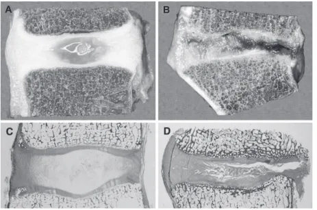
degeneration focuses on the NP because of its importance in maintaining a healthy and functioning IVD (Figure 1). Specifi cally, the ECM of the NP fails to maintain homeostasis for adequate collagen and proteoglycan synthesis (Sive et al 2002). The fi rst step in understanding this phenomenon begins with identifying the molecular phenotype of the NP cells. No marker currently exists to distinguish these cells from common hyaline cartilage cells, since both have similar ECM macromolecules. Furthermore, even if stem cells could be instructed to differentiate into hyaline cartilage, they would still lack the essential fl uid properties of the NP, and would fail to recreate normal function of the IVD. Also of tantamount importance is being able to separate the different cells. This could be accomplished fi rst by elucidating the ratio of proteoglycans and collagen within the NP (Mwale et al 2004). Eventually, both the repopulation of NP cells and the concomitant reproduction of adequate ECM, with a normal proteoglycan/collagen ratio, must be realized for stem cell regeneration of the IVD to be considered.
Among the culprits to be targeted when considering IVD degeneration are the issues of diffusion of nutrients,
cell viability, proteoglycan synthesis, and disruptions in collagen production leading to decreased proteoglycan and collagen II in the IVD disc matrix. Molecular mediators such as degrading enzymes, infl ammatory mediators, and growth factors are involved in all the above processes.
Matrix metalloproteinases (MMPs) are the best charac-terized matrix degrading enzyme, and are major players in IVD degeneration. MMPs degrade various collagens, but their activity is post-transitionally inhibited by the binding of inhibitor of matrix metalloproteinases (TIMPs). The contribution of these enzymes to IVD degeneration most likely results from an imbalance of normal ECM collagen degradation by MMPs and MMP inhibition by TIMPs (Le Maitre et al 2004). Proteoglycan and cathepsins degrading enzymes, such as aggrecanase-1 which targets aggrecan, have also been suspects in IVD degeneration.
Pain manifesting from IVD degeneration may be linked to participating molecular infl ammatory mediators. Though heterogeneous models have generally been used to investi-gate cytokines, they clearly play a role. Some interleukins identifi ed may even recruit MMPs. In the same way, growth
factors involved in IVD degeneration, such as transforming growth factor beta (TGF-B), insulin growth factor-1 (IGF-1), and fi broblast growth factor (FGF), have also been examined. These factors function as mitogens that increase rates of mitosis and proliferation of essential IVD components. For instance, TGF-B induces increased proteoglycan production and inhibits MMPs (leading to less ECM degradation) in disc cells (Pattison et al 2001). Bone morphogenic proteins (BMPs) produce similar effects as growth factors but are considered morphogenic since they are highly chondrogenic. However, though their function has been delineated, no clear correlation has been seen between factor levels and IVD degeneration. Effects on degeneration varied widely in the presence of different factor levels, although some of this confusion is probably attributable towards the heterog-enous assortment of investigations, some of which included extruded discs. Defi nitive studies remain outstanding, but many hypothesize that increased growth factor expression is a likely physiological response to disc herniation that encourages disc repair.
Transcription factors have also been implicated in degen-eration since ECM turnover is also under the direction of genetic regulation. SMADs and latent membrane protein-1 (LMP-1) are intracellular regulators that stimulate proteo-glycan and collagen II synthesis by upregulating BMP-2 and BMP-7 (Yoon et al 2004). Sox9 is another factor that promotes collagen II expression within the disc by increasing collagen II mRNA transcription (Li et al 2004).
The molecular pathways involved in IVD degeneration are not all the natural consequences of stress and aging, but can also be infl uenced by genetics. Gene polymorphisms for ECM proteins, like aggrecan, have been associated with disc and early multilevel degeneration (Doege et al 1997). A polymorphism in cartilage intermediate layer protein (CLIP) has also been recently correlated with susceptibility to disc degeneration (Seki et al 2005). Because of the limited innervations and blood supply of the intervertebral discs, the pathology of their degeneration hinges upon the interaction between a wide array of environmental, genetic, and molecu-lar factors. Therefore, the therapeutic strategy will have to be a concerted approach that addresses all these issues.
The aforementioned mechanisms for molecular and cellular degeneration are important components of the clini-cal presentation of disc degeneration. However, it should be noted that not every patient that has degenerative disc disease has back or leg pain (Jensen et al 1994). This underscores the fact that the mechanisms of pain are not well under-stood. The degenerated IVD is associated with progressive
dessication, loss of biomechanical properties, loss of disc height, and in some cases disc herniations (Haefeli et al 2006). These entities must be clinically appreciated because stem cell mediated IVD repair would not obviated the need to remove a herniated disc compressive a spinal root. Cur-rent experience and knowledge focus on stem cell mediated IVD repair with the objective of returning the disc ECM to its premorbid state and allowing for the imbibition of water and subsequent return of disc height. This may or may not be associated with pain relief, but currently offers the best cellular strategy for disc repair. In fact, a cellular strategy could be used for multiple aims, not just disc repair, but also the delivery of anti-infl ammatory agents to help with the ultimate clinical objective- reduction of pain.
MSCs and the IVD
Originally discovered 30 years ago, mesenchymal stem cells (MSCs) are the elusive vehicle for cell therapy in the IVD (Friedenstein et al 1976). They are valued for their multipotency, or ability to differentiate into cell types of mesenchymal origin (fat, bone, cartilage, etc.), and down the necessary cell lineage for regeneration or replacement of degenerated disc cells (Prockop et al 2003). There are two main strategies for acquisition of these desired somatic stem cells: through manipulation of embryonic stem cells (ESCs), or through extraction of MSCs from the fat or bone marrow of the patient.
ESCs are extracted from the inner cell mass of blastocyst stage embryos. The promise of ESCs lies in their plasticity and immortality. These cells are pluripotent, that is, through specialized culture techniques, ESCs can be induced to differentiate into cells of all three germ layers (endoderm, mesoderm, and ectoderm). Indeed, the plasticity of these cells is much greater than any one group of somatic stem cells such as bone marrow derived MSCs. Furthermore, once in culture, ESC lines can be maintained indefi nitely, providing an everlasting source of cells for implantation. There is still much to uncover about how to derive chondrocytes or even MSCs from human ESC lines, and the danger of teratoma formation is always present with ESCs (Trounson 2002). Instead of direct MSCs differentiation, recent investigations in the fi eld point towards neural crest stem cells (generated from ESCs) as a source of MSCs, presenting an alternate pos-sibility of a reliable and effi cient method of MSCs derivation from human ESCs (Lee et al 2007).
an autologous source of cells with low risk of infection and immunogeniticity. In addition, there is a long history of in vitro manipulation and clinical in vivo investigations for orthopedic trials with these cells (Horwitz et al 2002). Despite the fact that true MSC yield from bone marrow aspirate is less than 0.01%, their proliferative capacity of makes this a suffi cient amount for MSC or MSC derived chondrocyte mediated IVD regeneration (Pittenger et al 1999). Adipose tissue is also an attractive substitute source of MSCs because of the relative ease in procuring fat over bone marrow. However, some studies have found that the gene expression profi les of bone marrow derived MSCs match that of native cartilage more closely when differentiating into chondrocytes (Winter et al 2003). Bone marrow and adipose derived MSCs also have differing properties when evaluated for cell surface markers (De Ugarte et al 2003; Huang et al 2005). Thus, until adipose MSCs are more thoroughly investigated and understood, bone marrow MSCs remain the most effi cacious option for stem cell IVD therapy.
Preclinical studies of MSC transplantation exist with disc degeneration typically induced with a needle puncture method, and subsequent evaluation of degeneration before and after MSC transplantation. Though this does not accurately represent the complex disease and degeneration process that normally occurs in humans, it is successful in demonstrating the regenera-tive capabilities of MSCs in the presence of IVD dessication
(Leung et al 2006). Sakai and colleagues (2006) used a rabbit model of nucleus aspiration to induce degeneration. They injected MSCs embedded in an atelocollagen matrix where they persisted over a month, amplifying the proteoglycan content of targeted discs. Implantation of autogenic MSCs was shown to preserve annular structure, restablishe disc nuclei positive for glycosaminoglycan and keratin sulfate proteoglycans, and partially restore disc height and hydration in similar studies (Leung et al 2006).
MSC derivation
The limiting factor for exploiting stem cells for therapeutic use is obtaining well characterized cells for transplantation. Directing the appropriate differentiation of MSCs (and ESCs) is a complex molecular and cellular puzzle that is contingent upon not only the inherent properties of cells, but also the environment in which they are cultured.
The soluble factors TGF-B and BMP are necessary
components of culture media used to induce in vitro
chondrogenic differentiation of MSCs. In fact, careful use of soluble factors in media can lead to chondrogenesis with a genetic profi le more analogous to IVD tissue than articular cartilage (Figure 2) (Steck et al 2005). Another method of alter MSC microenvironment to trigger chondrogenic differentiation involves co-culturing with different cell populations to take advantage of cell-cell contact and molecular signal activation.
A
osteonectin osteonectin collagen II collagen II collagen I collagen I
prelp prelp
comp comp
chondroadherin chondroadherin
decorin decorin
osteopontin osteopontin
fibromodulin fibromodulin
biglycan biglycan
osteocalcin osteocalcin collagen III collagen III
aggrecan aggrecan
collagen XI collagen XI
clip clip
lumican lumican
mia mia
collagen X collagen X
0 250 500 750 0 250 500 750 1000 1250 1500 1750 2000
ind MSC
ind MSC cartilage
IVD tissue
B
nd
4633
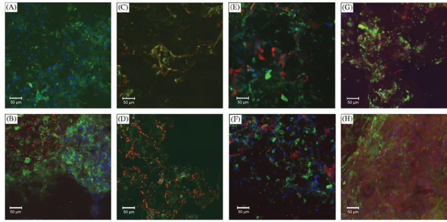
Utilizing the autocrine and paracrine factors secreted by one cell type leads to the activation of cell surface receptors on MSCs. Experiments culturing human NP cells and MSCs found that differentiation was reliant on cell-cell contact by looking at gene expression of Sox9, type II collagen, and aggrecan (Figure 3) (Richardson et al 2006).
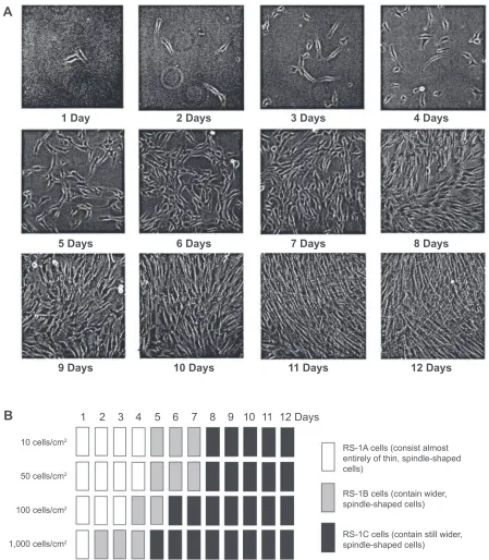
The three dimensional properties of the culture system have also shown to exercise substantial infl uence on the process of cell fate determination. MSCs are pelleted down into a dense micromass before addition of soluble factors to recreate the in vivo state that leads to cartilage formation. This structure helps direct the chrondrogenic cascade of MSC differentiation from micromass into cartilage. Mesenchymal condensation allows for extracellular signaling molecules such as Wnt glycoproteins and N-cadherin to form cadherin and connexin adhesion complexes for the beginning stages of ECM formation. Cartilage then begins to form on this three dimensional scaffold. Plating density of MSCs prior to soluble factor addition also infl uences the effi ciency of differentia-tion (Figure 4). This is because plating density can change the cell morphology; specifi cally, wider spindle shaped cells corresponding with denser MSCs plating. (Figure 5). Wider cells also have an increased propensity to differentiate after exposure to soluble factors in vitro (Sekiya et al 2002). In this way, employing density dependent culturing techniques can produce cartilage formation from MSCs in vitro, and increase
the effi ciency of MSC differentiation. Another technique described to direct MSCs toward the desired cell fate has been with co-culture systems, where the differentiated cell types provide the autocrine factors to increase the amount of nucleus pulposus cells in vitro (Yamamoto et al 2004). Clearly, there are many microenvironmental considerations to be made when designing an optimal in vitro MSC culturing system (Le Visage et al 2006). Exposure to a particular microenvironment may result in physiological variations that can be genetically perpetuated to daughter cells, epigenetically conditioning them to a particular cell fate (Gregory et al 2005).
MSC implantation to IVD
and its options
Stem cell therapy can easily be adapted in the clinical setting (with or without MHC typing) once an appropriate delivery “package” of cells and molecular adjuncts is devised. The objective, with our present understanding, would be to restore disc height by decreasing the dessication of degenerated discs. Although, disc degeneration is associated with back pain, it is clearly not the only contributor. As such, stem cell mediated disc repair would aim to remedy on component of a complex disease entity. Knowledge and experience gained, along with increasing understanding of multifacto-rial mechaisms of back pain would lay the foundation for further advances. With that noted, the delivery “package”
50 μm
50 μm
50 μm
50 μm
50 μm
50 μm
50 μm
50 μm
A 600
500
400
300
200
100
0
0 1 2 3 4 5 6 7 8 9 10 11 12 Days
0 0
1 2 3 4 5 6 7 8 9 10 11 12 Days
1,000 cells/cm2
100 cells/cm2
10 cells/cm2
50 cells/cm2
100 cells/cm2
1,000 cells/cm2
10 cells/cm2
50 cells/cm2
B
Fold increase
To
tal cells (million)/60-cm
2 dish
1.0 1.5
0.5
Figure 4 Relation between initial plating density and expansion of MSCs. Passage 3 MSCs (donor 89L) were plated on 60 cm2 dishes at 10, 50, 100, and 1,000 cells/cm2. The cells
were harvested and counted at 1 to 12 days. Fold increase A) and total cell numbers per 60 cm2 dish B) are shown. Data are expressed as mean ± standard deviation (n = 3).
would not only consist of growth factors for effective MSC engraftment, but could also include various biodegradable gels or grafts to provide initial biomechanical support and a structure for biological reconstruction of more severely degenerated discs. Allogenic stem cells could be implanted in minimally degenerated discs via percutaneous techniques.
If disc degeneration was more sever, stem cells could be transplanted with minimally invasive surgical and radio-logical techniques through a true lateral retroperitoneal approached guided by discogram. Employing this minimally invasive approach would also allow for co-implantation of a biodegradable interbody graft, that would be replaced by the
A
B
1 Day 2 Days 3 Days 4 Days
8 Days 7 Days
6 Days 5 Days
9 Days
12 Days
RS-1A cells (consist almost entirely of thin, spindle-shaped cells)
RS-1B cells (contain wider, spindle-shaped cells)
RS-1C cells (contain still wider, spindle-shaped cells)
1
10 cells/cm2
50 cells/cm2
100 cells/cm2
1,000 cells/cm2
2 3 4 5 6 7 8 9 10 11
10 Days 11 Days 12 Days
Figure 5 Initial cell density and time in culture affect cell morphology. Passage 3 MSCs were plated at 10, 50, 100, and 1,000 cells/cm2. A) Representative pictures of MSCs
plated at an initial cell density of 50 cells/cm2 at 1 to 12 days. B) Schematic diagram of MSC culture morphologies at four different initial cell densities from 1 to 12 days.
engrafted stem cells. Ultimately, the best clinical intervention would incorporate a personalized strategy for each patient and the degree and type of disc degeneration identifi ed. The tools for this strategy would include, but not limited to, cell transplants, biodegradable implants, and internal fi xation as needed.
A large number of NP like cells will need to be generated for profi cient IVD repair. The NP cell type is not well charac-terized, but NP cells identifi ed by their production of the req-uisite ECM components can suffi ce. Though autologous bone marrow derived MSCs are the ideal candidates for therapy, allogenic MSCs could serve as a more economical substitute. MSCs lack HLA class II receptors and sibling donors should not generate an immune response in recipients (Yang 2007). A cocktail of MSCs or differentiated NP cells would have to include molecules that promote the survival and engraftment of transplanted cells, and be rigorously tested in large animal models before clinical trials are undertaken. It will also be imperative to develop approved protocols of MSC culture and differentiation with strict quality controls to ensure safety before application in humans. Once the science and methods of MSC manipulation are better elucidated, IVD regeneration with stem cell therapy could be a viable treatment option for patients. In fact, osteogenesis imperfecta is already being treated effectively with MSCs and presents a model of the potential of MSCs for regenerative medicine. Developing this therapy to the unique cartilaginous tissue of the IVD could be the safest, most effi cient application of stem cell biology to disease processes treated by surgeons.
Disclosure
The authors report no confl icts of interest in this work.
References
De Ugarte D, Alfonso Z, Zuk P, et al. 2003. Differential expression of stem cell mobilization-associated molecules on multi-lineage cells from
adipose tissue and bone marrow. Immunol Lett, 89:267–70.
Doege K, Coulter S, Meek L, et al. 1997. A human-specifi c polymorphism in the coding region of the aggrecan gene. Variable number of tandem repeats produce a range of core protein sizes in the general population.
J Biol Chem, 272:13974–9.
Friedenstein A, Gorskaja J, Kulagina N. 1976. Fibroblast precursors in normal
and irradiated mouse hematopoietic organs. Exp Hematol, 4:267–74.
Gregory C, Ylostalo J, Prockop D. 2005. Adult bone marrow stem/progenitor cells (MSCs) are preconditioned by microenvironmental “niches” in
culture: a two-stage hypothesis for regulation of MSC fate. Sci STKE,
2005:pe37.
Haefeli M, Kalberer F, Saegesser D, et al. 2006. The course of macroscopic
degeneration in the human lumbar intervertebral disc. Spine,
31:1522–31.
Horwitz E, Gordon P, Koo W, et al. 2002. Isolated allogeneic bone marrow-derived mesenchymal cells engraft and stimulate growth in children with osteogenesis imperfecta: Implications for cell therapy
of bone. Proc Natl Acad Sci U S A, 99:8932–7.
Huang J, Kazmi N, Durbhakula M, et al. 2005. Chondrogenic potential of progenitor cells derived from human bone marrow and adipose tissue:
a patient-matched comparison. J Orthop Res, 23:1383–9.
Hunter C, Matyas J, Duncan N. 2003. The notochordal cell in the nucleus
pulposus: a review in the context of tissue engineering. Tissue Eng,
9:667–77.
Jensen M, Brant-Zawadzki M, Obuchowski N, et al. 1994. Magnetic resonance imaging of the lumbar spine in people without back pain.
N Engl J Med, 331:69–73.
Le Maitre C, Freemont A, Hoyland J. 2004. Localization of degradative enzymes and their inhibitors in the degenerate human intervertebral
disc. J Pathol, 204:47–54.
Le Visage C, Kim S, Tateno K, et al. 2006. Interaction of human mesenchymal stem cells with disc cells: changes in extracellular matrix
biosynthesis. Spine, 31:2036–42.
Lee G, Kim H, Elkabetz Y, et al. 2007. Isolation and directed differentiation of neural crest stem cells derived from human embryonic stem cells.
Nat Biotechnol, 25:1468–75.
Leung V, Chan D, Cheung K. 2006. Regeneration of intervertebral disc by mesenchymal stem cells: potentials, limitations, and future direction.
Eur Spine J, 15(Suppl 3):S406–13.
Li Y, Tew S, Russell A, et al. 2004. Transduction of passaged human articular chondrocytes with adenoviral, retroviral, and lentiviral
vectors and the effects of enhanced expression of SOX9. Tissue Eng,
10:575–84.
Mwale F, Roughley P, Antoniou J. 2004. Distinction between the extracellular matrix of the nucleus pulposus and hyaline cartilage: a
requisite for tissue engineering of intervertebral disc. Eur Cell Mater,
8:58–63; Discussion, 63–4.
Paesold G, Nerlich A, Boos N. 2007. Biological treatment strategies
for disc degeneration: potentials and shortcomings. Eur Spine J,
16:447–68.
Pattison S, Melrose J, Ghosh P, et al. 2001. Regulation of gelatinase-A (MMP-2) production by ovine intervertebral disc nucleus pulposus cells grown in alginate bead culture by transforming growth factor-beta(1)and
insulin like growth factor-I. Cell Biol Int, 25:679–89.
Pittenger M, MacKay A, Beck S, et al. 1999. Multilineage potential of adult
human mesenchymal stem cells. Science, 284:143–7.
Prockop D, Gregory C, Spees J. 2003. One strategy for cell and gene therapy:
harnessing the power of adult stem cells to repair tissues. Proc Natl
Acad Sci U S A, 100(Suppl 1):11917–23.
Richardson S, Curran J, Chen R, et al. 2006. The differentiation of bone marrow mesenchymal stem cells into chondrocyte-like
cells on poly-L-lactic acid (PLLA) scaffolds. Biomaterials,
27:4069–78.
Sakai D, Mochida J, Iwashina T, et al. 2006. Regenerative effects of transplanting mesenchymal stem cells embedded in atelocollagen to
the degenerated intervertebral disc. Biomaterials, 27:335–45.
Seki S, Kawaguchi Y, Chiba K, et al. 2005. A functional SNP in CILP, encoding cartilage intermediate layer protein, is associated with
susceptibility to lumbar disc disease. Nat Genet, 37:607–12.
Sekiya I, Larson B, Smith J, et al. 2002. Expansion of human adult stem cells from bone marrow stroma: conditions that maximize the
yields of early progenitors and evaluate their quality. Stem Cells,
20:530–41.
Sive J, Baird P, Jeziorsk M, et al. 2002. Expression of chondrocyte markers
by cells of normal and degenerate intervertebral discs. Mol Pathol,
55:91–7.
Steck E, Bertram H, Abel R, et al. 2005. Induction of intervertebral
disc-like cells from adult mesenchymal stem cells. Stem Cells,
23:403–11.
Trounson A. 2002. Human embryonic stem cells: mother of all cell and
tissue types. Reprod Biomed Online, 4(Suppl 1):58–63.
Winter A, Breit S, Parsch D, et al. 2003. Cartilage-like gene expression in differentiated human stem cell spheroids: a comparison of bone
marrow-derived and adipose tissue-derived stromal cells. Arthritis
Yamamoto Y, Mochida J, Sakai D, et al. 2004. Upregulation of the viability of nucleus pulposus cells by bone marrow-derived stromal cells:
signifi cance of direct cell-to-cell contact in coculture system. Spine,
29:1508–14.
Yang X. 2007. Immunology of stem cells and cancer stem cells. Cell Mol
Immunol, 4:161–71.
Yoon S, Park J, Kim K, et al. 2004. ISSLS prize winner: LMP-1 upregulates intervertebral disc cell production of proteoglycans and BMPs in vitro