Copyright © 2003, American Society for Microbiology. All Rights Reserved.
Three Mating Type-Like Loci in
Candida glabrata
Thyagarajan Srikantha, Salil A. Lachke, and David R. Soll*
Department of Biological Sciences, The University of Iowa, Iowa City, Iowa 52242Received 23 September 2002/Accepted 6 January 2003
Candida glabrata, the second most prevalent Candidaspecies colonizing humans, possesses three mating
type-like (MTL)loci (MTL1,MTL2, andMTL3). These loci contain pairs ofMTLgenes with their respective
coding regions on complementary Crick and Watson DNA strands. Each pair of genes is separated by a shared
intergenic promoter region, the same configuration found at the mating type loci ofSaccharomyces cerevisiae.
Two of theMTLloci,MTL1andMTL2, contain either theMTLa1/MTLa2 configuration or theMTL␣1/MTL␣2
configuration in different strains. All but one of the 38 testedC. glabratastrains were either aa␣or a␣␣. One
test strain was␣␣␣. Based on the mating type genotype, theMTLgenes at theMTL1orMTL2loci, and the
size of theXbaI fragment harboringMTL1orMTL2, four classes ofC. glabratastrains (I, II, III, and IV) were
distinguished. Northern analysis revealed that strains were either a-expressors or ␣-expressors and that
expression always reflected the genotype of either the MTL1 orMTL2 locus, depending on the class. The
expression pattern in each class, therefore, is similar to that observed inS. cerevisiae, which harbors two silent
cassette loci,HMRandHML, and the expression locusMAT. High-frequency phenotypic switching between
core phenotypes in an␣-expressing, but not in an a-expressing, strain modulated the level ofMTLexpression,
suggesting a possible relationship between core phenotypic switching and mating.
Candida glabratais the second most prevalentCandida spe-cies in humans (15, 17, 32, 38, 41). Although it is genetically far more related toSaccharomyces cerevisiaethan toCandida al-bicans(3, 42, 48), it mimics in many respects the pathogenic capabilities ofC. albicans, the most prevalentCandidaspecies, residing as a commensal in healthy individuals and causing vaginitis (15, 43, 44) and bloodstream infections (40). Recently, it was demonstrated that in the elderly, C. glabrata has emerged as the major commensal (32). The prominence ofC. glabrataas a pathogen is of particular clinical concern because it is naturally resistant to azole drug therapy (4, 16, 20, 36).
Until recently, it was generally assumed thatC. glabratadid not undergo the bud-hypha transition, and no reports had been published on phenotypic switching, two developmental pro-grams that contribute to the pathogenic success ofC. albicans. However, recent studies have demonstrated that C. glabrata forms pseudohyphae (12, 26, 27, 39), forms noncompartmen-talized tubes distinct from true hyphae (27), and undergoes high-frequency phenotypic switching (26, 27). C. glabrata, therefore, possesses developmental programs at least as com-plex as those ofC. albicans. Recently, it was demonstrated that C. albicans, which is diploid, possesses the mating type-like (MTL) lociMTLaandMTL␣(21) and that homozygousMTLa
and homozygousMTL␣strains will mate both in vivo (22) and in vitro (35). It was subsequently demonstrated that while most strains ofC. albicansare heterozygous for mating type at the MTL locus and do not undergo white-opaque switching, ho-mozygous strains do undergo the transition (33, 37), and the opaque phenotype dramatically facilitates mating (34, 37). Given the pathogenic similarities ofC. albicansandC. glabrata and the developmental correlates between the two species, we
have searched for and identified the mating type genes ofC. glabrata. We demonstrate that C. glabrataharbors three loci containing mating type-like genes, that two loci, MTL1 and MTL2, can contain either an MTLa1-MTLa2 or an MTL␣ 1-MTL␣2 configuration in different strains, and that based on expression patterns,C. glabratastrains can be distinguished as either a mating type or ␣ mating type. Our results further indicate that in any one class, only one of the threeMTLloci serves as the mating type expression locus.
MATERIALS AND METHODS
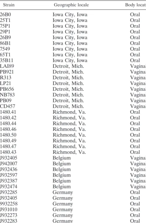
Maintenance and growth of strains.Thirty-eight different clinical isolates ofC. glabratapreviously genetically fingerprinted with theC. glabrataDNA finger-printing probe Cg12 (30) are described in Table 1. All strains were maintained on YPD agar slants (1.5% [weight/vol] agar, 2% [weight/vol] Bacto Peptone, 2% [weight/vol] glucose, 1% [weight/vol] yeast extract). For experimental purposes, cells were streaked on fresh YPD agar plates and incubated for 3 to 4 days at 25°C prior to use. For monitoring phenotypic switching, cells were plated on YPD agar containing 1 mM CuSO4(26).
Isolation and sequence analysis ofMTLloci.Based on homology comparisons with the MAT␣2p or MTL␣2p from the speciesS. cerevisiae,Kluyveromyces lactis, andC. albicans by using the multiple-alignment editor of Clustal W software developed by Michele Clamp (www.cmbi.kur.nl/bioinf/tools/clustalw .shtml), two highly conserved amino acid sequences containing homeobox do-mains were identified (1, 2, 21, 29). These sequences, WFAKKNIENPY and WVSNRRRKEK, were used to design the forward primer MP2F2 and the reverse primer MP2R1 (Table 2), respectively. These two degenerate primers were used to amplify similar homeobox-containing regions withTaqpolymerase (Life Tech/Invitrogen Inc., Gaithersburg, Md.) by PCR amplification ofC. gla-bratagenomic DNA. PCR products were purified by using the Wizard PCR Clean-up kit (Promega Corp, Madison, Wis.) and cloned into pGEM-T Easy (Promega Corp.). Plasmids pE30.8, pE30.9, and pE30.10, containing 440-, 125-, and 240-bp inserts, respectively, were sequenced in both directions with an ABI model 373A automatic sequencing apparatus and fluorescent Big Dye terminator chemistry (PE-ABI Inc., Foster City, Calif.). Protein-coding regions were deter-mined by the Wu-BLAST/BEAUTY Search algorithm-based program (49).
Sequence analysis and strain comparison.To obtain the full-lengthMTL1
locus and its flanking sequences, 105plaques of aC. glabratagenomic library of
strain 7549 (30) were screened with the 125-bp DNA fragment, which spanned the MAT␣2p homeodomain. Plaque lysates from 55 positive primary clones were used as a template for PCR, using MP2F1 and MP2R1 as primers (Table 2). Five * Corresponding author. Mailing address: Room 302 BBE,
Depart-ment of Biological Sciences, University of Iowa, Iowa City, IA 52242. Phone: (319) 335-1117. Fax: (319) 335-2772. E-mail: david-soll@uiowa .edu.
328
on September 8, 2020 by guest
http://ec.asm.org/
lambda clones, MP1,MP2, MP5, MP16, andMP18, which generated 125-bp PCR products, were chosen for secondary screening.MP1.1, which contained a 3.4-kb insert, was used to characterize the sequence of theMTL1
region and flanking sequences by lambda clone walking, using custom primers and the ABI sequencing apparatus. Similarly,MP16.1 was used to characterize theMTL2locus, andMP5.1 andMP18.1 were used to characterize theMTL3
locus. Based on the DNA sequence of MTL1, two primers, MLFLF2 and MLFLR2 (Table 2), spanning the flanking region of theMTL1open reading frames (ORFs) were designed and used in PCRs to generate homologous DNA regions from two additional strains, 1480.47 and PB921. The derived PCR prod-ucts were cloned into pGEM-T Easy and sequenced. Comparisons and align-ments of protein sequences were performed by using the multiple alignment editor of Clustal W/Jalview software (www.embi.kun.nl.bioinf/tools/clustalw .shtml). Promoter analysis was performed with the Matrix method for identifying putative regulatory protein binding sites in theS. cerevisiaePromoter Database (http://ctsigma.cshl.org/jiar/).
Southern and Northern analyses.Southern and Northern analyses were per-formed according to procedures previously described (26, 45, 46). In Southern analyses in which more than one probe was used, the originally hybridized blots were stripped at 75°C according to the protocols of Church and Gilbert (7). For Southern analysis, the hybridization probes forMTLa2/␣2,MTLa1, andMTL␣1 contained ORF regions derived from the primer pairs P1RACE1-CGPF1, A1RACE1-CGAR1, and CGPIF1-CGPIR2, respectively (Table 2). For South-ern analysis involving oligonucleotide-based hybridization to distinguishMTLa2 andMTL␣2 sequences among threeMTLloci, 500 ng of FuncP2 (Table 2) was end labeled with [32P]ATP by using T4 polynucleotide kinase as described by
sequences would also be conserved inC. glabratabased on the high level of genetic relatedness between it andS. cerevisiae(3, 42), degenerate primers that encompassed approximately 125 bp of theMAT␣2 ORF ofS. cerevisiaewere designed. Using these primers andC. glabrataDNA, three distinct PCR prod-ucts of 450, 240, and 125 bp were identified. Sequence analysis of the recombinant plasmids containing the PCR products revealed that the 125- and 450-bp inserts contained uninter-rupted ORFs flanked by highly conserved sequences. A BLAST-based sequence similarity search revealed that the 450-bp ORF did not encode a MAT␣2p but rather encoded a protein homologous to a JUN activation domain binding pro-tein or morphogenetic factor in humans and plants, respec-tively (6, 8). However, the BLAST search of the 125-bp PCR product revealed that it was derived from a locus homologous toMAT␣2. The deduced amino acid sequence of the 125-bp PCR product suggested that the ORF encoded 41 amino acids with identities of 69, 57, and 50% and similarities of 82, 77, and 65% to the MAT␣2p or MTL␣2p ofS. cerevisiae,K. lactis, and C. albicans, respectively.
Isolation and characterization of the MTL1a and MTL2a
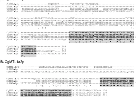
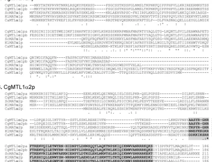
loci.The 125-bp fragment was used in turn as a probe to screen a C. glabrata EMBL3a library constructed from genomic DNA of strain 7549 (30) for the gene locus. Fifty-five putative clones were identified and tested for the presence of a 125-bp MAT␣2-like insert by PCR. One of these clones,MP1.1, was used to determine the sequence of a 2,587-bp insert by cus-tomized primer walking in both directions (Fig. 1A and 2A). A BLAST-type search of the sequence identified a 312-bp S. cerevisiaeMATa1p-like protein-coding sequence and a 510-bp S. cerevisiae MATa2p/MAT␣2p-like protein-coding sequence separated by a 180-bp putative promoter region. The locus containing these two coding sequences and intergenic se-quence will be referred to as mating type-like locus 1 (MTL1). TheS. cerevisiae MATa1-like coding sequence inMTL1, which we will refer to as MTL1a1, was 126 bp shorter than the S. cerevisiae MATa1 coding region. The deduced 104-amino-acid sequence had 28% overall similarity withS. cerevisiaeMATa1p (Fig. 3A). The-carboxy terminal 57 amino acids showed a high level of similarity with the carboxy-terminal regions ofC. albi-cans MTLa1p (21), K. lactis MATa1p (1), andS. cerevisiae MATa1p (2) (Fig. 3A). TheS. cerevisiae MATa2/MAT␣2-like coding sequence inMTL1was 150 bp longer than theS. cer-evisiae MATa2 coding region. The deduced 170-amino-acid sequence had an overall identity of 26% with bothS. cerevisiae MATa2p andS. cerevisiaeMAT␣2p. The carboxy-terminal 123 amino acids exhibited 31% identity with the carboxy-terminal 119 amino acids of bothS. cerevisiaeMATa2p andS. cerevisiae MAT␣2p (Fig. 3B). The carboxy-terminal two-thirds of MAT␣2p is identical to the full-length MATa2p inS. cerevisiae(2).
Align-LAI89 Detroit, Mich. Vaginal
PB921 Detroit, Mich. Vaginal
R313 Detroit, Mich. Vaginal
LP21 Detroit, Mich. Vaginal
PB656 Detroit, Mich. Vaginal
NB783 Detroit, Mich. Vaginal
PB09 Detroit, Mich. Vaginal
CD457 Detroit, Mich. Vaginal
1480.41 Richmond, Va. Oral
1480.42 Richmond, Va. Oral
1480.44 Richmond, Va. Oral
1480.46 Richmond, Va. Oral
1480.50 Richmond, Va. Oral
1480.49 Richmond, Va. Oral
1480.47 Richmond, Va. Oral
1480.43 Richmond, Va. Oral
J932405 Belgium Vaginal
J942007 Belgium Vaginal
J932436 Belgium Vaginal
J932597 Belgium Vaginal
J932387 Belgium Vaginal
J932474 Belgium Vaginal
J932285 Germany Oral
J932405 Germany Oral
9932258 Germany Oral
J931010 Germany Oral
J932273 Germany Oral
J932283 Germany Oral
J941814 The Netherlands Blood
aAll isolates used in this study were confirmed asC. glabrataby DNA
finger-printing with the probe,Cg12 (22). Each isolate was recovered from a separate human subject.
on September 8, 2020 by guest
http://ec.asm.org/
ment of MTL1a2p withS. cerevisiaeMATa2p and MAT␣2p re-vealed a region of highest similarity in the carboxy-terminal end, spanning 60 amino acids (Fig. 3B). This region includes a home-odomain signature sequence (WVXNRRR) (Fig. 3B) that is a near match with that ofC. glabrataandS. cerevisiae. This region has been implicated in ternary complex formation with DNA and MATa1p inS. cerevisiae(29). The two protein-coding regions in MTL1ofC. glabratastrain 7549 will be referred to asMTL1a1 andMTL1a2, respectively (Fig. 1A).
No potential methionine initiation amino acid could be iden-tified in frame in either MTL1a1p or MTL1a2p. Sequencing of three independent 5⬘ rapid amplification of cDNA ends (5⬘ -RACE)-derived PCR products ofMTLa1 mRNA revealed an additional 21-bp sequence upstream of the first isoleucine codon (Fig. 2A). 5⬘-RACE-derived PCR products ofMTL1a2 mRNA revealed an additional 15-bp sequence upstream of the first glutamine codon (Fig. 2B). 5⬘-RACE analysis, therefore, did not identify any typical AUG-type initiation codons or distinguish whetherMTL1a1 orMTL1␣2 transcripts are trans-lationally functional. The MTL1a1 andMTL1a2 ORFs were positioned on complementary Crick and Watson DNA strands, suggesting divergent transcription from the intervening pro-moter region, a configuration similar to that in S. cerevisiae (Fig. 1A) (2, 18).
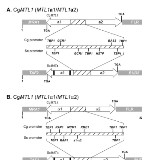
The 3⬘end ofMTL1a1 was flanked by the geneMRA1(Fig. 1A), which encodes a multicopy suppressor of RAS1 in S. cerevisiae (http://genome-www.stanford.edu/saccharomyces/). The 3⬘end ofMTL1a2 was flanked by the undefined sequence FLR (Fig. 1A). In S. cerevisiae, the functionalMATalocus is flanked byBUD5at the 3⬘end ofMATa1 and byTAF2at the 3⬘ end ofMATa2 (Fig. 1A), while the silentHMR andHML loci are flanked by YCRWDDta12/YCR097W-a and YCL068C/ HCl065W, respectively (http://genome-www.stanford.edu /saccharomyces/). The intergenic promoter region between MTL1a1 andMTL1a2 was similar in size to that forMATain S. cerevisiae(Fig. 1), but the sequence of theMTL1a1-MTL1a2 intergenic region was dissimilar to that ofS. cerevisiae MATa 1-MATa2, except for the presence of two putative binding sites forTBP1, one at the 3⬘end of each of the presumed overlap-ping promoters forMTL1a1 andMTL1a2 (Fig. 1A).
To identify a DNA fragment containing a second MTLa
locus in strain 7549, a negative PCR selection strategy, involv-ing two primer pairs for theMTL1flanking regions, was used to screen 55 primary lambda clones. The two primer pairs used, MLFLF3-MLFLR3 and FLR2-MLFLR2, represented the 3⬘ends ofMTL1a1 and MTL1a2, respectively (Table 2). Lambda clone16.1 was selected for sequencing, and a total of 1,960 nucleotides were determined by primer walking. Analysis of the nucleotide sequence revealed that the DNA fragment harbored anMTLa1 ORF and anMTLa2 ORF on Crick and Watson strands and that they were flanked by sequences dis-tinct from those flankingMTL1. We therefore designated this DNA fragmentMTL2.
The full-lengthMTL2a1 ORF, which included 472 nucleo-tides flanking the 3⬘end, was identical toMTL1a1. A stretch of 347 nucleotides at the 3⬘end ofMTL2a1 represented a unique flanking sequence of MTL2a. The full-length ORFs of both MTL1a2 and MTL2a2 were identical except for the last 11 codons of the former and 20 codons of the latter, which are unique prior to the same TGA stop codon. Thus, MTL2a2p is 9 amino acids longer than MTL1a2p. The MTL2a flanking region, which spans the 3⬘end ofMTL2a1, contained a short sequence homologous to S. cerevisiaeandC. albicans Nep1p
FIG. 1. Configurations of the C. glabrata MTL1 locus (A) The
MTL1locus containingMTL1a1 andMTL1a2 in strain 7549. (B) The
MTL1locus containingMTL1␣1 andMTL1␣2 in strains PB921 and 1480.47. In both cases comparisons are made to comparableS. cerevi-siaelociMATaandMAT␣.MRA1, 3⬘flanking region ofMTL1a1;FLR, 3⬘ flanking region of MTL1a2; TAF2, 5⬘ flanking region of the
MATa1/␣1 locus;BUD5, 3⬘flanking region ofMATa2/␣2. a1 and a2,
MTL1a1 andMTL1a2, respectively, orMATa1 orMATa2, respectively; ␣1 and␣2,MTL1␣1 andMTL1␣2, respectively, orMAT␣1 orMAT␣2, respectively. The thick black bars in MATa1 and MAT␣1 represent introns. Hatched portions of loci represent intergenic promoter re-gions. Arrow directions reflect orientation of transcription on comple-mentary Crick and Watson DNA strands.
TABLE 2. Primers used in this study
Primer Sequence
MP2F2 ...5⬘-TGGTTTGCAAAGAAYANNGAGAAYCCNTA-3⬘ MP2R1...5⬘-TTTTTCTTTTCTTCTTCTATTCGANACCCA-3⬘ PIRACE1 ...5⬘-GCCATCAAGGTAGGTCTGAAT-3⬘
CGPF1...5⬘-TAACCAACTTATATCATTGTGTTCCA-3⬘ AIRACE1 ....5⬘-GAACTTGATTGGTGGTGATCCCA-3⬘ CGAR1...5⬘-CAACACGGTAGGTTTACGATA-3⬘ CGPIF1 ...5⬘-ATGAAGTATACTGCCACAAAA-3⬘ CGPIR2...5⬘-CTGAGAGAATGACGGAGAGTGTA-3⬘ MLFLR2 ...5⬘-TATGTCTTGCGCGTCCAATTGCT-3⬘ MLFLF2...5⬘-ATAAGCAATCAGTATGTGTA-3⬘ FLR2...5⬘-ACATCACCAACATACGCACCGCT-3⬘ 12.36F1 ...5⬘-ATGTCAGTCTGAACTAGTGAATA-3⬘ MLFLF3...5⬘-AGGGACATCGCTGAGGCCAGA-3⬘ MLFLR3 ...5⬘-ATTGACCCAAGAAGTGGTGAGA-3⬘ MTLOCR1...5⬘-GCTTGCAATCAAAGTGTTCTG-3⬘ MTLOCF1 ...5⬘-GTGCTGATCACTATCGAATGC-3⬘
FuncP2...5⬘-TATTGGATTAAGTAGATGCGTAATACTAATT CTTGATTTCTTTGACAT-3⬘
on September 8, 2020 by guest
http://ec.asm.org/
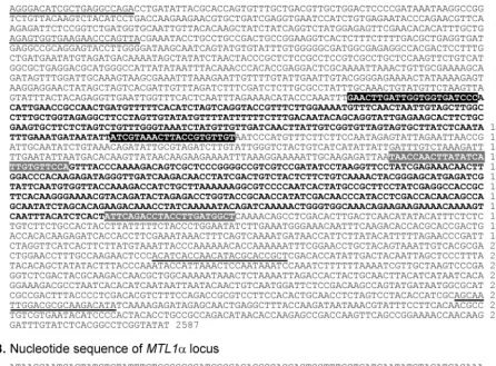
FIG. 2. Nucleotide sequences of theMTL1alocus derived from strain 7549 and theMTL1␣locus derived from strain PB621. (A) The ORFs forMTL1a1 andMTL1a2 are shown in boldface. The primer pairs forMTL1a1 (AIRACE1 and CGAR1) are shown as black boxes with boldface white print, and those forMTL1a2 (CGPF1 and PIRACE1) are shown as grey boxes with boldface white print. The primer pairs which flank the 3⬘end of theMTL1a1 ORF and the 3⬘end of theMTL1a2 ORF are underlined. (B) The ORFs forMTL1␣1 andMTL1␣2 are shown in boldface print. The primer pairs forMTL1␣1 (CGPIR1 and CGPIF1) are shown as black boxes with white print, and those forMTL1␣2 are shown as grey boxes with white print. The primers used to linkMTL1␣1 withMTL1␣2 are underlined. Forty-eight nucleotides at the 5⬘end of theMTL1␣2 ORF that are common toMTL1␣2 andMTL3␣2 are show by dashed lines. The rest ofMTL␣2, excluding the open-box sequence, is common toMTLa2. The nucleotide sequence unique to the 3⬘end of theMTL1␣2 ORF is shown in an open box.
on September 8, 2020 by guest
http://ec.asm.org/
(14). The 3⬘end ofMTL2a2 was flanked by the threonyl-tRNA and a short sequence homologous to the Fadd death effector domain (13).
Isolation and characterization of an MTL1 locus and an
MTL3locus containingMTL␣genes.To test whether the
con-figuration of theMTL1locus of strain 7549 was common to all C. glabratastrains, we cloned and sequenced anMTLlocus in two additional strains ofC. glabrata, PB921 and 1480.47. The primers MLFLF2 and MLFLR2 (Table 2), which represented the 3⬘ ends of the flanking regions ofMTL1a1 andMTL1a2, respectively, of strain 7549, were used to generate PCR prod-ucts from genomic DNA. While these primers generated a PCR product of 2,075 bp for strain 7549, they generated a PCR product of 2,269 bp from strains PB921 and 1480.47 (Fig. 1B). A BLAST-type search identified anS. cerevisiae MAT␣1-like coding sequence and anS. cerevisiae MATa2/MAT␣2-like cod-ing sequence (23). Because theMAT␣1-like andMAT␣2-like coding sequences were similar to those ofS. cerevisiae MAT␣1 andMAT␣2, respectively, the paired coding regions were des-ignatedMTL1␣1 andMTL1␣2. The flanking sequences of the MTL1␣ locus in strains PB921 and 1480.47 were identical to
those ofMTL1ain reference strain 7549. BothMTL1␣2 and MTL1␣1 contained uninterrupted ORFs beginning with codons for the initiation amino acid methionine. As in the case of theMTLa2-MTLa1 configuration of theMTL1andMTL2 loci in strain 7549,MTL1␣2 andMTL1␣1 were positioned on complementary Crick and Watson DNA strands, with an in-tergenic promoter (Fig. 1B and 2B). The MTL1␣ promoter encompassed 333 bp, which was 70 bp longer than theMAT␣
promoter ofS. cerevisiae. TheMTL␣promoter contained three TATA box binding protein sites and a putative binding site for the repressor-activator protein Rap1p, both found in the MAT␣2 promoter of S. cerevisiae(Fig. 1B). TheMTL␣ pro-moter of C. glabrata also contained putative Mcm1p and Rme1p binding sites, neither of which was present in the MAT␣promoter ofS. cerevisiae(Fig. 1B). There was no rec-ognizable binding site for the MATa1p-MAT␣2p heterodimer (a1-␣2) (Fig. 1B and 2B) found in the promoter ofS. cerevisiae (Fig. 1B).
TheMTL1␣1 ORF encodes a deduced protein of 184 amino acids with an overall identity of 29% and similarity of 51% with S. cerevisiae MAT␣1p (Fig. 4A). The carboxy-terminal 88 FIG. 3. Sequence comparison of MTL1a1p and MTL1a2p ofC. glabratastrain 7549 with MATa1ps and MATa2ps of other yeast species. (A) Aligned sequences ofC. glabrata(Cg) MTL1a1p and MATa1ps or MTLa1ps ofS. cerevisiae(Sc),C. albicans(Ca), andK. lactis(Kl).B. Aligned sequences ofC. glabrataMTL1a2p andS. cerevisiaeMATa2p and MAT␣2p. The shaded sequences in both panels represent the highly conserved amino acid residues (boldface) involved in the interaction betweenS. cerevisiaeMATa1p andS. cerevisiaeMAT␣2p in the formation of thea1-␣2 repressor complex. Note thatS. cerevisiaeMATa2p and MAT␣2p share identical amino acid residues in this region, but the physiological role of
S. cerevisiaeMATa2p is not known. Asterisks indicate identical residues, two stacked dots indicate conservative substitutions based on similar functional groups, and one dot indicates conservative substitutions based on similar effects on secondary structure. The accession numbers for sequences other than those ofC. glabrataare as follows: ScMATa1p, PO1366; CaMATa1p, AAD51404.1; KlMATa1p, AAG21094.1; ScMATa2p, CAA246201.1; and ScMAT␣2p, AAA34762.1. The accession numbers forC. glabrata MTL1aandMTL2aare AY191461 and AY191464, respectively.
on September 8, 2020 by guest
http://ec.asm.org/
amino acids showed 50% identity with MAT␣1p (Fig. 4A). The MTL1␣2 ORF encoded a deduced protein of 187 amino acids with an overall identity of 42% and similarity of 70% withS. cerevisiaeMAT␣2p (Fig. 4B) (18). The highly conserved car-boxy-terminal end, spanning 62 amino acids, exhibited 60% identity (Fig. 4B). MTL1␣2p was identical to MTL1a2p inC. glabratastrain 7549, except that the former was 16 amino acids longer at the amino-terminal end and contained an initial methionine. Since the primers used to clone theMTL1loci in strains PB921 and 1480.47 represented the flanking regions of MTL1in strain 7549, these results indicate that the sameMTL locus,MTL1, harbors alternativeMTL1aorMTL1␣genes in different strains.
A preliminary Southern analysis ofXbaI-digested DNA of strain 7549 hybridized withMTL1a2-␣2 revealed three bands, but the same Southern blot probed withMTL1a1 revealed only two of these bands, suggesting a third locus containing an MTL␣ ORF. To identify this third locus in strain 7549, a positive PCR selection strategy employing theMTL1␣1 ORF-specific primer pair CGP1F1-CGP1R2 (Table 2), was used to screen 55 primary lambda clones. Two clones, MP5.1 and
MP18.1, containing theMTL␣1 ORF were selected. The se-quence of 2,169 nucleotides was determined by a primer walk-ing strategy. This locus was designatedMTL3␣. Sequence anal-ysis revealed that this locus harbored divergently transcribed MTL3␣1 andMTL3␣2 ORFs, both organized similarly to the MTL1␣ locus (see Fig. 6B). The sequence of the MTL3␣1 ORF was identical to that of theMTL1␣1 ORF, except that the alanine codon at bp 152 was replaced by a serine codon. Al-though both theMTL1␣2 andMTL3␣2 ORFs contained iden-tical stretches of 164 amino acids at their amino-terminal ends, their carboxy termini were dissimilar. MTL3␣2p contained a unique stretch of 22 amino acids at the carboxy terminus that was not shared by MTL1␣2p (see Fig. 6B). The flanking re-gions ofMTL3contained unique sequences with no significant homology to any sequence in the database.
C. glabratacontains three independentMTLloci.To exam-ine the distribution of the three MTL loci among unrelated strains, Southern blot analyses were initially performed on the genomic DNAs of four independent strains digested with BamHI,EcoRI,PstI, orXbaI and probed with the full-length ORF ofMTL1a1,MTL1a2, orMTL1␣1. The four test strains FIG. 4. Sequence comparison of MTL1␣1p and MTL1␣2p ofCandida glabratastrains PB921 (CgMTL1␣1pa and CgMTL1␣2pa) and 1480.47 (CgMTL1␣1pb and CgMTL1␣2pb) with MAT␣1ps or MTL␣2ps and MAT␣2ps or MTL␣2ps of other yeast species. (A) Aligned sequences ofC. glabrataMTL␣1pa and MTL␣1pb with MAT␣1p ofS. cerevisiae(Sc), MAT␣1p ofK. lactis(Kl), and MTLa1p ofC. albicans(Ca). (B) Aligned sequences ofC. glabrataMTL1␣2pa with MAT␣2p ofS. cerevisiae, MAT␣2p ofK. lactis, and MTL1␣2p ofC. albicans. The shaded sequences in panel B and the method used to indicate conserved amino acid sequences are described in the legend to Fig. 3. The accession numbers for the sequences other than those ofC. glabrataare as follows: ScMAT␣1p, AAA34763.1; KlMAT␣1p, AAG21092.1 (note that the carboxy-terminal 42 amino acids of thisK. lactissequence are not shown); CaMAT␣1p, AAD51411.1; ScMAT␣2p, AAA34762.1; KlMAT␣2p, AAG21091.1; and CaMAT␣2p, AAD51408.1. The accession numbers forC. glabrata MTL1␣a,MTL1␣b, andMTL3␣are AY191462, AY191463, and AY207368, respectively.
on September 8, 2020 by guest
http://ec.asm.org/
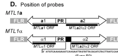
were isolated from different geographic locales and proved to be genetically unrelated when DNA fingerprinted with the complex species-specific probe Cg12 (data not shown). The Southern blot hybridization patterns of the four strains probed with a DNA sequence common toMTL1␣2 andMTL1a2 (re-ferred to asMTLa2-␣2) included two or three bands, depend-ing on the restriction enzyme (Fig. 5A and D). The XbaI patterns of the four test isolates probed with MTLa2-␣2 all contained 15-, 7- and 5.8-kb bands, which were designated A, B, and C, respectively (Fig. 5A). The A, B, and C bands representMTL3,MTL1, andMTL2loci, respectively. An anal-ysis of 34 additionalC. glabrataisolates revealed that all con-tained three bands either at 15, 7, and 5.8 kb or at 15, 5.8, and 5.2 kb (Table 3). SinceMTL1a2 was identical to the 414 bp of the 3⬘ end of MTL1␣2, Southern blot hybridization with MTLa2-␣2 could not distinguish between bands containing MTLa2 orMTL␣2 sequences.
In order to distinguish between MTLa2 and MTL␣2 se-quences, Southern blots ofXbaI-digested genomic DNAs of 38 strains were probed with the end-labeled antisense oligonucle-otide primer FuncP2 (Table 2; Fig. 5D), which is unique to the 5⬘ends ofMTL␣2 ORFs. The hybridization identified among the strains three fragments (A, B, and C) containingMTL␣2 ORF sequences. The total number of bands in any one strain was either one or two, with the exception of strain 1480.47, which contained three bands (Table 3). Fifty-eight percent of all strains exhibited only one band, and in those cases, it was always the 15-kb A band. Forty-one percent of strains con-tained a combination of A plus B or A plus C. Hybridization of the exceptional strain 1480.47 with FuncP2 showed that all three DNA fragments, A, B, and C, contained the MTL␣2 ORF (Table 3).
The Southern blot hybridization patterns ofXbaI-digested DNAs of the four test strains probed withMTLa1 (Fig. 5C) contained no bands, one band (band B), or two bands (bands B and C) (Fig. 5B). Southern blots of the remaining 34 isolates of the test collection probed withMTLa1 revealed patterns of one band (band B or C) or two bands (bands B and C) only (data not shown). In only one unique strain in the entire collection, strain 1480.47,MTLa1 did not hybridize to any band (Fig. 5B), although theMTL␣2-specific primer hybridized to three bands (Fig. 5C).MTLa1 did not hybridize to band A in any of the 38 tested strains, in contrast to theMTL␣2-specific primer (Table 3).
The Southern blot hybridization patterns ofXbaI-digested DNAs of the four test strains probed withMTL␣1 (Fig. 5D) contained either one band (band A), two bands (bands A and C), or three bands (bands A, B and C) (Fig. 5C). In the 34 remaining strains in the collection,MTL␣1 hybridized to one band (band A) or two bands (bands A and B or bands A and C) (data not shown), the same pattern observed with the FuncP2 probe, suggesting that theMTL␣1 ORF always pairs with the MTL␣2 ORF. The Southern blot hybridization pat-terns of all 38 test strains included band A (Fig. 4C; Table 3). In 58% of strains,MTL␣1 hybridized only to band A, the same group of strains in whichMTLa1 hybridized to bands B and C. We designate strains in this group class I (Table 3). In 18% of strains,MTL␣1 hybridized to both A and B; this is the same group of strains in whichMTLa1 hybridized only to band C. In this class, the B band exhibited a polymorphism of either 7 or
5.2 kb. Half of the strains in this class contained the 7-kb B band, and the remaining half contained the 5.2-kb B band. We designate strains in this group class II (Table 3). In 5% of strains,MTL␣1 hybridized to both A and C; this is the same group of strains in which MTLa1 hybridized only to band B. We designate strains in this group class III (Table 3). The distinguishing feature between class II and class III is the composition of MTL1and MTL2. In class II isolates,MTL1 includes MTL1␣1 and MTL1␣2, while in class III isolates, MTL2 contains this configuration. In class IV, there was no hybridization toMTLa1;MTL␣1 hybridized to bands A, B, and C (Fig. 5C). If we distinguish loci as aor␣ by whether they harborMTLa1 orMTL␣1, respectively, then these results sug-gest that class I strains possess twoMTLaloci and oneMTL␣
locus (aa␣), class II strains possess twoMTL␣ loci and one MTLalocus (a␣␣), class III strains possess twoMTL␣loci and one MTLa locus (a␣␣), and class IV strains possess three MTL␣loci (␣␣␣) (Tables 3 and 4).
To test the relationship between the bands containingMTL ORFs and the threeMTLloci,XbaI-digested DNAs of the 38 test strains were probed sequentially with the DNA fragments flanking the 3⬘ends of MTL1a1 andMTL1a2 of strain 7549. With strain 7549 DNA as template, the primer pair MLFLR2 and FLR2 (Table 2) generated a 395-bp PCR product that spanned the 3⬘flanking region ofMTLa2 inMTL1, and primer pair MLFLR3 and MLFLF3 (Table 2) generated a 246-bp PCR product that spanned the 3⬘flanking region ofMTLa1 in MTL1(Fig. 1A). Southern blots ofXbaI-digested DNAs from the 38 test strains probed with either the 395- or 246-bp PCR product revealed that both flanking regions of MTL1 were always associated with the B band, either 7 or 5.2 kb, in all strains (Table 3), indicating that eachC. glabratagenome con-tained only oneMTL1locus in the same relative position. All strains that exhibited bands B and C when probed withMTLa1 and only band A when probed with eitherMTL␣1 or FuncP2 (class I) exhibited only the 7-kb band B when probed with the 3⬘ flanking regions (Table 3), demonstrating that in these strains only one of the two MTLa1 genes is located in the MTL1locus. A majority of strains that exhibited bands A and B when probed withMTL␣1 exhibited hybridization with only the 5.2- or 7-kb band B (class II) when probed with the 3⬘
flanking regions (Table 3), demonstrating that in these strains only one of the twoMTL␣1 genes is located in theMTL1locus. Surprisingly, two strains that exhibited bands A and C when probed withMTL␣1 and FuncP2 (class III) hybridized only to the 7-kb band B when probed with the 3⬘ flanking regions, demonstrating that in these strains theMTL␣1 genes are lo-cated at the MTL2 locus. Southern blots of XbaI-digested DNAs from the 38 test strains probed with the 280-bp PCR product flanking the 3⬘end of theMTL2a1 ORF, which was generated with the primer pair MTLOCF1-MTLOCR1 (Table 2), revealed multiple hybridization bands, suggesting that the flanking region harbors a repeat element that is dispersed throughout the genome, in addition to its linkage to the C DNA fragments (data not shown). Interestingly, the complex hybridization pattern was identical among strains in each class, suggesting close genetic relatedness of strains within a class.
PCR analysis of MTL size and distribution. Although
Southern analysis revealed threeMTLloci (MTL1,MTL2, and MTL3) in each strain and discriminated four classes, it did not
on September 8, 2020 by guest
http://ec.asm.org/
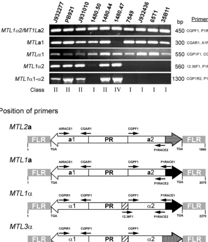
demonstrate that theMTLgenes were similar in size between strains and between loci. A PCR-based approach was used to investigate this question. To test for any variability in the se-quence common toMTL1a2 andMTL1␣2 at theMTL1locus, the primer pair CGPF1 and P1RACE1 (Table 2) was used to generate a single PCR product of 450 bp (Fig. 6B). This 450-bp PCR product was obtained in each of 10 test strains (Fig. 6A), which included five class I strains, four class II strains, and one class IV strain, and in each of the additional 28 isolates in the test collection (data not shown). These results demonstrate that the region common toMTLa2 andMTL␣2 is highly con-served inC. glabrata. Since the nucleotide sequences encom-passing PIRACE1 is present inMTL1a2andMTL1␣2 but not in the MTL3(band A) orMTL2(band C) locus, the results clearly suggest that the 450-bp PCR product must have been generated fromMTL1in all classes. To discriminate between (i)MTLa2 andMTL␣2 and (ii)MTL1␣2 andMTL3␣2, the 5⬘
primer 12.36F1, which is unique to the full-length MTL␣2 ORFs of either MTL1␣ or MTL3␣ loci, and the 3⬘ primer P1RACE1, which is common toMTL1a2 andMTL1␣2 but not
toMTL3␣2 (Fig. 6B), were employed. This primer pair gen-erated a 560-bp PCR product for the four class II strains and one class IV strain but produced no product for the five class I strains (Fig. 6A). These results therefore suggest that class II and IV strains contain an MTL1␣2 gene at theMTL1locus, while class I strains contain noMTL1␣2 gene. Southern anal-ysis revealed that class I isolates containedMTL␣1 andMTL␣2 solely at theMTL3locus in the A fragment (Tables 3 and 4). These PCR results (Fig. 6A) confirm that in class I isolates, MTL3␣1 is paired with anMTL3␣2-type sequence and not an MTL1␣2-type sequence in the A fragment.
To investigate further the linkage between MTL1␣1 and MTL1␣2, we employed the primer pair CGPIR2, which is common to both MTL3␣1 and MTL1␣1 (and probably MTL2␣1) and PIRACE1, which is unique only to the 3⬘end of MTL1␣2 (Fig. 6B) but is absent inMTL3␣2 and probably in MTL2␣2. These primers generated a 1,300-bp PCR fragment which included full-length MTL1␣1 and MTL1␣2 ORFs for the four class II strains and one class IV strain but not for any of the class I strains (Fig. 6A). These results demonstrate that in class II and IV strains,MTL1␣1 paired withMTL1␣2 at the MTL1locus. For two strains, PB921 and 1480.47, sequencing data confirmed thatMTL1␣1 was paired withMTL1␣2 in the B fragment (Fig. 1B and 2B). These results also confirm that MTL3␣1 is paired withMTL3␣2 and not with anMTL1␣2-type ORF in band B in class II strains.
To investigate whether the MTLa1 ORFs present at the MTL1 or MTL2 locus are the same size, the primer pair CGAR1 and A1RACE1 (Table 2), which should generate a single 300-bp PCR product of MTL1a1 (Fig. 6B), was em-FIG. 5. Southern blot analysis of theMTLloci ofC. glabrata. Ap-proximately 2.5g of total genomic DNA from strains J932285, 7549, 35B11, and 1480.47 were individually digested with the four restriction enzymes BamHI (B),EcoRI (E),PstI (P), andXbaI (X). Digested DNA was resolved in an agarose gel and transferred to a Hybond N nylon membrane. Triplicate Southern blots were individually hybrid-ized with the ORFs for MTL1 genes. (A) Hybridization with the 450-bp PCR fragment (primers CGPF1 and P1RACE1) containing sequences identical to bothMTL1a2 andMTL1␣2. (B) Hybridization with the 300-bp PCR fragment (primers CGAR1 and A1RACE1) containing theMTL1a1 ORF. (C) Hybridization with the 550-bp PCR fragment (primers CGPIF1 and CGPIR1) containing the MTL1␣1 ORF. Three distinct hybridizable fragments inXbaI-digested genomic DNA are designated A (15 kb), B (7 kb) and C (5.8 kb). (D) Schematic representation ofMTL1aandMTL1␣loci, showing the positions of probes. The primer pairs used to generate theMTL1ORF probes are described in Fig. 2. The sequence of theMTL␣2-specific oligonucleo-tide FuncP2 is also shown. The molecular sizes of the expected frag-ments are shown to the right and left of panels A and C and to the left of panel B.
on September 8, 2020 by guest
http://ec.asm.org/
ployed. This primer pair generated a 300-bp PCR product in the nine test isolates in classes I and II but not in isolates in class IV (Fig. 6A), which was consistent with the results of the Southern analysis, indicating that all strains but 1480.47 con-tained at least one copy ofMTLa1. These primers also gener-ated a 300-bp PCR product in the 28 additional isolates in the collection. Finally, to test whetherMTL␣1 ORFs present in all three loci are of the same size, the primer pair CGPIF1 and CGPIR1 (Table 2), which generated a single 550-bp PCR product containing theMTL␣1 ORF (Fig. 6B), was employed. This primer pair generated a 550-bp PCR product for all 10 test strains (Fig. 6A), as well as the remaining 28 isolates of the collection; these data are consistent with the results of the Southern analysis demonstrating that all strains contain at least one copy of MTL␣1. Since the sequences of MTL1␣1 and MTL3␣1 were identical, it was not possible to identify the origin of PCR products in class II, III, and IV strains.
Strain-specific transcription ofMTLgenes.InS. cerevisiae,
mating type genes are present at three loci (MAT,HML, and HMR) but are expressed only at one locus (MAT) (18). S. cerevisiaestrains can therefore be discriminated phenotypically asMAT␣orMATa, depending upon the genotype of theMAT locus. To test whether similar distinctions can be made forC. glabrata, and as a strategy for identifying a possible expression locus, 12 isolates were analyzed by Northern blot hybridization for expression of MTLa1 and MTL␣1, using the respective ORFs as probes. Three of the test strains were class I (J932283, J932436, and 35B11), eight were class II (PB921, LP21, PB656, CD457, 1480.46, 1480.41, J932474, and 1480.44), and one was class IV (1480.47). While class II and IV strains expressed MTL␣1, no class I isolates expressed it (Fig. 7). Conversely, while all tested isolates in class I expressedMTLa1, no class II or IV isolates expressed it (Fig. 7). Therefore, class II and class IV isolates can be classified as␣-expressors, while class I
iso-lates can be classified asa-expressors. Predicated on the cas-sette model inS. cerevisiae, predictions of theMTLexpression locus can be made based on the duplication of eitherMTL␣1 orMTLa1 in two of the threeMTLloci. While all class II and IV isolates possibly carry identicalMTL␣1 ORFs at both the MTL1andMTL3loci, class III isolates possibly carry identical MTL␣1 ORFs at both theMTL2andMTL3loci. Therefore, eitherMTL1orMTL3may represent the expression locus in class II, while either MTL2or MTL3may represent the ex-pression locus in class III isolates. Class I isolates, on the other hand, carried identicalMTLa1 ORFs at both the MTL1and MTL2loci. Therefore, in class I isolates, eitherMTL1orMTL2 may represent the expression locus.
Transcription of MTL genes during phenotypic switching.
Since the differential expression of MATa and MAT␣ genes confers cell type specificity inS. cerevisiae(2, 18, 23) and since white-opaque switching inC. albicansis intimately involved in the mating process (33, 34, 37), we compared expression of MTL␣1 andMTLa1 among the phenotypes of the reversible high-frequency core switching system ofC. glabrata (26, 27). Most strains ofC. glabrata switch reversibly and at high fre-quency between the following four core phenotypes, graded in color on agar containing CuSO4: very dark brown, dark brown
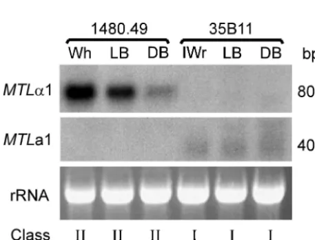
(DB), light brown (LB), and white (Wh) (26, 27).C. glabrata also switches reversibly between core phenotypes and an irreg-ular wrinkle (IWr) phenotype composed primarily of pseudohy-phae (27). Cells ofC. glabratastrain 1480.49, an␣-expressor of class II exhibiting the LB phenotype on agar containing CuSO4, were clonally plated, and Wh, LB, and DB colonies
were analyzed.MTL␣1 was expressed by cells with the tested core phenotypes in a graded fashion that correlated with color gradation (Wh⬎LB⬎DB) (Fig. 8). None of cells with the switch phenotypes expressedMTLa1 (Fig. 8). Cells ofC. gla-bratastrain 35B11, ana-expressor exhibiting the LB phenotype on agar containing CuSO4, were clonally plated, and LB, DB,
and IWr colonies were analyzed. Neither LB, DB, nor IWr cells expressedMTL␣1, but all three expressedMTLa1 at sim-ilar low levels (Fig. 8). These results demonstrate that while general MTL expression is dictated by the genotype of the MTL1orMTL2locus, the core switching system influences the level ofMTL␣1 expression in a graded fashion.
DISCUSSION
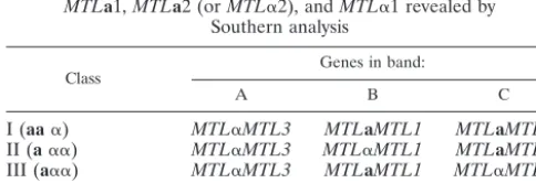
AlthoughC. glabratarepresents the second most prevalent Candidaspecies involved in human disease (15, 17, 32, 38, 41), TABLE 3. Classification of 38C. glabratastrains based on the distribution ofMTLa1,MTLa2 (orMTL␣2), andMTL␣1 revealed by
Southern analysis
Class isolatesNo. of isolates% of
Band pattern with probe:
MTL1a1 MTLa2-␣2a FuncP2b MTL1␣1 MTL1 3⬘and 5⬘
flanking regions
I 22 58 A, B, C A, B, C A A B
II 14 36 C A, B, C A, B A, B B
III 2 5 B A, B, C A, C A, C B
IV 1 A, B, C A, B, C A, B, C B
V NAc NA A, B, C A, B, C B
aThis probe does not distinguish betweenMTLa2 andMTL␣2. bThis probe distinguishes betweenMTLa2 andMTL␣2. cNA, not applicable.
TABLE 4. Models for the six classes based on the distribution of
MTLa1,MTLa2 (orMTL␣2), andMTL␣1 revealed by Southern analysis
Class Genes in band:
A B C
I (aa␣) MTL␣MTL3 MTLaMTL1 MTLaMTL2
II (a␣␣) MTL␣MTL3 MTL␣MTL1 MTLaMTL2
III (a␣␣) MTL␣MTL3 MTLaMTL1 MTL␣MTL1
IV (␣␣␣) (1480.47) MTL␣MTL3 MTL␣MTL1 MTL␣MTL2
V (aaa) MTLaMTL3 MTLaMTL1 MTLaMTL2
on September 8, 2020 by guest
http://ec.asm.org/
research into its basic biology has been minimal compared to that forC. albicans, primarily because it has been assumed that infor-mation gathered for C. albicanswould be transferable to other Candida species. However, genetic comparisons of the major Candidaspecies have revealed thatC. glabratais far more related toS. cerevisiaethan it is toC. albicans(3, 42, 49). Here we provide
evidence for the first time thatC. glabratapossesses three mating type-like loci with configurations similar to that ofS. cerevisiae. Expression of mating type genes inC. glabratamay be restricted to a single mating type-like locus that can contain eitheraor␣
genes, suggesting that a cassette system similar to the one basic to S. cerevisiaemating may exist inC. glabrata(18).
FIG. 6. PCR analysis of theMTLloci ofC. glabrata.(A) PCR assays were performed with genomic DNAs of 10C. glabratastrains. Five separate PCRs were performed with each strain to test the presence or absence of the full-length ORFs. Note that the primers CGPF1 and P1RACE1 do not discriminate betweenMTLa2 andMTL␣2, and the primers CGPIR2 and PIRACE1 generate a fragment containingMTL␣1,MTL␣2 and the intergenic promoter region. (B) Schematic representation ofMTL2a,MTL1a, andMTL3␣from strain 7549 andMTL1␣from strains PB621 and 1480.47. Positions of key primer pairs used in the PCR analysis are shown.
on September 8, 2020 by guest
http://ec.asm.org/
Configuration of the threeMTLloci.C. glabratapossesses three independent mating type-like loci (MTL1, MTL2, and MTL3), each containing pairs of mating type genes. All three have been characterized. In our test strain 7549, bothMTL1 andMTL2were demonstrated to contain anMTLa1 ORF and anMTLa2 ORF on complementary Crick and Watson DNA strands. Although in-frame AUG codons were not evident in eitherMTLa1 or MTLa2, one out-of-frame AUG codon was present 39 and 15 nucleotides upstream of an isoleucine or glutamine codon inMTLa1 andMTLa2, respectively. It is not
clear whether the hybridizableMTLa1 transcript identified in Northern blots or the 5⬘-RACE product represents the trans-latable mRNA. Interestingly,MATa1, the homolog ofMTLa1, has no known function in haploidacells ofS. cerevisiae(32). MTLa1 and MTLa2 were separated by an intergenic pro-moter region. This configuration is similar to that of the three mating type loci in S. cerevisiae (19, 25, 47), suggesting that regulation may also be similar. However, key regulatory se-quences in the intergenic promoter ofS. cerevisiae MATa1 and MATa2 were not shared with the comparable promoter ofC. glabrata, suggesting differences in regulation. Characterization of theMTL1 locus in two additional strains revealed that in these strains theMTL␣1 ORF and theMTL␣2 ORF resided in theMTL1locus on cDNA strands in a configuration similar to that ofMTLa1 andMTLa2. Again, while theMTL␣1-MTL␣2 configuration included an intergenic promoter, like the one separating and regulatingMAT␣1 andMAT␣2 inS. cerevisiae, the identified regulatory sequences differed. The intergenic promoter region ofC. glabratacontained Rap1p, Mcm1p, and Rme1p binding sites (http://cgsigma.cshl.org/jian/), which are not present in the comparable S. cerevisiae promoter, and lacked ana1-␣2 binding site present in the S. cerevisiae pro-moter (24). The latter observation suggests that inacells ofC. glabrata, transcription ofMTL␣1 may not be repressed by an MTLa1p-MTL␣2p heterodimer as in a cells in S. cerevisiae. Instead, another transacting factor such as Rme1p, a repressor of meiotic gene expression inS. cerevisiae(16, 17), may sub-stitute in the repression ofMTL␣1 transcription inastrains of C. glabrata. Alternatively, Rme1p, which can act as either an activator or a repressor (5, 10, 11), depending on thecis-acting element, may function as an activator ofMTL␣1 expression in
␣-type strains. Functional characterization of the MTL pro-moters will be the first step in revealing the unique mecha-nisms of regulation inC. glabrata.
Genetic models of the threeMTLloci in class I, II, and III
isolates.Using a combination of Southern analysis, PCR
anal-ysis, and sequencing, we were able to type each of the three MTLloci asaor␣for a number ofC. glabratastrains based on the presence of MTLa1-MTLa2 or MTL␣1-MTL␣2 pairs, which could be discriminated unambiguously at each of the three loci. Our results first demonstrate that the great majority of strains are eithera␣␣ oraa␣. Only one of the 38 isolates tested, 1480.47, deviated from the two patterns, exhibiting an
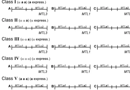
␣␣␣ genotype. Based on these studies, the 38 tested strains (97%) separated into four classes based on genotype and the size of the XbaI fragment harboring MTL1 or MTL2 (i.e., fragments B or C) (Fig. 9; Table 4). Class I strains were␣aa
and contained the combination ofMTLa1 andMTLa2 at the MTL1andMTL2loci in bands B and C, respectively (Fig. 9). Class I strains also harbored the combinationMTL␣1-MTL␣2 at theMTL3locus in band A (Fig. 9). Class II strains were␣␣a
and contained the combinations ofMTL1␣1 andMTL1␣2 at theMTL1locus in band B,MTL2a1 andMTL2a2 at theMTL2 locus in band C, andMTL3␣1 andMTL3␣2 at theMTL3locus in band A (Fig. 9). Class III strains were also ␣␣a but con-tained MTL2␣1 and MTL2␣2 at the MTL2 locus in band C rather than in band B (Fig. 9). Class III strains also harbored the combinationMTL3␣1-MTL3␣2 at theMTL3locus in band A, like the other two classes, and MTL2a1 and MTL2a2 in band B (Fig. 9). Based on theMTLgenotypes in fragment B or FIG. 7. Northern analysis of the expression ofMTL␣1 andMTLa1
in 12 strains ofC. glabrata. Approximately 20g of total cellular RNA was applied to each lane. Duplicate Northern blots were probed with either the 300-bp PCR fragment (primers CGAR1 and A1RACE1) containing theMTL1a1 ORF or the 550-bp PCR fragment (primers CGPIF1 and CGPIR2) containing theMTL1␣1 ORF. To assess load-ing, ethidium bromide-stained 28S rRNA patterns are included. The approximate molecular sizes of transcripts are shown to the right of the hybridization patterns. The class andMTL1expression pattern of each strain are noted.
FIG. 8. Northern analysis of the expression ofMTL␣1 andMTLa1 in the switch phenotypes of anMTL␣-expressing strain (1480.49) and an MTLa-expressing strain (35B11). Cells of the two strains were plated on agar containing 1 mM CuSO4, which discriminates between
the switch phenotype in the core switching system ofC. glabrata(26, 27). Cells from Wh, LB, and DB colonies of strain 1480.49 and from IWr, LB, and DB colonies of strain 35B11 were picked and replated before they were analyzed. Duplicate blots were probed with either the 550-bp PCR fragment (primers CGPIF1 and CGPIR1) containing the
MTL1␣1 ORF or the 300-bp PCR fragment (primers CGAR1 and A1RACE1) containing theMTL1a1 ORF. To assess loading, ethidium bromide-stained 28S rRNA patterns are included. The class andMTL1
expression pattern of each strain are noted.
on September 8, 2020 by guest
http://ec.asm.org/
C and theMTLflanking sequence, it is likely that the class IV isolate 1480.47 was derived from either a class II or class III isolate. Since our collection included only a limited number of strains, there is the possibility that additional configurations, such asaaa, exist (Fig. 9).
Expression patterns: suggestion of a cassette system like
that inS. cerevisiae.Northern analysis revealed thatC. glabrata,
like S. cerevisiae, could be categorized based on mating type gene expression. Strains expressed eitherMTLa1 or MTL␣1, never both, just as haploid S. cerevisiae expressesMATa1 or MAT␣1 but never both. In the case ofS. cerevisiae, genes in HMRandHMLloci are silent, while genes in theMATlocus are expressed (18). InC. glabrata, the expression pattern sug-gested a similar scenario. While class II and IV isolates ex-pressedMTL␣1, class I strains expressedMTLa1. The former classes of isolates possessed twoMTL␣ loci, while the latter class possessed two MTLa loci. Northern analysis demon-strated a direct correlation between the expression ofMTLa1 orMTL␣1 and the presence of two corresponding ORFs at two MTLaand twoMTL␣loci, respectively. However, our data do not distinguish which of the two similar loci in each case is the expression locus.
If only one of the two loci inC. glabratais the expression locus and a correlate to theMATlocus ofS. cerevisiaeand the other twoMTLloci are correlates to the silent lociHMRand HML, then the threeC. glabrata MTLloci may also function in
a mobile cassette system similar to that ofS. cerevisiae, in which copies of the silentMTLloci recombine with theMTL expres-sion locus to switch mating type. Experiments to test this hy-pothesis are now in progress.
Expression patterns and phenotypic switching.Our results
indicate thatMTLexpression correlates with the genotype of the MTL1 or MTL2 locus. The ␣-expression strain 1480.49 expressedMTL␣1 exclusively in all three tested phenotypes in the core switching system (Wh, LB, and DB), while thea -ex-pressing strain 35B11 expressed MTLa1 exclusively in both core switch phenotypes (LB and DB) and the IWr phenotype. However, in the␣-expressing strain, the levels ofMTL␣1 tran-script differed between the three core switch phenotypes ac-cording to the hierarchy Wh ⬎ LB ⬎ DB, suggesting that MTL␣1 expression is modulated in a graded fashion by the core switching system, just like transcription of the methalo-thionine geneMTII, for pigmentation and phloxine B staining (26, 27). Interestingly, the intergenic promoter that controls transcription ofMTL␣1 contains an Mcm1p binding site, which has been implicated in the regulation of phase-specific gene expression during white-opaque switching inC. albicans(31). The relationship between mating and switching inC. glabrata, therefore, deserves further investigation.
ACKNOWLEDGMENTS
This research was supported by grants AI 2392 and DE014219. FIG. 9. A model of theMTLloci in the four classes ofC. glabratastrains. The threeMTLloci were distinguished asXbaI fragments A, B, and C in Southern analyses (see Fig. 5). The genotypes of the loci in the different classes were interpreted from the combined results of Southern, PCR, and sequencing data. The strains are distinguished as␣aa(class I),␣␣a(classes II and III), or␣␣␣(class IV). A hypothesized class V isaaa. The expression patterns are distinguished as eitheraexpression or␣expression, as determined by Northern analysis.
on September 8, 2020 by guest
http://ec.asm.org/
We thank Claude Pujol, Shawn Lockhart, and Rui Zhao for helpful discussions.
REFERENCES
1. Astrom, S. U., A. Kegel, J. O. Sjostrand, and J. Rine.2000.Kluyveromyces lactisSir2p regulates cation sensitivity and maintains a specialized chromatin structure at the cryptic alpha-locus. Genetics156:81–91.
2. Astell, C. R., L. Ahlstrom-Jonasson, M. Smith, K. Tatchell, K. A. Nasmyth, and B. D. Hall.1981. The sequence of the DNAs coding for the mating-type loci ofSaccharomyces cerevisiae. Cell27:15–23.
3. Barns, S. M., D. J. Lane, M. L. Sogin, C. Bibeau, and W. G. Weisburg.1991. Evolutionary relationships among pathogenicCandidaspecies and relatives. J. Bacteriol.173:2250–2255.
4. Blaschke-Hellmessen, R.1996. Fluconazole and itraconazole susceptibility testing with clinical yeast isolates and algae of the genusProtothecaby means of the Etest. Mycoses39(Suppl. 2):39–43.
5. Blumental-Perry, A., W. Li, G. Simchen, and A. P. Mitchell.2002. Repres-sion and activation domains of RME1p structurally overlap, but differ in genetic requirements. Mol. Biol. Cell13:1709–1721.
6. Carninci, P., Y. Shibata, N. Hayatsu, Y. Sugahara, K. Shibata, M. Itoh, H. Konno, Y. Okazaki, M. Muramatsu, and Y. Hayashizaki.2000. Normalization and subtraction of cap-trapper-selected cDNAs to prepare full-length cDNA libraries for rapid discovery of new genes. Genome Res.10:1617–1630. 7. Church, G. M., and W. Gilbert.1984. Genomic sequencing. Proc. Natl. Acad.
Sci. USA81:1991–1995.
8. Claret, F. X., M. Hibi, S. Dhut, T. Toda, and M. Karin.1996. A new group of conserved coactivators that increase the specificity of AP-1 transcription factors. Nature383:453–457.
9. Conner, B. J., A. A. Reyes, C. Morin, K. Itakura, R. L. Teplitz, and R. B. Wallace.1983. Detection of sickle cell beta S-globin allele by hybridization with synthetic oligonucleotides. Proc. Natl. Acad. Sci. USA80:278–282. 10. Covitz, P. A., I. Herskowitz, and A. P. Mitchell.1991. The yeastRME1gene
encodes a putative zinc finger protein that is directly repressed by a1-alpha 2. Genes Dev.5:1982–1999.
11. Covitz, P. A., and A. P. Mitchell.1993. Repression by the yeast meiotic inhibitorRME1. Genes Dev.7:1598–1608.
12. Csank, C., and K. Haynes.2000.Candida glabratadisplays pseudohyphal growth. FEMS Microbiol. Lett.189:115–120.
13. Eberstadt, M., B. Huang, Z. Chen, R. P. Meadows, S. C. Ng, L. Zheng, M. J. Lenardo, and S. W. Fesik.1998. NMR structure and mutagenesis of the FADD (Mort1) death-effector domain. Nature392:941–945.
14. Eschrich, D., M. Buchhaupt, P. Kotter, and K. D. Entian.2002. Nep1p (Emg1p), a novel protein conserved in eukaryotes and archaea, is involved in ribosome biogenesis. Curr. Genet.40:326–338.
15. Fidel, P. L. Jr., J. A. Vazquez, and J. D. Sobel.1999.Candida glabrata: review of epidemiology, pathogenesis, and clinical disease with comparison toC. albicans. Clin. Microbiol. Rev.1:80–96.
16. Fortun, J., A. Lopez-San Roman, J. J. Velasco, A. Sanchez-Sousa, E. de Vicente, J. Nuno, C. Quereda, R. Barcena, G. Monge, A. Candela, A. Hon-rubia, and A. Guerrero.1997. Selection ofCandida glabratastrains with reduced susceptibility to azoles in four liver transplant patients with invasive candidiasis. Eur. J. Clin. Microbiol. Infect. Dis.16:314–318.
17. Hazen, K. C.1995. New and emerging yeast pathogens. Clin. Microbiol. Rev. 4:462–478.
18. Herskowitz, I., J. Rine, and J. N. Strathern.1992. Mating-type determination and mating-type interconversion inSaccharomyces cerevisiae, p. 583–656.In
E. W. Jones, J. R. Pringle, and J. R. Broach (ed.), The molecular and cellular biology of the yeastSaccharomyces. Cold Spring Harbor Laboratory Press, Cold Spring Harbor, N.Y.
19. Hicks, J., J. N. Strathern, and A. J. Klar.1979. Transposable mating type genes inSaccharomyces cerevisiae. Nature282:478–483.
20. Hitchcock, C. A., G. W. Pye, P. F. Troke, E. M. Johnson, and D. W. Warnock. 1993. Fluconazole resistance inCandida glabrata. Antimicrob. Agents Che-mother.37:1962–1965.
21. Hull, C. M., and A. D. Johnson.1999. Identification of a mating type-like locus in the asexual pathogenic yeastCandida albicans. Science285:1271–1275. 22. Hull, C. M., R. M. Raisner, and A. D. Johnson.2000. Evidence for mating of the
“asexual” yeastCandida albicansin a mammalian host. Science289:307–310. 23. Jacquet, M., J. M. Buhler, F. Iborra, M. C. Francingues-Gaillard, and C.
Soustelle.1991. TheMATlocus revisited within a 9.8 kb fragment of chro-mosome III containingBUD5and two new open reading frames. Yeast 7:881–888.
24. Johnson, A. D.1995. Molecular mechanisms of cell-type determination in budding yeast. Curr. Opin. Genet. Dev.5:552–558.
25. Klar, A. J., J. McIndoo, J. B. Hicks, and J. N. Strathern.1980. Precise mapping of the homothallism genesHMLandHMRinSaccharomyces cer-evisiae. Genetics96:315–320.
26. Lachke, S. A., T. Srikantha, L. K. Tsai, K. Daniels, and D. R. Soll.2000. Phenotypic switching inCandida glabratainvolves phase-specific regulation of the metallothionein geneMT-IIand the newly discovered hemolysin gene
HLP. Infect. Immun.68:884–895.
27. Lachke, S. A., S. Joly, K. Daniels, and D. R. Soll.2002. Phenotypic switching and filamentation inCandida glabrata. Microbiology148:2661–2674. 28. Landsman, D., O. W. McBride, N. Soares, M. P. Crippa, T. Srikantha, and
M. Bustin.1989. Chromosomal protein HMG-14. Identification, character-ization, and chromosome localization of a functional gene from the large human multigene family. J. Biol. Chem.264:3421–3427.
29. Li, T., M. R. Stark, A. D. Johnson, and C. Wolberger, C.1995. Crystal structure of the MATa1/MAT alpha 2 homeodomain heterodimer bound to DNA. Science270:262–269.
30. Lockhart, S. R., S. Joly, C. A. Pujol, J. D. Sobel, M. A. Pfaller, and D. R. Soll. 1997. Development and verification of fingerprinting probes forCandida glabrata. Microbiology143:3733–3746.
31. Lockhart, S. R., M. Nguyen, T. Srikantha, and D. R. Soll.1998. A MADS box protein consensus binding site is necessary and sufficient for activation of the opaque-phase-specific geneOP4ofCandida albicans. J. Bacteriol.180: 6607–6616.
32. Lockhart, S. R., S. Joly, K. Vargas, J. Swails-Wenger, L. Enger, and D. R. Soll.1999. Natural defenses againstCandidacolonization breakdown in the oral cavities of the elderly. J. Dent. Res.78:857–868.
33. Lockhart, S. R., C. A. Pujol, K. Daniels, M. G. Miller, A. D. Johnson, M. A. Pfaller, and D. R. Soll.2002. InCandida albicans, white-opaque switchers are homozygous for mating type. Genetics162:737–745.
34. Lockhart, S. R., K. Daniels, R. Zhao, D. Wessels, and D. R. Soll.2003. The cell biology of mating inCandida albicans. Eukaryot. Cell2:49–61. 35. Magee, B. B., and P. T. Magee.2000. Induction of mating in Candida albicans
by construction of MTLa and MTLalpha strains. Science289:310–313. 36. Marichal, P., H. Vanden Bossche, F. C. Odds, G. Nobels, D. W. Warnock, V.
Timmerman, C. Van Broeckhoven, S. Fay, and P. Mose-Larsen.1997. Mo-lecular biological characterization of an azole-resistantCandida glabrata
isolate. Antimicrob. Agents Chemother.41:2229–2237.
37. Miller, M. G., and A. D. Johnson.2002. White-opaque switching inCandida albicansis controlled by mating-type locus homeodomain proteins and al-lows efficient mating. Cell110:293–302.
38. Odds, F. C.1988.Candida and candidiasis. Bailliere. Tendall, London, United Kingdom.
39. Odds, F. C., M. G. Rinaldi, C. R. Cooper, Jr., A. Fothergill, L. Pasarell, and M. R. McGinniss.1997.CandidaandTorulopsis: a blinded evaluation of use of pseudohypha formation as basis for identification of medically important yeasts. J. Clin. Microbiol.35:313–316.
40. Pfaller, M. A.1996. Nosocomial candidiasis: emerging species, reservoirs, and modes of transmission. Clin. Infect. Dis.22(Suppl. 2):S89–S94. 41. Pfaller, M. A., R. N. Jones, S. A. Messer, M. B. Edmond, and R. P. Wenzel.
1998. National surveillance of nosocomial blood stream infection due to
Candida albicans: frequency of occurrence and antifungal susceptibility in the SCOPE Program. Diagn. Microbiol. Infect. Dis.31:327–332. 42. Santos, M. A., T. Ueda, K. Watanabe, and M. F. Tuite.1997. The
non-standard genetic code ofCandidaspp.: an evolving genetic code or a novel mechanism for adaptation? Mol. Microbiol.26:423–431.
43. Sobel, J. D.1996.Candidavulvovaginitis. Semin. Dermatol.15:17–28. 44. Spinillo, A., E. Capuzzo, R. Gulminetti, P. Marone, L. Colonna, and G.
Piazzi.1997. Prevalence of and risk factors for fungal vaginitis caused by non-albicans species. Am. J. Obstet. Gynecol.176:138–141.
45. Srikantha, T., L. K. Tsai, K. Daniels, and D. R. Soll.2000.EFG1null mutants ofCandida albicansswitch but cannot express the complete pheno-type of white-phase budding cells. J. Bacteriol.182:1580–1591.
46. Srikantha, T., L. Tsai, K. Daniels, A. J. Klar, and D. R. Soll.2001. The histone deacetylase genesHDA1andRPD3play distinct roles in regulation of high-frequency phenotypic switching inCandida albicans. J. Bacteriol. 183:4614–4625.
47. Strathern, J. N., A. J. Klar, J. B. Hicks, J. A. Abraham, J. M. Ivy, K. A. Nasmyth, and C. McGill.1982. Homothallic switching of yeast mating type cassettes is initiated by a double-stranded cut in theMATlocus. Cell31:183–192. 48. Wong, S., G. Butler, and K. H. Wolfe.2002. Gene order evolution and
paleopolyploidy in hemiascomycete yeasts. Proc. Natl. Acad. Sci. USA99: 9272–9277.
49. Worley, K. C., B. A. Wiese, and R. F. Smith.1995. BEAUTY: an enhanced BLAST-based search tool that integrates multiple biological information resources into sequence similarity search results. Genome Res.5:173–184.