Bosn J Basic Med Sci 2013; 13 (3): 186-191Abstract
Development of novel therapeutic modalities is crucial for the treatment of oral squamous cell carcinoma (OSCC). Recent scientifi c studies have been focused on herbal medicines as potent anti-cancer drug candidates. Th is study is the fi rst to investigate the cytotoxic eff ects and the mechanism of cell death induced by grape seed extract (GSE) in oral squamous cell carcinoma (KB cells). MTT (-(,-dimetylthiazol--yl)-, diphenyltetrazolium bromide) and trypan blue assays were performed in KB cells as well as human umbilical vein endothelial cells (HUVEC) were used to analyze the cytotoxic activity of GSE. Furthermore, the apoptosis-inducing action of the extract was determined by TUNEL, DNA fragmentation and cell death analysis. Statistical signifi cance was determined by analysis of variance (ANOVA), followed by Duncan’s test at a signifi cance level of P≤.. Th e results showed apoptotic potential of GSE, confi rmed by signifi cant inhibition of cell growth and vi-ability in a dose- and time- dependent manner without inducing damage to non-cancerous cell line HUVEC. Th e results of this study suggest that this plant contains potential bioactive compound(s) for the treatment of oral squamous cell carcinoma.
© Association of Basic Medical Sciences of FB&H. All rights reserved
KEY WORDS: cytotoxicity, apoptosis, Anticancer, oral squamous cell carcinoma, grape seed extract
vinifera) in oral squamous cell carcinoma
Amirala Aghbali1, Sepideh Vosough Hosseini1, Abbas Delazar2, Nader Kalbasi Gharavi1,
Fatemeh Zare Shahneh3, Mona Orangi3, Ali Bandehagh4, Behzad Baradaran2*
1Department of Oral and Maxillofacial Pathology, Faculty of Dentistry, Tabriz University of Medical Sciences, Daneshgah street 51666-14766, Tabriz, Iran. 2 Drug Applied Research Center, Tabriz University of Medical Sciences, Daneshgah street 51666-14766, Tabriz, Iran. 3Immunology Research Center, Tabriz University of Medical Sciences, Daneshgah street 51666-14766, Tabriz, Iran.
INTRODUCTION
Oral squamous cell carcinoma (OSCC) is one of the most common oral cancers with increasing risk in younger people less than years of age, especially in de-veloped countries. Principal methods for treatment of OSCC are radiotherapy and surgery but recurrences are common with further resistance to therapy [-]. Nowadays complementary and alternative medicine (CAM) provides treatments for various cancers using herbal medi-cine derivatives from natural products. Recent scientific studies have been focused on herbal medicine as potent anti-cancer drug candidates []. Taxol from Taxus brevi-folia L, as a plant-derived compound, is extensively used in the clinical practice. Cytotoxic effects of medicinal plant extracts can be mediated through the induction of apopto-sis []. Apoptoapopto-sis, programmed cell death, occurs in
physi-ological and pathphysi-ological conditions and is characterized by morphological alterations such as cell shrinkage, mem-brane blebbing, chromatin condensation and DNA frag-mentation [, ]. Morphological hallmarks of apoptosis in the nucleus are chromatin condensation and nuclear frag-mentation. Apoptosis plays a key role in eliminating dam-aged or abnormal cells without harming normal cells []. Grapes (Vitis vinifera) are indigenous to southern Europe and Western Asia cultivated worldwide. The grape vine is rich in flavonoids, polyphenols, anthocyanin, proan-thocyanidins, procyanidins and trans-resveratrol []. The seeds and the leaves of the grape vine are used in herbal medicines, whilst fruit is consumed as a dietary supple-ment. Recent studies have revealed that grape seed extract (GSE) has antioxidant and free radical scavenging, anti-diabetic, cardioprotective, hepatoprotective, anti-carcino-genic, anti-microbial, and anti-viral activities [-]. In the present study, cytotoxic effects of grape seed extract (GSE) were investigated for the first time and its possible cell death induction potential on oral squamous cell carci-noma (KB cell line) was determined. Furthermore, the pos-sible side effects of the extracts on human umbilical vein endothelial cells (HUVEC), as normal cells, were evaluated.
* Corresponding author: Behzad Baradaran,
Drug Applied Research Center, Tabriz University of Medical Sciences, Daneshgah street, 51666-14766 Tabriz, Iran Phone: (+98) 411 3364665; Fax: +98-411-3364665 e-mail: [email protected]
Bosn J Basic Med Sci 2013; 13 (3): 187-191
MATERIALS AND METHODS
Reagents
Grape (Vitis vinifera) plant was collected from Eastern Azerbaijan Province, Iran, in April . A voucher speci-men was deposited at the Herbarium of Faculty of Phar-macy, Tabriz University of Medical Sciences. KB cell (oral squamous cell carcinoma cell line) and HUVEC (human umbilical vein endothelial cells) were purchase from the National Cell Bank of Iran (Pasteur Institute, Tehran, Iran). RPMI- medium, fetal bovine serum (FBS), penicillin, and streptomycin were purchased from Sigma (Germany), dimethylsulfoxide (DMSO) and trypan blue from Merck (Germany), trypsin/EDTA solution from Gibco (U.K), and -(,-dimetylthiazol--YL)- ,- diphenyltetrazolium bro-mide (MTT reagent), Cell Death Detection ELISA plus kit, and In Situ Cell Death Detection Kit, POD from Roche Diag-nostics GmbH (Germany) and DAB (, ’-diaminobenzidine).
Preparation of extracts
Th e grape seeds from Vitis Vinifera were washed, dried and ground to powder using a blender. Extractions were per-formed in a Soxhlet apparatus with methanol. Dry powder of the grape seeds were extracted with N-hexane, Dichlo-romethane and methanol in Soxhlet extractor, the obtained extract dried in a rotary evaporator in low power and ºC temperature, then dried powders were stored in a covered glass container in refrigerator until usage, so the mentioned extract was completely dry and did not contain any metha-nol and its cytotoxic eff ects are because of the pure extract itself.Th e grape seed extract (GSE) was concentrated by a rotary evaporator (Heildolph, Germany) at about ºC and then dried under very low pressure. Th e dried extracts were stored at –ºC; mg of each extract were dissolved in μL of DMSO and diluted with RPMI- medium to the defined concentrations as indicated. Then, test solutions were sterilized using .-μm syringe filters (Nunc, Den-mark) and used as stock solution for further experiments.
Cell culture
The KB and HUVEC cells were grown in RPMI- me-dium supplemented with FBS, U/mL penicillin and μg/mL streptomycin. The cells were incubated in a humidified incubator containing CO at ºC. Af-ter reaching an confl uence, the cells were rinsed with PBS/. EDTA and harvested from cm flasks
us-ing . trypsin/EDTA solution. Then, the cells were sub-cultured into -cm flasks, -well plates or -well
plates (Nunc, Denmark) according to experiments. All applied measurements were performed in triplicate.
MTT assay
Cytotoxicity of GSE was assessed on KB cells as well as HU-VEC using the MTT reagent according to manufacturer's protocol. Th e cells were seeded in -well plates ( cells/
well) and incubated for h at °C with CO. Th e cells were treated with different concentrations of solvent ex-tracts (, , , , , , μg/mL) and . (v/v) DMSO as a negative control. After h of treatment, μL of MTT reagent ( mg/mL) labeling reagent was added to each well. Th e plates were incubated at °C with CO for hours. Th en, μL of the solubilizing solution was added to each well, followed by incubation overnight at °C to dissolve formazan crystals. Finally, absorbance was read us-ing an ELISA plate reader (Bio Teck, Germany) at a wave-length of nm []. Th e percentages of cytotoxicity and cell viability were calculated using the following equations: cytotoxicity = - [means absorbance of treated cells/mean ab-sorbance of negative control], viability = - cytotoxicity.
Trypan blue assay
Cell membrane integrity was evaluated using trypan blue dye exclusion. Trypan blue passes through the membranes of dead cells and thus stains them, unlike live cells. KB cells ( cells/well) in -well plates were exposed against
dif-ferent concentrations of GSE and . (v/v) DMSO for h. After medium was removed from the wells, cells were washed with μL of PBS. The cells were detached by adding μL of . trypsin/EDTA. RPMI- medium supplement with FBS ( μL) and . trypan blue ( μL) was added to each well, and the plates were incubated for min. Th en, -μL aliquot was removed and placed on a Neubauer hemacytometer. Finally, the number of viable cells was counted under light microscopy. Th e number of vi-able cells was calculated according to the following formula: [Th e unstained cell count × the dilution of the cell suspension × ]= viable cells.
Th e percentage of viability was calculated as: [viable cells/the total cell count] × = viability
Assessment of apoptosis
Apoptotic cells death were measured using the Cell Death Detection ELISA Plus kit, which quantifies histone-asso-ciated DNA fragments (mono- and oligo-nucleosomes). KB cells ( cells/well) were treated against diff erent
Bosn J Basic Med Sci 2013; 13 (3): 188-191 TUNEL assayDNA fragmentation was detected by terminal deoxytransfer-ase (TdT)-mediated dUTP nick- end labeling (TUNEL) with the In Situ Cell Death Detection Kit, POD (Roche Diagnos-tics GmbH, Germany) as described by manufacturer’s proto-col. Briefl y, (.×) KB cells were sub-cultured into -well
plates and incubated for h at °C and CO. Th e cells were treated with GSE at concentrations required for in-hibition of growth of KB cells (IC) for h. Negative con-trol cells were treated with the same fi nal concentration of DMSO present in treated wells [. (v/v)]. After treatment, the cells were fi xed with (w/v) paraformaldehyde in PBS (pH .) for hour at room temperature and rinsed twice with PBS. Th en, the fi xed cells were incubated with blocking solution ( HO in methanol) for min and rinsed with PBS. Th e cells were then incubated in permeabilizing solution (. Triton X- in . sodium citrate) for min on ice. Subsequently, μL of reaction mixture containing TdT en-zyme and nucleotide were added to the cells and incubated for h at °C. After washing three times with PBS, the slides were incubated with μL converter-POD sterptavidin HRP solution for min, and rinsed three times with PBS. Finally, the cells were incubated with DAB. Substrate solution and the stained cells were analyzed using invert microscopy [].
DNA Fragmentation assay
Biochemically, apoptosis is characterized by the activation of a nuclear endonuclease that cleaves DNA into multimers of - base pairs and can be visualized as an oligosomal ladder by standard agarose gel electrophoresis. KB cells were seeded in -well plates and kept in a CO incubator. KB cells were treated by GSE in IC concentrations for hours. At the end of incubation period, the cells were centrifuged for rpm for min at °C. Th e pellet was suspended in a lysis buff er ( mM Tris-HCI, pH=., mM NaCl, I mM EDTA, mg/mL Proteinase K, SDS), and incu-bated at °C. Th e pellet was dissolved in TE buff er (. M
Tris-HCl, pH=., mM EDTA). DNA samples were elec-trophoretically separated on . agarose gel, containing ethidium bromide (. μg/mL). DNA was visualized by a UV transilluminator []. Untreated cells were used as control.
Statistical analysis
Data are presented as means ± SEM. Statistical signifi-cance was carried out by ANOVA, followed by Dun-can’s test. Statistical significance was defined at p≤.. IC values were derived from prohibit analysis. SPSS program was used for statistical analysis of data.
RESULTS
The cytotoxic effects of grape seed extract (GSE) on the growth of oral squamous cell carcinoma (KB cell line) were determined by MTT and trypan blue assays as shown in Figures and . As shown in Figure , the cells treated with hydroalcoholic extract exhibited significant decline in viability in comparison to the untreated con-trol cells, whereas no cytotoxic activity was observed on normal human umbilical vein endothelial cells (HUVEC). Moreover, treatment of KB cells with GSE showed cell growth inhibition in a time- and dose-dependent man-ner. The most significant cytotoxicity was achieved in the higher concentration and longer time of the GSE treatment on KB cells. Data analysis of cytotoxicity as-say showed that IC (dose required for inhibition) of GSE on KB cells was . μg/mL for hours. Direct counting for viable cells using the trypan blue exclusion test showed that of GSE-treated KB cells with the highest concentration ( μg/mL) absorbed the dye at h (Figure ). Th e results showed that dose-dependent apoptosis of KB cells occurred under the incubation with diff erent concentration of GSE.. Th e highest apoptotic eff ect of GSE was .. One of the hallmarks of apoptotic KB cells (treated with GSE)was the presence of nucleosomal DNA fragments, confi rmedby
FIGURE 1. Eff ects of GSE on proliferation of KB and HUVEC cells for 24 h.
Bosn J Basic Med Sci 2013; 13 (3): 189-191
TUNEL assay. As shown in Figure , after the treatment of KB cells with hIC concentration of GSE, the apoptotic cells produced dark brown-stained nuclei, whereas the
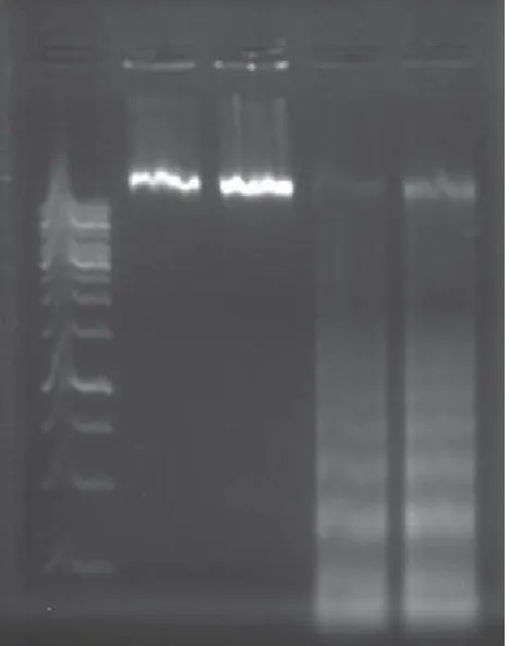
non-apoptotic cells were not stained; a simi-lar observation was noted in the nega-tive control cells treated with . (v/v) DMSO. To confirm the effect of ex-tracts on the induction of apoptosis in KB cells, the GSE was examined on the internucleosomal DNA fragmentation as a characteristics feature of apoptosis. As shown by agarose gel electro-phoresis in Figure , increased DNA fragmentation was apparent in KB cells after treatment with μg/mL (near to IC) of GSE. Fragmented DNA was clearly observed in KB cells, whereas untreated cells did not ex-hibit ladders. Therefore, it is possible that GSE causes apoptosis in KB cells.
DISCUSSION
Oral squamous cell carcinoma (OSCC) poses an important health concern all over the world. Standard treatment plan for oral squamous cell carcinoma depends on the stage of the disease. Early-stage tumors are treated primarily by surgery or radiotherapy, with both modalities resulting in similar local control and sur-vival rates. More advanced carcinomas often require multi-modality therapy with surgery, radiation and chemotherapy, which can result in very high morbidity []. Treatment mo-dalities of oral SCC have numerous side eff ects. Radical sur-gery can result in disfi gurement and functional impairment. Common side eff ects of radiotherapy include mucositis, oral candidiasis, loss of taste and xerostomia, which may be per-manent due to the detrimental eff ect of radiation on salivary glands []. Osteoradionecrosis of bones within the radia-tion fi eld (most commonly the mandible) may occur as a re-sult of damage to the bone vasculature and osteocytes and is one of the most serious complications of radiotherapy []. Current chemotherapeutic drugs exert many cyto-toxic effects on normal cells, resulting in adverse sys-temic and cytotoxic effects and development of resis-tance to therapy in OSCC patients. Despite continuing research and advances in treatment, the clinical outcomes for HNSCC have not improved significantly over the last several decades, with the overall -year survival rate as low as [-]; therefore, development of new treatment modalities is crucial for prevention and to reduce mor-tality. Herbal medicines (plant-derived compounds), in combination with routinely implemented treatment mo-dalities, such as radiotherapy and surgery, can reduce mor-bidity and mortality in OSCC patients. Recent studies focus
FIGURE 4. DNA laddering: DNA fragmentation on 1.8% agarose gel electrophoresis; Lane 1, 1000 bp marker; Lanes 2 and 3, un-treated cells; Lanes 4 and 5,GSE-un-treated cells.
Bosn J Basic Med Sci 2013; 13 (3): 190-191on herbal medicines as potent anti-cancer drug candidates. Previous studies have revealed the anticancer activities of herbal extracts against oral squamous cell carcinoma cell lines. These include Tamoxifen in combination with Cis-platin, -Fluorouracil, Cordycepin, Scutellariabaicalensis, Quercetin and Artemisinin []. In a study by Ghashmet et al, time- and dose-dependent inhibitory effect of anti-proliferative activity of local honey (Tualang) on OSCC and HOS cell lines was investigated by MTT assay, light and fluorescent microscope, and Annexin V-FITC Apop-tosis Detection Kit []. In a study by Hseu et al, ethanol () extracts of Alpinia pricei rhizome could induce G/M phase of the cell cycle arrest and apoptosis in KB cells []. Grapes (Vitisvinifera) are rich in fl avonoids, polyphenols, an-thocyanin, proanthocyanidins, trans-resveratrol, and procy-anidins. Recent studies have revealed that GSE has antioxi-dant and free radical scavenging activities, and anti-diabetic, cardioprotective, hepatoprotective, carcinogenic, anti-microbial, and anti-viral eff ects. Dinicola et al showed that GSE has anti-proliferative and apoptotic eff ects on CaCO and HCT- colon cancer cell lines []. Sun et al. [] indicat-ed that GSEs have the ability to inhibit HNSCC cell invasion by targeting the expression of EGFR and activation of NF-κ B as well as inhibiting the epithelial-to-mesenchymal transition In the present study, for the first time the cytotoxic ef-fects of the grape seed extract was investigated and their possible effects on cell death properties in KB cell line were determined. Furthermore, the probable side ef-fects of the grape seed extract on HUVEC were evaluated. The results showed that grape seed extract inhibited the growth of oral squamous cell carcinoma through the in-duction of apoptosis. The IC values strongly indicated that the GSE had a potent cytotoxic effect on KB cells. The crucial results of our study addressed to selectivity of GSE eff ects on KB vs. HUVEC cells (eff ect was observed on KB, but HUVEC cells) indicate possibility for its promising use for its promising use for the treatment oral squamous cell carcinoma. Th e diff erent sensitivities of human oral squa-mous cell carcinoma and normal cells to GSE suggested that used GSE may be used as a natural chemotherapeutic drug. Many chemotherapeutic agents can trigger the apoptosis of cancer cells. Morphological changes in apoptosis are chroma-tin condensation and nuclear fragmentation. Apoptotic DNA fragmentation is a key feature of apoptosis, characterized by the activation of endogenous endonucleases with subsequent cleavage of chromatin DNA into internucleosomal frag-ments of approximately - base pairs (bp) [, ]. Th e results of our study showed induction of apoptosis in the can-cer cells as a result of treatment with the GSE. Cell death as-say, TUNEL and DNA fragmentation analyses were used to reveal if induction of apoptosis was achieved by GSE. It was
shown that DNA fragmentation happened in the late stage of apoptosis. Th erefore, results of our in vitro study have shown an anticancer potential of the GSE and selectivity to inhibit growth and induce apoptosis in KB, unlike HUVEC cells. However, additional experiments should be done to test grape seed extracts effect on other cancer cell lines
in vitro and in vivo (animal studies) conditions to estab-lish their therapeutic ability as well as its adverse effects.
CONCLUSION
Th e present study demonstrated the in vitro cytotoxic and apoptotic activity of GSE in human KB cell line, possibly sug-gesting a new potential chemotherapeutic agent for the treat-ment of oral squamous cell carcinoma.
ACKNOWLEDGEMENTS
Th is study was supported by a grant from the Drug Applied Research Center of Tabriz University of Medical Sciences.
DECLARATION OF INTEREST
Th e authors declare that there is no confl icting interest.
REFERENCES
[] Bagan JV, Scully C. Recent advances in oral oncology : epide-miology, aetiopathogenesis, diagnosis and prognostication. Oral Oncol ; ():-.
[] Warnakulasuriya S. Global epidemiology of oral and oropharyngeal cancer. Oral Oncol ; (-):-.
[] Mehrotra R, Yadav S. Oral squamous cell carcinoma: Etiology, pathogenesis and prognostic value of genomic alterations. Indian J Cancer ; :-.
[] Saraste A, Pulkki K. Morphologic and biochemical hallmarks of apoptosis. Cardiovasc Res ; ():-.
[] Mehta RG, Murillo G, Naithani R, Peng X. Cancer chemopreven-tion by natural products: How far have we come? Pharm Res ; ():-.
[] Ziegler U, Groscurth P. Morphological features of cell death. News Physiol Sci ; :-.
[] Galluzzi L, Maiuri MC, Vitale I, Zischka H, Castedo M, Zitvogel L, et al. Cell death modalities: classifi cation and pathophysiological implications. Cell Death Diff er ; ():-.
[] Elmore S. Apoptosis: a review of programmed cell death. Toxicol Pathol ; ():-.
[] Roy AM, Baliga MS, Elmets CA, Katiyar SK. Grape seed proantho-cyanidins induce apoptosis throughp, Bax, and Caspase path-ways. Neoplasia ; :-.
[] Kaur M, Singh RP, Gu M, Agarwal R, Agarwal C. Grape seed ex-tract inhibits in vitro and in vivo growth of human colorectal carci-noma cells. Clin Cancer Res ; :-.
[] Ye X, Krohn RL, Liu W, Joshi SS, Kuszynski CA, McGinn TR, et al. Th e cytotoxic eff ects of a novel IH grape seed proanthocy-anidin extract on cultured human cancer cells. Mol Cell Biochem ; :-.
Bosn J Basic Med Sci 2013; 13 (3): 191-191
FEBS J. ;():-.
[] Mossman T. Rapid colorimetric assay for cellular growth and sur-vival: application to proliferation and cytotoxicity assays. J Immu-nol Methods ; (-):-.
[] Frankfurt OS, Krishan A. Enzyme-linked immunosorbent assay (ELISA) for the specifi c detection of apoptotic cells and its appli-cation to rapid drug screening. J Immunol Methods ;(-):-.
[] Rogakou EP, Nieves-Neira W, Boon C, Pommier Y, Bonner WM. Initiation of DNA fragmentation during apoptosis induces phos-phorylation of HAX histone at serine . J Biol Chem ; (): -
[] Basnakian A G, James S. A rapid and sensitiveassay for the detec-tion of DNA fragmentadetec-tion during early phases of apoptosis. Nu-cleic Acids Res ; ():-.
[] Posner MR. Integrating systemic agents into multimodality treat-ment of locally advanced head and neck cancer. Ann Oncol ; (Suppl ) vii:-
[] Vissink A, Jansma J, Spigkervet FKL, Spijkervet FK, Burlage FR, Coppes RP. Oral sequelae of head and neck radiotherapy. Crit Rev Oral Biol Med ; :-.
[] Th orn JJ, Hansen HS, Specht L, Bastholt L. Osteoradionecrosis of the jaws: clinical characteristics and relation to fi eld of irradiation. J Oral Maxillofac Surg ; ():-.
[] Stell PM. Survival times in end-stage head and neck cancer. Eur J Surg Oncol ; ():-.
[] Vokes EE, Weichselbaum RR, Lippman SM, Hong WK. Head and neck cancer. N Engl J Med ; :-.
[] Argiris A, Brockstein BE, Haraf DJ, Stenson KM, Mittal BB, Kies MS, et al. Competing causes of death and second primary tumors in patients with locoregionally advanced head and neck cancer treated with chemoradiotherapy. Clin Cancer Res ; :-.
[] Ghashm A, Othman N, Khattak M, Ismail N, Saini R. Antiprolifera-tive eff ect of Tualang honey on oral squamous cell carcinoma and osteosarcoma cell lines. BMC Complement Altern Med ; : .
[] Hseu YC, Chen CS, Wang SY. Alpinia pricei rhizome extracts in-duce cell cycle arrest in human squamous carcinoma KB cells and suppress tumor growth in nude mice. Evid Based Complement Al-ternat Med ;:
[] Dinicola S, Cucina A, Pasqualato A, D’Anselmi F, Proietti S, Lisi E, et al. Antiproliferative and Apoptotic Eff ects Triggered by Grape Seed Extract (GSE) versus Epigallocatechin and Procyanidins on Colon Cancer Cell Lines. Int J Mol Sci. ; (): –. [] Sun Q, Prasad R, Rosenthal E, Katiyar SK. Grape seed
proanthocy-anidins inhibit the invasiveness of human HNSCC Cells by target-ing EGFR and reverstarget-ing the epithelial-to-Mesenchymal Transition. PLoS ONE ; (): e.
[] Johnstone RW, Ruefl i AA, Lowe SW. Apoptosis: a link between cancer genetics and chemotherapy. Cell ; :‒. [] Brown JM, Attardi LD. Th e role of apoptosis in cancer