A rare case of unilateral variations of forearm
arteries: anatomy, embryology and clinical
implications
Myroslava Kumka,
MD, PhD1Sheila Purkiss,
BSc, MSc11 Department of Anatomy, Canadian Memorial Chiropractic College, Ontario, Canada. Corresponding author: Myroslava Kumka
Department of Anatomy, Canadian Memorial Chiropractic College, 6100 Leslie Street, Toronto, ON M2H 3J1 T: (416) 482-2340 ext 175
© JCCA 2015
This study documents the existence and topographic anatomy of a rare case of variant forearm arteries found in the left upper limb of a 68-year-old male cadaver. The arteries of the arm followed typical courses, but both the radial and ulnar arteries in the forearm followed a superficial course. The common interosseous artery and recurrent ulnar arteries branched from the radial, not the ulnar, artery. The radial artery was larger than the ulnar artery and was the major source of blood supply to the forearm. Clinical implications for single superficial forearm arteries are reviewed. A person with both superficial radial and superficial ulnar arteries would be at a substantially increased risk of injury or iatrogenic effects that could compromise blood supply to the hand. This study will enhance clinician’s awareness of potential arterial variations, so they can provide
Introduction
Variations in upper limb arteries are a source of great interest since they provide insight into individual de-velopment and can affect both diagnosis and treatment.1,2 Reviewing the literature, we found the variations in the arterial pattern of the upper limb are common and have long received attention from anatomists and clinicians.3-14 It is important that surgeons, chiropractors, and other medical professionals are aware of variations in the course of the forearm arteries that can affect both symptoms and diagnoses. These variations can affect the interpretation of morphological and functional findings, or lead to dif-ficulty interpreting angiographic images.15-20 They can directly affect the success, and complication rates of pro-cedures, such as cannulation, radial forearm flap surgery, arterial grafting, fasciotomy for compartment syndrome, cardiac catheterization, angioplasty, and orthopaedic sur-gery.6,7,10,12,16,17,19-31
It is especially important to understand these variations in order to avoid misdiagnosing forearm pathology, as de-scribed by McWilliams et al27, when a variant superficial ulnar artery was clinically mistaken for phlebitis. Also of note is the increased vulnerability of superficial arteries to injury and laceration.14,20,32
The purpose of the presented study is to document the existence and topographic anatomy of a case of variant forearm arteries. We hope our study helps to enhance clinician’s awareness of potential arterial variations, so they can then provide adequate assessment, diagnosis and treatment of upper limb lesions.
Materials and Methods
During a routine dissection of the upper limbs of a 68-year-old male cadaver, atypical courses and branch-ing pattern of the left forearm arteries were encountered.
These variant arteries were followed and their features documented and photographed.
Results
Both variant forearm arteries in the left upper limb arose from a typical brachial artery. As is usual, the brachial ar-tery was at first medial to the humerus, and then gradually spiralled anterior to it, reaching the midpoint of the cubital fossa, lateral to the median nerve. Within the cubital fossa the brachial artery was located centrally and divided near the neck of the radius into its terminal branches, the radial and ulnar arteries. We observed a variant course for the ulnar artery, and a variant course and unusual branching pattern for the radial artery (Figure 1A).
Ulnar artery
The ulnar artery, as one of the two terminal branches of the brachial artery, was smaller than is commonly seen. In typical cases, the ulnar artery is the larger terminal branch of the brachial artery.4,14 The variant ulnar artery des-cended through the entire forearm superficially, covered only by the skin, subcutaneous tissue and the antebrachial fascia. For this reason, we classified it as a superficial ul-nar artery (SUA).
The course of the SUA with respect to the median nerve was also of note. Distal to the elbow, the SUA fol-lowed the usual course lateral to the median nerve, then crossed superficial to the median nerve, but was separ-ated from it by the humeral head of the pronator teres muscle (Figure 1B). In the typical course, the ulnar artery crosses deep to the median nerve and is separated from it by the ulnar head of the pronator teres muscle.4,14 On its course from the cubital fossa to the medial side of the forearm midway between the elbow and wrist, the SUA descended superficial to the pronator teres, flexor
digitor-limb lesions.
(JCCA. 2015;59(3):253-260)
k e y w o r d s : radial artery, ulnar artery, arterial variations, arterial development, ultrasonography, diagnosis, chiropractic
lésions des membres supérieurs.
(JCCA. 2015;59(3):253-260)
um superficialis and flexor carpi radialis muscles (Figure 1A). In typical cases, the ulnar artery passes deep to these muscles.4,14
The distal half of the SUA, from the level of the mid-forearm to wrist, follows a typical course for the ul-nar artery: at the wrist it was accompanied medially by
the ulnar nerve and the tendon of the flexor carpi ulnaris, then traversed the superficial part of the flexor retinacu-lum, and continued across the palm as the superficial pal-mar arterial arch. The comparison of the courses of the variant SUA and the typical ulnar artery is provided in Table 1.
LEGEND
1 – brachial artery 2 – median nerve 3 – pronator teres
muscle: humeral head
4 – ulnar artery 5 – radial artery 6 – brachioradialis
muscle 7 – flexor digitorum
superficialis muscle 8 – flexor carpi radialis
muscle 9 – common
interosseous artery
Figure 1A. Illustration of the superficial course of the variant ulnar
and radial arteries in the anterior forearm region. The variant ulnar artery (4) is the smaller terminal branch of the brachial artery (1) and descends superficial to the forearm flexors. The variant radial artery (5) is the larger terminal branch of the brachial artery and runs superficial along its course in the forearm.
Figure 1.
Topography of the variant radial and ulnar arteries.
Figure 1B.
Radial artery
Contrary to what is typically seen, the radial artery in this case was the main branch of the brachial artery, and was therefore larger than usual. The course of a typical radial artery in the proximal forearm is deep to the belly of the brachioradialis muscle, and in the distal forearm it is more superficial, covered only by skin and antebra-chial fascia.4,14 In our subject the radial artery was covered only by skin and antebrachial fascia along its course in the forearm and did not run deep to the brachioradialis (Fig-ure 1A). For this reason, we classified it as a superficial radial artery (SRA). Unlike most of the reported super-ficial radial arteries,4,6,14,18,33-35 the superficial radial artery in our subject followed a typical course in the wrist by running deep to the extensor tendons at the level of the anatomical snuff box.
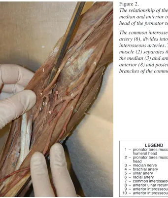
We also found an atypical branching pattern of the ra-dial artery. The common interosseous artery, which usual-ly arises from the ulnar artery, in this case emerged as a short branch of the radial artery distal to the radial tube-rosity (Figure 1B). While passing toward the interosseous membrane, the common interosseous artery was separat-ed from the mseparat-edian and anterior interosseous nerves by the ulnar head of the pronator teres muscle (Figure 2). The
common interosseous artery gave off the anterior interos-seous artery and the anterior and posterior ulnar recurrent arteries before continuing across the interosseous mem-brane as the posterior interosseous artery. All of these branches followed typical courses and supplied the major part of the posterior and anterior muscular compartments of the forearm region.4,14
Discussion
Since the presence of both a superficial ulnar and a super-ficial radial artery in one arm is extremely rare, we will first discuss the incidence of the better documented singly occurring superficial forearm arteries. We will then dis-cuss the two studies in which both forearm arteries were superficial, and the clinical implications of this type of variation.
Superficial ulnar arteries are relatively rare, 0.7% to 9.4%.6,14,30 However, they usually branch much high-er, either from the axillary artery or the brachial artery as it courses in the arm, and are classified as superficial brachioulnar arteries.6 The superficial ulnar artery in our subject originated in the cubital fossa, so would not meet the definition of a brachioulnar artery.
Superficial radial arteries are even more rare, at an
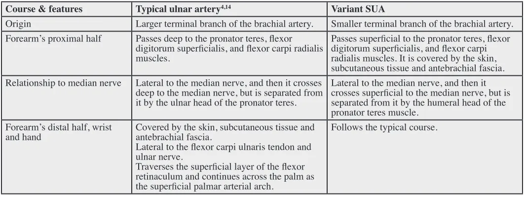
Comparison of the courses of the variant superficial ulnar artery (SUA) and the typical ulnar artery.
Course & features Typical ulnar artery4,14 Variant SUA
Origin Larger terminal branch of the brachial artery. Smaller terminal branch of the brachial artery. Forearm’s proximal half Passes deep to the pronator teres, flexor
digitorum superficialis, and flexor carpi radialis muscles.
Passes superficial to the pronator teres, flexor digitorum superficialis, and flexor carpi radialis muscles. It is covered by the skin, subcutaneous tissue and antebrachial fascia. Relationship to median nerve Lateral to the median nerve, and then it crosses
deep to the median nerve, but is separated from it by the ulnar head of the pronator teres.
Lateral to the median nerve, and then it crosses superficial to the median nerve, but is separated from it by the humeral head of the pronator teres muscle.
Forearm’s distal half, wrist
and hand Covered by the skin, subcutaneous tissue and antebrachial fascia. Lateral to the flexor carpi ulnaris tendon and ulnar nerve.
Traverses the superficial layer of the flexor retinaculum and continues across the palm as the superficial palmar arterial arch.
incidence rate of ~0.5%, although this varies by popula-tion.6,36,37 They most often occur at the level of the ana-tomical snuff box, with the artery passing superficial to the tendons that form the borders of the snuff box, rather than deep to them.4,6,14,33 The superficial radial artery in our subject differed from this pattern since it originated in the cubital fossa, was superficial along the forearm, and followed a typical course deep to the tendons that border the anatomical snuff box.
Rodriguez-Niedenfuhr et al.6, described a superficial brachioulnoradial artery as a superficial brachial artery branching at the elbow into radial and ulnar arteries and coexisting with a typical brachial artery that continues as
the common interosseous trunk. This is a different varia-tion than in our study where the brachial artery followed a typical course with just the radial and ulnar arteries fol-lowing a superficial course.
The incidence of a combined superficial radial artery and superficial ulnar artery is far less than the incidence singly. We have only found two case reports where both a super-ficial radial and a supersuper-ficial ulnar artery occurred in the forearm, and only one of these subjects had an abnormal branching pattern of the radial artery.32,38 In both of these studies the subjects had a superficial brachial artery with many branches, but did not have a typical brachial artery. In these cases the superficial radial and superficial ulnar
LEGEND
1 – pronator teres muscle: humeral head
2 – pronator teres muscle: ulnar head
3 – median nerve 4 – brachial artery 5 – ulnar artery 6 – radial artery
7 – common interosseous artery 8 – anterior ulnar recurrent artery 9 – anterior interosseous artery 10 – anterior interosseous nerve
Figure 2.
The relationship of the common interosseous artery with the median and anterior interosseous nerves and with the ulnar head of the pronator teres muscle.
artery in the cubital fossa. This was different than in our study, where the SRA and SUA arose from a typical bra-chial artery. Similar to our study, the superficial radial and superficial ulnar arteries rejoined their “common textbook” position in the distal forearm. In one subject the common interosseous artery arose from the radial artery, but this subject also had a median artery originating from the com-mon interosseous artery. In both of these studies there were variant arteries along most of the upper limb, both in the arm and forearm. Our study demonstrates that it is possible to have variant forearm arteries occurring with typical arm arteries. Despite this being a rare variant, the risk of injury and iatrogenic consequences that could impact blood sup-ply to the hand are substantially higher than the risks asso-ciated with the more frequently reported single superficial forearm arteries. The clinical implications relating to a sin-gle SUA or SRA will be combined to discuss the clinical implications of the variants described in this study.
An awareness of these variations is essential in order to prevent difficulty in performing physical exams, or inter-preting physical findings. McWilliams et al.27, presented a case in which a superficial ulnar artery was misdiagnosed as phlebitis. Chin et al.15, described the difficulty even trained anaesthesiologists have in differentiating between an artery and vein in a case when a superficial ulnar artery is present. In the current study this difficulty would be compounded by the difference in size of the radial and ulnar arteries, with the radial being larger than usual, and therefore having a relatively stronger pulse.
Clinical difficulties associated with superficial brachioulnar arteries and superficial radial arteries have been separately reported. These include inadvertent cannulation and difficulty interpreting angiographic im-ages.2,9,19,30 Due to its position close to the cephalic vein, a superficial radial artery is at greater risk of being ac-cidentally cannulated than a superficial ulnar artery.15 Surgery for radial forearm flaps, coronary bypass and compartment syndrome could lead to accidental division of the artery, which could jeopardize blood flow to the hand.1,6,15,16,18,23-29,30,35 On the contrary, if the superficial brachioulnar artery is identified, it could actually be of benefit for plastic surgeons performing reconstructive surgery with skin flaps, since it potentially has lower complication rates and better cosmetic outcome than ra-dial forearm flaps.6,30
forearm flap procedures makes preoperative identification of the arterial path by Doppler ultrasound or angiography important.9,17,18,20,22,27,31 With the variant arteries described in this study this would be even more vital, since the ra-dial artery was the main source of blood supply to the hand.
The clinical implications that were discussed above with regard to superficial brachioulnar and superficial ra-dial arteries would also apply to our subject. The argu-ment could be made that since both forearm arteries are superficial, a person with these variations is at much greater risk for an injury that could compromise blood supply to the hand.
Different theories about arterial development in the upper limb have been a source of controversy for many years.39 When we combine the information from studies that analysed embryonic development using 3D recon-structive imaging40 with information from molecular and genetic studies41, a more comprehensive picture of arterial development emerges.
Vasculogenesis occurs when signalling pathways cause hemangioblasts and endothelial cells to assemble into primitive tubular networks. The cells involved further differentiate into arterial and venous endothelial cells, creating the primary capillary plexus. This primary capil-lary plexus is transformed into a complex network by a remodelling and sprouting process called angiogenesis. A delicate balance between activators and inhibitors in the signalling pathways controls vessel formation during angiogenesis. The capillary plexus develops in a proximal to distal pattern and is present in the entire upper limb by the 28th day of human embryonic development.
vessel formation sometime between the 41st and 44th days of human embryonic development.
Conclusions
We present an unusual case of superficial radial and ulnar arteries. Typically variations in forearm arteries are asso-ciated with early branching from the brachial artery, or, in the case of the radial artery, a superficial course distally in the forearm and wrist. In the presented case the brachial artery bifurcates as usual in the cubital fossa, but both ra-dial and ulnar arteries are superficial for the length of the forearm, then they resume typical paths as they cross the wrist. Also of note in this case, the common interosseous artery emerged as a branch of the radial artery, not the ulnar. To our knowledge, this combination of variations has not previously been reported. A person with these variations is at much greater risk of injury affecting blood supply to their hands, and of iatrogenic complications if invasive procedures are undertaken without identifying the variations beforehand.
References
1. Bergman RA, Thompson SA, Afifi AK. Catalog of Human variation. Baltimore-Munich: Urban & Schwazzenberg, 1984: 103-112.
2. Ciervo A, Khan M, Pangilinan AJ, Dardik H. Absence of the brachial artery: Report of a rare human variation and review of upper extremity arterial anomalies. J Vasc Surg. 2001; 33(1): 191-194.
3. McCormack LJ, Cauldwell EW, Anson J. Brachial and antebrachial arterial patterns. Surg Gynecol Obstet. 1953;96:43-54.
4. Williams PL, ed. Gray’s Anatomy. The Anatomical basis of Medicine and Surgery.38th ed. Edinburgh: Churchill Livingstone, 1995; 1538-1544.
5. Mordick TG. Vascular variation of the radial forearm flap: a case report. J Reconstr Microsurg. 1995; 11(5): 345-346. 6. Rodriquez-Niedenfuhr M, Vazquez T, Nearn L, Ferreira B,
Parkin I, Sanudo JR. Variations of the arterial pattern in the upper limb revisited: a morphological and statistical study, with review of the literature. J Anat. 2001; 199(Pt 5): 547-566.
7. Gabe ED, Figal JC, Wisner JN, Laguens R. Radial artery graft vasospasm. Eur J Cardiothorac Surg. 2001; 19: 102-104.
8. Porter CJ, Mellow CG. Anatomically aberrant forearm arteries: an absent radial artery with co-dominant median and ulnar arteries. Br J Plast Surg. 2001; 54(8): 727-728. 9. Connel DA, Koulouris G, Thorn DA, Potter HG.
Contrast-enhanced MR angiography of the hand. Radiographics. 2002; 22(3): 583-599.
10. Patterson RB, Smail D. Radial artery as conduit for distal renal artery reconstruction. J Vasc Surg. 2003; 38(3): 609-612.
11. Loukas M, Curry B. A case of an atypical radial artery. Clin Anat. 2006; 19(8): 706-707.
12. Kohonen M, Teerenhovi O, Terho T, Laurikka J, Tarkka M. Forearm vessel atherosclerosis. A harbinger of carotid disease? Scand Cardiovasc J. 2009; 43(1): 69-71. 13. Klimek-Piotrowska W, Pacholczak R, Walocha J.
The multiple variations of the arterial pattern in upper extremities: A case report and embryological pathogenesis. Clin Anat. 2012; 26: 1031-1035.
14. Moore KL, Dalley FD, Agur AMR. Clinically oriented anatomy.7th. ed. Philadelphia: Wolters Kluwer/Lippincott Williams & Wilkins, 2014: 670-819.
15. Chin KJ, Singh K. The superficial ulnar artery-a potential hazard in patients with difficult venous access. Br J Anaesth. 2005; 94(5): 692-693.
16. Pelin C, Zagyapan R, Mas N, Karabay G. An unusual course of the radial artery. Folia Morphol. 2006; 65(4): 410-413. 17. Roca-Tey R, Rivas A, Samon R, Ibrik O, Martinez-Cercos R,
Viladoms J. Noninvasive assessment of forearm vessels by color Doppler ultrasonography (CDU) before and after creation of radiocephalic fistula (RCF). Nefrologia. 2007; 27(4): 489-495.
18. Shen S, Hong MK. A rare case of bilateral variations of upper limb arteries: brief review of nomenclature, embryology and clinical applications. Surg Radiol Anat. 2008; 30(7): 601-603.
19. Shetty SD, Nayak BS, Madhav NV, Abhinitha P. The abnormal origin, course and the distribution of the arteries of the upper limb: a case report. J Clin Diagn Res. 2012; 6(8): 1414-1416.
20. Salunke AA, Nambi GL, Dhanwate AD, Siriwardana HA. Superficial ulnar artery: Clinical recommendations to avoid iatrogenic complications due to variation in arterial system. Niger Med J. 2014; 55(3): 276-277.
21. Poteat WL. Report of a rare human variation: absence of the radial artery. Anat Rec. 1986; 214(1): 89-95.
22. Nunoo-Mensah J. An unexpected complication after harvesting of the radial artery for coronary bypass grafting. Ann Thorac Surg. 1998; 66(3): 929-931.
23. Aftabuddin M, Islam N, Jafar M, Hague E,
Alimuzzaman M. Management of isolated radial and ulnar arteries at the forearm. J Trauma. 1995; 38(1): 149-151. 24. Devansh MS. Superficial ulnar artery flap. Plast Reconstr
Surg. 1996; 97: 420-426.
25. Yuksel M, Yuksel R, Weinfeld AB, Shenaq SM. Superficial ulnar artery: embryology, case report and clinical
significance in reconstructive microsurgery. J Reconstr Microsurg. 1999; 15: 415-420.
superficial ulnar artery. Eur J Ultrasound. 2000; 13(2): 155-157.
28. Greene MA, Malias MA. Arm complications after radial artery procurement for coronary bypass operation. Ann Thorac Surg. 2001; 72(1): 126-128.
29. Valantine RG, Modrall JG, Clagett GP. Hand ischemia after radial artery cannulation. J Am Coll Surg. 2005; 201(1): 18-22.
30. Dartnell J, Sekaran P, Ellis H. The superficial ulnar artery: Incidence and calibre in 95 cadaveric specimens. Clin Anat. 2007; 20: 929-932.
31. McCartney CJ, Xu D, Constantinescu C, Abbas S, Chan VW. Ultrasound examination of peripheral nerves in the forearm. Reg Anesth Pain Med. 2007; 32(5): 434-439. 32. D’Costa S, Shenoy BM, Narayana K. The incidence of a
superficial arterial pattern in the human upper extremities. Folia Morphol. 2004; 63(4): 459-463.
33. Wood SJ, Abrahasm PH. Bilateral superficial radial artery at the wrist associated with radial origin of a unilateral median artery. J Anat. 1997; 189: 691-693.
34. Sasaki K, Nozaki M, Aiba H, Isono N. A rare variant of the radial artery: clinical considerations in raising a radial forearm flap. Br J Plast Surg. 2000; 53(5): 445-447. 35. Yagain VK, Dave MR, Anadkat S. Unilateral high origin
71(2): 121-124.
36. Lo RN, Leung MP, Lau KC, Yeung CY. Abnormal radial artery in Down’s syndrome. Arch Dis Child. 1986; 61: 885-890.
37. Nie B, Zhou Y, Li G, Shi D, Wang J. Clinical study of arterial anatomic variations for transradial coronary procedure in Chinese population. Chin Med J. 2009; 122(18): 2097-2102.
38. Kachlik D, Konarik M, Baca V. Vascular patterns of upper limb: an anatomical study with accent on superficial brachial artery. Bos J Basic Med Sci. 2011; 11(1): 4-10. 39. Natsis K, Papadopolou AL, Paraskevas G, Totlis T,
Tsikaras P. High origin of the superficial ulnar artery arising from the axillary artery: anatomy, embryology, clinical significance and a review of the literature. Folia Morphol. 2006;65(4): 400-405.
40. Rodriquez-Niedenfuhr M, Burton GJ, Deu J, Sanudo JR. Development of the arterial pattern in the upper limb of staged human embryos: normal development and anatomic variations. J Anat. 2001; 199(Pt 4): 407-417.