N O T E
Extractives of
Quercus crispula
sapwood infected
by the pathogenic fungus
Raffaelea quercivora
II: isolation
and identification of phenolic compounds from infected sapwood
Kayoko Imai• Kosei Yamauchi•Tohru Mitsunaga
Received: 17 June 2013 / Accepted: 23 August 2013 / Published online: 20 September 2013 ÓThe Japan Wood Research Society 2013
Abstract There has been a mass mortality of oak trees in the area along the coast of the Japan Sea. This phenomenon is caused by the ambrosia beetle Platypus quercivorus, which carries the ambrosia fungus Raffaelea quercivora. Extractives of a necrotic brownish coloration formed in the infected sapwood of Quercus crispula were investigated. The methanol extract of the damaged sapwood of
Q. crispulawas concentratedin vacuo and centrifuged to yield precipitates and the supernatant. The precipitates were subjected to Sephadex LH-20 column chromatogra-phy and preparative HPLC to describe a novel ellagic acid derivative (1). The 10 % methanol water-soluble part of the supernatant was subjected to medium-pressure ODS col-umn chromatography and preparative HPLC, respectively, to analyze a known lignan (2). Sulfuric acid hydrolysis of (1)yielded an ellagic acid and a gallic acid. NMR and LC-TOF/MS indicated that an ellagic acid and a gallic acid bonded to a xylose with glycosidic and ester bonds, respectively. Compound (1) was identified as 4,5-dihy- droxy-6-(3,7,8–trihydroxy-5,10-dihydro–chromeno[5,4,3-cde]chromen-2-yloxy)-tetrahydro-pyran-3-yl ester, and compound (2) was identified as (–)-lyoniresinol. The pre-sence of (–)-lyoniresinol from damaged sapwood indicated that infection ofR. quercivoramay cause the formation of a pseudo-heartwood in the sapwood ofQ. crispula. Keywords Quercus crispulaRaffaelea
quercivora(–)-lyoniresinol InfectionTannin
Introduction
The mass mortality of oak trees has been prevalent along the eastern coast of the Japan Sea since the late 1980s [1]. This large-scale forest mortality is caused by the ambrosia fungus, Raffaelea quercivora, which is a symbiotic microorganism carried by the ambrosia beetle, Platypus quercivorusMurayama [2]. The beetle carries the fungus in a specialized fungal storage organ called a mycangium for breeding its larva. The ambrosia beetle bores into the trunk of a dead or wilting oak tree and a brownish yellow col-oration is generated around the beetle gallery in the sap-wood. The colored part could contain several substances, such as repellents against the beetle, because beetles newly attacking a tree have an inclination to avoid colored parts of the trunk. However, knowledge about the chemical components and the biological function of the colored part is limited.
Sapwood coloration caused by enzymatic reactions has been reported in Douglas fir,Pseudotsuga menziesii[3,4]. Compounds existing in the living cells of Douglas-fir sapwood, such as o-dihydroxy phenols, (–)-epicatechin, and dihydroquercetin, react with enzymes naturally occurring in the trees and form brown-colored materials in the exposed surface of lumber from Douglas fir. Bauch et al. [5] elucidated that the yellow coloration inQuercus roburL. and Q. petraeais due to the reaction products of hydrolyzable tannins caused by the fungusPaecillomyces variotii. The tannase of P. variotii [6] would play the important role of releasing the reactants from wood com-ponents for the coloration.
In our previous report, tannase and laccase activities were detected in a culture of R. quercivora, and they decomposed hydrolyzable tannin to gallic acid followed by oxidization to purprogallin carboxylic acid (PGCA).
K. ImaiK. YamauchiT. Mitsunaga (&)
Department of Applied Life Science, Faculty of Applied Biological Science, Gifu University, Yanagido 1-1, Gifu 510-1193, Japan
Furthermore, we suggested that PGCA is one of the oration substances or precursors of the unextractable col-ored substances of Q. crispula sapwood infected by R. quercivora[7].
This paper deals with the isolation and identification of a lignan and hydrolyzable tannins from yellowedQ. crispula
sapwood infected by the pathogenic fungusR. quercivora.
Experimental
General
HPLC equipped with a reversed phase column (InertsilÒ ODS-3 V, 4.6 mm i.d.9250 mm, GL Science, Japan) and a diode array detector (SPD-M10A vp, SHIMADZU) was used for analysis of the components. The following solvent system was used: a linear gradient elution for 45 min from 5 to 100 % of solvent A, (solvent A: methanol; solvent B: water including 0.05 % trifluoroacetic acid). The flow rate was 1 mL/min, monitored at 280 nm.
Semi-preparative HPLC was performed on an InertsilÒ ODS-3 column (10u9250 mm) at room temperature. The mobile phase was a mixture of solvent B and 100 % ace-tonitrile (solvent C) at a flow rate of 3 mL/min using a gradient elution of 0–30 % of solvent C by a linear decrease for 0–60 min. 1H and 13C nuclear magnetic res-onance (NMR) spectra were recorded on a JEOL ECA-500 (500 MHz), and chemical shifts are given ind (ppm) val-ues relative to that of the solvent [methanol-d4(dH3.35;dC
49.0)].
FAB-MS analysis was performed with a JMS-700/GI (JEOL) spectrometer operated in positive ion mode with 3-Nitrobenzyl alcohol (NBA) as the matrix. LC-TOF/MS analysis was performed with a WatersÒXevoTMQTof MS spectrometer operated in negative ion mode with a spray source type ES, capillary voltage of 3.3 kV, cone voltage of 30.0 V, collision energy of 21.0 V, and calibration temperature of 500°C. The mass range was 50–1000 Da. Optical rotations were measured using a JASCO P-2300 system.
Plant material
Quercus crispulainfected byR. quercivora was collected in Toyama Forest and Forestry Products Research Center in November 2007.
Extraction and isolation
Air-dried plant material was separated into bark, unaffected sapwood, affected sapwood and heartwood. The affected sapwood was ground by a ultra centrifugal mill and the
powder (725.6 g) was extracted with 70 % aqueous ace-tone (11.0 L). The aliquot of the extract was analyzed using HPLC. The decoction was concentrated in vacuo and centrifuged (3500 rpm for 10 min at 4°C) to yield a pre-cipitate (5.25 g) and supernatant (15.4 g). The ethanol soluble part of the precipitate was pre-fractionated by Sephadex LH-20 column chromatography (CC) (u2.4 cm920 cm) eluting with ethanol (500 mL), 50 % aqueous ethanol (200 mL), 50 % aqueous methanol (200 mL), and 70 % aqueous acetone (200 mL) to give 8 fractions. Fraction 5 (eluted with 50 % aqueous ethanol) was purified by preparative HPLC to give (1) (5.5 mg).
The 10 % methanol soluble part of the supernatant was subjected to medium-pressure CC with ODS (C18 120A 20/40lm) eluting with 10, 20, 30, 35, 40, and 50 % methanol to give 6 fractions. The 35 % methanol eluate was subjected to preparative HPLC to give (2)(106.7 mg).
4,5-dihydroxy-6-(3,7,8-trihydroxy-5,10-dihydro- chromeno[5,4,3-cde]chromen-2-yloxy)-tetrahydro-pyran-3-yl ester (1)
Brown oil; 5.5 mg; LC-TOF/MS: m/z 584.9888 [M -H]-, (calcd. for C26H18O16), m/z 300.9526 [M-ellagic
acid]-,C12H13O9; 1H NMR (500 MHz, CD3OD) d 7.79,
7.49 (2H in total, each s, ellagic acid unit protons), 7.09 (2H, s, galloyl-H), 3.62, 3.73, 3.88, 4.22, 5.00, 5.11 (6H in total, sugar protons), see Table1; 13C NMR (150 MHz, CD3OD) d166.4 (galloyl C-7), 159.8 (ellagic acid unit
C-7,70), 148.8 (ellagic acid unit C-40), 146.9 (ellagic acid unit C-4), 145.2 (galloyl C-3,5), 141.4 (ellagic acid unit C-3), 139.6 (ellagic acid unit C-30), 138.7 (galloyl C-4), 136.7 (ellagic acid unit C-20), 136.4 (ellagic acid unit C-2), 119.7 (galloyl C-1), 115.4 (ellagic acid unit C-1), 112.4 (ellagic acid unit C-5), 112.0 (ellagic acid unit C-10), 110.6 (ellagic acid unit C-50), 109.0 (galloyl C-2,6), 108.8 (ella-gic acid unit C-60), 108.0 (ellagic acid unit C-6), 102.9 (xylose C-1), 73.3 (xylose C-2), 73.1 (xylose C-3), 71.5 (xylose C-4), 62.5 (xylose C-5).
(–)-lyoniresinol (2)
White amorphous crystal; FAB-MS (positive mode) m/z 421.0 [M?H]? (C22H28O8); 1H NMR (CD3OD,
600 MHz) d 1.60–1.61 (1H, m, H-80), 1.94–1.96 (1H, m, H-8), 2.55 (1H, dd,J=11.5 and 15.0 H-70) 2.67 (1H, dd,
J=4.8 and 15.1 H-70), 3.35 (3H, s, 30-OMe), 3.47 (1H, m, H-90), 3.57 (1H, dd, J=4.8 and 10.3 H-90), 3.48 (2H, d,
J=2.76, H-9), 3.71 (6H, s, 3-OMe and 5-OMe), 3.82 (3H, s, 50-OMe), 4.29 (1H, d, J=5.52, H-7), 6.36 (2H, s, H-2,6), 6.56 (1H, s, H-20);13C NMR (CD3OD, 150 MHz):
65.5 (C-90), 105.6 (C-2, 6), 106.5 (C-20), 124.9 (C-10), 128.9 (C-60), 133.2 (C-4), 137.5 (C-40), 138.0 (C-1), 146.4 (C-50), 147.3 (C-30), 147.7 (C-3,C-5); [a]D
23.3
= -9.5°
(c=0.21, MeOH).
Results and discussion
A concentrated 70 % aqueous acetone decoction was obtained from the dried sapwood ofQ. crispulainfected by
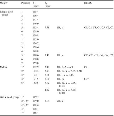
Raffaelea quercivora. When the concentrated solution was sitting at room temperature, a precipitate was observed. After having been centrifuged, the precipitate (brownish-red colored, 5.25 g) and supernatant (khaki colored, 15.4 g) were separated. The precipitate was subjected to Sephadex LH-20 column chromatography and preparative HPLC to yield (1). The chemical shifts and coupling constants of1H and13C NMR spectra of (1) are shown in Table1. Acid hydrolysis of (1) gave an ellagic acid and a gallic acid, indicating that it possesses these phenolic acids with ester binding and/or glycosidic linkage as shown in ellagitannins such as tellimagrandin II [8]. The negative ion mode of the
LC-TOF/MS for (1) by ESI gave 2 main peaks at m/z 584.9888 and m/z 300.9526 with an intensity of 80 and 100 %, respectively. In our previous paper, ester bonds of gallic acid and hexahydroxy diphenoic acid (HHDP) to a sugar of tercheblin gave only molecular ions without any fragments using the conditions described in this paper for the negative ion mode of the LC-TOF/MS [9]. In addition, pentagalloylglucose gave 2 main peaks for the molecular ion and fragment ion corresponding to [M-galloyl]?(data not shown). These results mean that an ester binding in hydro-lyzable tannins is not cleaved, while a glycosidic linkage is easily cleaved by the ESI mode of the LC-TOF/MS. If the fragment ion m/z 300.9526 could be obtained by loosing 1 mol of ellagic acid from molecular ion m/z 584.9888, an ellagic acid should be connected at the C1 position of the sugar moiety as a glycosidic linkage. Thus, a mole of gallic acid could be connected at another position of the sugar as an ester bond, which appears at m/z 300.9526. According to the mass number of the molecular ion and the fragment ion peaks, the sugar moiety of (1) is considered to be xylose.
The glycoside was determined to be xylose through the chemical shifts and coupling patterns of its proton signals
Table 1 1H and13CNMR spectral data and key HMBC correlations of (1)
Moiety Position dC
(ppm)
dH
(ppm)
HMBC
Ellagic acid group
1 115.4
2 136.4
3 141.4
4 146.9
5 112.4 7.79 1H, s C1, C2, C3, C4, C5, C6, C7
6 108.0
7 159.8
10 112.0
20 136.7
30 139.6
40 148.8
50 110.6 7.49 1H, s C10, C20, C30, C40, C60, C70
60 108.8
70 159.8
Xylose 100 102.9 5.11 1H, d,J=6.9 C4
200 73.3 3.73 1H, dd,J=6.85, 8.60
300 73.1 3.88 1H, t,J=9.15
400 71.5 5.00 1H, m C7000
500 62.5 3.62 1H, dd,J=9.75,
11.45
4.22 1H, dd,J=5.70, 12.00
Gallic acid group 1000 119.7
2000, 6000 109.0 7.09 2H, s 3000, 5000 145.2
(6 protons, d 3.62–5.11), which were connected by a
1H-1H-COSY spectrum. HMBC (evolution time (D)=
200 ms) shows that the H-100 proton on xylose (d 5.11 ppm) was correlated with the C-4 of the ellagic acid moiety. From here onwards, the presence of the connection of the C-4 of ellagic acid to the C-100of xylose was indi-cated. Furthermore, the chemical shift of the H-400 proton
of the xylose residue appeared downfield compared to xylose, indicating a connection of the C-7000of gallic acid to the C-400of xylose. On the basis of these data, the structure of (1) was identified as 4,5-dihydroxy-6-(3,7,8-trihydroxy-5,10-dihydro-chromeno[5,4,3-cde] chromen-2-yloxy)-tet-rahydro-pyran-3-yl ester (Fig. 1), a novel ellagic acid derivative. A paper suggested that hydrolyzable tannins such as pedunculgin, 1(b)-O-galloylpedunculagin, casu-ariin, and casuarinin in the wood ofPlatycarya strobilacea
were decreased by the charring which causes radical oxi-dation. However, the concentration of 30-O-methyl-(1) in the wood was increased by charring [10]. Interestingly, (1) was newly isolated in the yellowed sapwood ofQ. crispula, so a similar phenomenon may occur in infected sapwood oxidized by the tannase and laccase in the pathogenic fungusR. quercivora. These studies indicated that radical oxidation by charring or infection may change the hydro-lyzable tannins into unique compounds like (1).
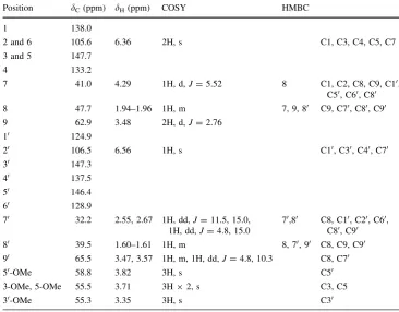
The 10 % methanol soluble part of the supernatant was subjected to Sephadex LH-20 CC and preparative HPLC, respectively, to afford (2) as a known lignan. The molec-ular formula of (2) was found to be C22H28O8by FAB-MS
(positive mode) m/z 421.0 [M?H]? (C22H28O8). The
results of1H and13C NMR spectra were shown in Table2. Two-dimensional NMR, HMQC, HMBC, and COSY were performed to determine the structure of (2). The results identified it as (–)-lyoniresinol showing -9.5° of optical rotation (Fig. 1).
O O
O
OH O
O H
OH O H
O H
O O O H
O
O
OH OH 1
2 3
4
5 6
1'
2' 3'
4' 5' 6' 1''
2'' 3'' 4''
5''
1''' 2''' 3'''
4'''
5''' 6'''
7'''
7 7'
1: 4,5-dihydroxy-6-(3,7,8-trihydroxy-5,10-dihydro-chromeno
[5,4,3-cde]chromen-2-yloxy)-tetrahydro- pyran-3-yl ester
1 2
3 4
5 6 1' 2' 3'
4'
5' 6'
O H H3CO
OCH3
OH
OCH3
H3CO
OH
OH H
H
7 8
7' 8'
9'
9
2: (-)-lyoniresinol
Fig. 1 Structures of1and2
Table 2 1H and13CNMR spectral data and key COSY and HMBC correlations of (2)
Position dC(ppm) dH(ppm) COSY HMBC
1 138.0
2 and 6 105.6 6.36 2H, s C1, C3, C4, C5, C7
3 and 5 147.7
4 133.2
7 41.0 4.29 1H, d,J=5.52 8 C1, C2, C8, C9, C10,
C50, C60, C80
8 47.7 1.94–1.96 1H, m 7, 9, 80 C9, C70, C80, C90
9 62.9 3.48 2H, d,J=2.76
10 124.9
20 106.5 6.56 1H, s C10, C30, C40, C70
30 147.3
40 137.5
50 146.4
60 128.9
70 32.2 2.55, 2.67 1H, dd,J=11.5, 15.0,
1H, dd,J=4.8, 15.0
70,80 C8, C10, C20, C60, C80, C90
80 39.5 1.60–1.61 1H, m 8, 70, 90 C8, C9, C90
90 65.5 3.47, 3.57 1H, m, 1H, dd,J=4.8, 10.3 C8, C70
50-OMe 58.8 3.82 3H, s C50
3-OMe, 5-OMe 55.5 3.71 3H92, s C3, C5
Several papers have been published on the isolation of lyoniresinol, including on the mixture of (?)- and (–)-lyoniresinol [11], its glycoside [12–14], (?)-lyoniresinol from the heartwood ofQ. crispula [15], and (–)-lyonires-inol from the root ofKirkia acuminata[16]. Interestingly, (–)-lyoniresinol was isolated from the sapwood of Q. crispulainfected byR. quercivorain the present study, in spite of the fact that lyoniresinol was not contained in the non-infected sapwood of Q. crispula. This experimental result might show the formation of pseudo-heartwood from sapwood byR. quercivorainfection.
On the other hand, resulting coloration parts in sapwood infected byR. quercivorawould be observed as a repellent part against the ambrosia beetle Platypus quercivorus as mentioned in the Introduction. Therefore, the substances, including two compounds isolated in this paper, consisted in the colored sapwood may have repellent properties against the beetle. In the next paper, the evidence would be made clear to solve the mystery of the mass mortality of oak tree.
Acknowledgments The authors are grateful to Dr. Koji Takabata (Toyama Forestry and Forest Products Research Center) for providing the infectedQ. crispulaplants. This research was partially supported by the Ministry of Education, Culture, Sports, Science and Technol-ogy Japan, and a Grant-in-Aid for Scientific Research (B), 16380096.
References
1. Ito S, Kubono T, Sahashi N, Yamada T (1998) Associated fungi with the mass mortality of oak trees (in Japanese). J Jpn For Soc 80:170–175
2. Kubono T, Ito S (2002)Raffaelea quercivorasp. nov. associated with mortality of Japanese oak and the ambrosia beetle (Platypus quercivorus). Mycoscience 43:255–260
3. Murray LL, Durria AAM (1996) An enzyme extract from Douglas-fir sapwood and its relationship to brown staining. Wood Fiber Sci 28(1):2–6
4. Laver ML, Durria AAM (1997) Chemical brown staining of Douglas-fir wood: characterization of a wood enzyme extract. Forest Prod J 47(4):93–97
5. Bauch J, Hundt H, Weissmann G, Lange W, Kubel H (1991) On the causes of yellow discolorations of oak heartwood (Quercus sect.robur) during drying. Holzforschung 45:79–85
6. Battestin V, Macedo GA (2006) Tannase production by Paec-illomyces variotii. Bioresour Technol 98(9):1832–1837 7. Imai K, Mitsunaga T, Takemoto H, Yamada T, Ito S, Ohashi H
(2009) Extractives of Quercus crispula sapwood infected by the pathogenic fungi Raffaelea quercivora (I): comparison of sapwood extractives non-infected and infected. J Wood Sci 55:126–132
8. Lee SH, Tanaka T, Nonaka GI, Nishioka I (1990) Hydrolysable tannins fromEuphorbia thymifolia. Phytochemistry 29(11):3621– 3625
9. Muddathir AM, Yamauchi K, Mitsunaga T (2013) Anti-acne activity of tannin related compounds isolated from Terminalia laxiflora. J Wood Sci. doi:10.1007/s10086-013-1344-4
10. Maeda H, Kakoki N, Ayabe M, Koga Y, Oribe T, Matsuo Y, Tanaka T, Kouno I (2011) ent-Eudesmane sesquiterpenoids, galloyl esters of the oak lactone precursor, and a 3-O -methylel-lagic acid glycoside from the wood ofPlatycarya strobilacea. Phytochemistry 72:796–803
11. Kaneda N, Dinghorn AD, Farnsworth NR, Tuchinda P, Udcha-chon J, Santisuk T, Reutrakul V (1990) Two diarylheptanoids and a lignan fromCasuarina junchuhniana. Phytochemistry 29:3366– 3368
12. Yuasa K, Ide T, Totsuka H, Ogimi C, Hirata E, Takushi A, Takeda Y (1997) Lignan and neolignan glycosides from stems of Alangium premnifolium. Phytochemistry 45:611–615
13. Latte´ KP, Kaloga M, Sha¨fer A, Kolodziej H (2008) An ellagitannin, n-butyl gallate, two aryltetralin lignans, and an unprecedented diterpene ester from Pelargonium reniforme. Phytochemistry 69:820–826
14. Jong-Anurakkun N, Bhandari MR, Kawabata J (2007)a -Gluco-sidase inhibitors from Devil tree (Alastronia scholaris). Food Chem 103:1319–1323
15. Omori S, Nishimoto F, Taneda K (1991) The extractive compo-nents of oak heartwood and commercial whiskey (In Japanese). Mokuzai Gakkaishi 37:82–87