C A S E R E P O R T
Open Access
Metastatic colonic and gastric polyps from
breast cancer resembling hyperplastic
polyps
Yoshiya Horimoto
1,2*, Tetsuro Hirashima
3, Atsushi Arakawa
4, Hiroyoshi Miura
5and Mitsue Saito
1Abstract
Breast cancer metastasis to the gastrointestinal tract is relatively rare and is generally found when patients complain of symptoms such as gastrointestinal obstruction. Herein, we report a case with metastatic colonic and gastric lesions from breast cancer, with the formation of mucosal polyps which resembled typical hyperplastic polyps.
A 47-year-old woman underwent curable surgery for breast cancer and received standard systemic treatments. Her primary tumor was composed of a mix of invasive lobular and ductal carcinomas. During adjuvant endocrine therapy, she developed multiple colonic metastases, identified by colonoscopy performed as part of a general health check-up. She had no symptoms. Small elevated sessile polyps in the transverse colon and rectum showed histological features of signet-ring cell type adenocarcinoma, similar to the invasive lobular component of the primary breast cancer. During treatments for recurrent disease, she also developed multiple gastric metastases, with the same endoscopic and pathological features as the colonic lesions. Her treatment regimen was switched to oral chemotherapy, and she has since maintained stable disease for nearly 3 years. Multiple bone metastases eventually developed, and she was again switched to another systemic treatment but, to date, has remained free of symptoms.
We emphasize that the endoscopic findings of the metastatic lesions in the colon and stomach in this case highly resembled hyperplastic polyps. Since biopsy is not always performed for hyperplastic polyps in the gastrointestinal tract, we believe that this case report may encourage endoscopists to offer biopsies to the patient who has a history of breast cancer.
Keywords:Breast cancer, Lobular carcinoma, Colonic metastasis, Mucosal metastatic polyp
Background
Breast cancer rarely metastasizes to the gastrointestinal (GI) tract, and only 5% of patients with recurrent dis-ease have GI metastasis [1]. Invasive lobular carcinoma (ILC), which is characterized by minimal cell-cell adhe-sion, is known to more often metastasize to the GI tract than invasive ductal carcinoma (IDC), and this is especially true of ILC of the signet-ring cell type [2,3].
In making the differential diagnosis of metastatic dis-ease, several immunohistochemical (IHC) markers are useful for identifying breast cancer as the primary
tumor. Estrogen receptor (ER) and human epidermal growth factor receptor 2 (HER2) are often examined, but their expressions are detected in 12–25% and 20% of gastric cancers [4–6], and in 30–56% and 4–14% of colon cancers [7–10], respectively. Thus, these exami-nations are not sufficient, in terms of tissue specificity, for identifying the origin of metastasis. CK7 is often observed in the epithelial layers of breast ducts and the lungs, while intestinal tissues show no expression of this protein [11, 12]. On the contrary, CK20 is often expressed in the large intestine and bile ducts, while it is rarely seen in the breast [11, 12]. Thus, metastatic disease from breast cancer is expected to be CK7-positive and CK20-negative, while the opposite is gen-erally seen in primary colon cancer. GCDFP-15 and mammaglobin are also widely used. GCDFP-15, one of * Correspondence:yoshiyahorimoto@hotmail.com
1
Department of Breast Oncology, Juntendo University School of Medicine, 2-1-1 Hongo, Bunkyo-ku, Tokyo 113-0033, Japan
2Department of Pathology and Oncology, Juntendo University School of
Medicine, 2-1-1 Hongo, Bunkyo-ku, Tokyo 113-0033, Japan Full list of author information is available at the end of the article
mucosal polyps from breast cancer, which had an entirely hyperplastic appearance, and produced no symptoms. Metastasis localized only in the GI mucosa, verified endoscopically [18] or surgically [19], is very rare.
Case presentation
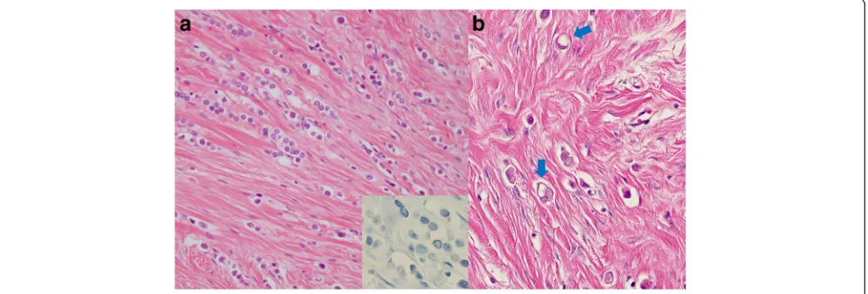
A 47-year-old woman felt a lump in her left breast and came to our hospital. We diagnosed invasive breast cancer (cT2N1M0 Stage IIB) and administered primary systemic chemotherapy (FEC × 4 cycles followed by Do-cetaxel × 4 cycles). Clinically, a complete response was obtained and she then underwent mastectomy (Bt+Ax). Pathological findings of the operative specimen were a mix of ILC and IDC. The ILC component had lost E-cadherin expression, and some cells showed signet-ring cell carcinoma features (Fig. 1a,b). Pathologically, the tumor was 105 mm in diameter and metastasis was de-tected in one lymph node; the histological features were consistent with ILC, while no lymphovascular involve-ment was detected (pT3N1aM0 Stage IIIA). The chemo-therapy effect was grade 2, according to the general rules for Clinical and Pathological Recording of Breast Cancer (17th edition) by the Japanese Breast Cancer Society. ER and progesterone receptor (PR) were both
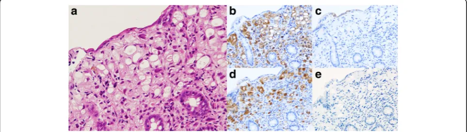
(Fig.2a) and was removed endoscopically. Histologically, adenocarcinoma (signet-ring cell type) was observed in the mucosa of the transverse colon (Fig.3a). Because the histological type was not one commonly seen in colon cancer cases, biopsy specimens were reviewed at our hospital and compared with that of the primary breast cancer. Immunohistochemical examination revealed the polyp to be ER positive, PR negative, HER2 negative, E-cadherin negative, Cytokeratin (CK) 7 positive, CK20 negative, gross cystic disease fluid protein (GCDFP-15) positive, and mammaglobin negative (Fig. 3a–e). Based on histological similarity and the results of additional IHC examinations, we concluded that the polyp repre-sented metastasis from the original breast cancer. We performed computed tomography and positron emission tomography at the time of diagnosis of recurrent disease, but no distant metastases, including the colonic polyp, were detected by these examinations.
Since the metastases were diagnosed 8 months after starting SERM, which had been preceded by AI as adjuvant therapy, the patient restarted treatment with the initial AI. Colonoscopy, performed 2 months later, revealed that the numbers of small polyps had increased and were present throughout the large intes-tines (Fig. 2b). Five biopsies in total were taken from
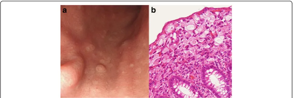
the ascending and transverse portions of the colon and the rectum, and all showed the same histological features as the signet-ring cell carcinoma polyp. How-ever, on re-examination 4 months later, the numbers of polyps had obviously decreased, indicating the endocrine treatment to be effective. The patient main-tained a stable disease state with this treatment for 1 year after the initial diagnosis of metastases. How-ever, she eventually developed multiple small white elevated lesions (Fig. 4a), which had not been present at the time of initial diagnosis of the recurrent disease. We confirmed these gastric lesions to have the same pathological features, signet-ring cell type adenocar-cinoma (Fig.4b), as the colonic lesions, despite having the appearance of hyperplastic polyps. The gastric polyps showed the same expression pattern, with CK7, CK20, GCDFP-15, and mammaglobin being positive, negative, positive, and negative, respectively. She started treatment with capecitabine, and the gastric lesions remained stable for 2 years and 7 months,
while the colonic lesions had almost disappeared within 10 months of starting this drug. She eventually developed multiple bone metastases, and her systemic treatment was switched to another chemotherapeutic regimen.
Throughout her clinical course, she has remained free of symptoms, suffering neither abdominal pain nor melena.
Conclusions
We could not ascertain whether the metastases repre-sented mucosal lesions if the patient had not undergone a routine health check-up. Otherwise, she might have progressed to a severe condition, such as GI perforation or obstructive ileus. Instead, this patient has been free of symptoms for more than 5 years with adequate systemic therapies against recurrent diseases.
We emphasize that, based on the endoscopic find-ings, these colonic and gastric metastatic lesions essentially had the appearance of hyperplastic polyps.
Since biopsy is not consistently performed for
Fig. 2Metastatic mucosal polyp in transverse colon.aA small elevated sessile polyp (4 mm in diameter) was found in the transverse colon. Polypectomy was endoscopically performed and pathological examination revealed it to be a signet-ring cell carcinoma.bMultiple metastatic polyps in the transverse colon: re-examination by colonoscopy, performed 4 months after the diagnosis of metastasis, showed an increase in the numbers of metastatic polyps throughout the large intestine. Five biopsies were taken from the ascending colon, transverse colon, and rectum and all were confirmed to be signet-ring cell carcinoma
hyperplastic polyps in the GI tract, this case report may encourage endoscopists to offer biopsies to patients with a history of breast cancer. Whether or not this endoscopic appearance indicates early GI metastasis needs to be determined based on further case reports.
Abbreviations
AI:Aromatase inhibitor; CK: Cytokeratin; ER: Estrogen receptor; GI: Gastrointestinal; HER2: Human epidermal growth factor receptor 2; IDC: Invasive ductal carcinoma; IHC: Immunohistochmical; ILC: Invasive lobular carcinoma; PR: Progesterone receptor; SERM: Selective estrogen receptor modulator
Authors’contributions
YH and TH identified the unusual features of this case. YH wrote the article. TH performed endoscopy and provided all endoscopic images for the article. AA was the principal pathologist and supplied all of the pathology images. YH, HM, and MS were responsible for overall care of the patient. MS critically revised the manuscript for content. All authors read and approved the final manuscript.
Ethics approval and consent to participate Not applicable.
Consent for publication
Written informed consent was obtained from the patient for publication of this case report and any accompanying images. A copy of the written consent is available for review by the Editor-in-Chief of this journal.
Competing interests
The authors declare that they have no competing interests.
Publisher’s Note
Springer Nature remains neutral with regard to jurisdictional claims in published maps and institutional affiliations.
Author details
1Department of Breast Oncology, Juntendo University School of Medicine,
2-1-1 Hongo, Bunkyo-ku, Tokyo 113-0033, Japan.2Department of Pathology
and Oncology, Juntendo University School of Medicine, 2-1-1 Hongo, Bunkyo-ku, Tokyo 113-0033, Japan.3Tamaplaza South Gastrointestinal Clinic, 3-14-12 Shinishikawa, Aoba-ku, Yokohama-shi, Kanagawa 225-0003, Japan.
4Department of Human Pathology, Juntendo University School of Medicine,
2-1-1 Hongo, Bunkyo-ku, Tokyo 113-0033, Japan.5Department of Surgery,
Koshigaya Municipal Hospital, 10-47-1 Higashikoshigaya, Koshigaya, Saitama 343-8577, Japan.
Received: 7 December 2017 Accepted: 13 March 2018
References
1. Mukaiyama T, Ogawa M, Horikoshi N, Inoue K, Inagaki J, Egaki K, Kasumi F, Nishi M, Sakamoto G. Analysis of metastatic behaviors and causes of death in 100 autopsied patients with breast cancer. Jpn. J. Breast Cancer. 1989;4:121–6.
2. Arpino G, Bardou VJ, Clark GM, Elledge RM. Infiltrating lobular carcinoma of the breast: tumor characteristics and clinical outcome. Breast Cancer Res. 2004;6:R149–56.
3. Merino MJ, Livolsi VA. Signet-ring cell carcinoma of the breast. Cancer. 1981; 48:1830–7.
4. Chandanos E, Rubio CA, Lindblad M, Jia C, Tsolakis AV, Warner M, Gustafsson JA, Lagergren J. Endogenous estrogen exposure in relation to distribution of histological type and estrogen receptors in gastric adenocarcinoma. Gastric Cancer. 2008;11:168–74.
5. Gan L, He J, Zhang X, Zhang Y, Yu G, Chen Y, Pan J, Wang J, Wang X. Expression profile and prognostic role of sex hormone receptors in gastric cancer. BMC Cancer. 2012;12:566.
6. Bang Y-J, Van Cutsem E, Feyereislova A, Chung HC, Shen L, Sawaki A, Lordick F, Ohtsu A, Omuro Y, Satoh T, Aprile G, Kulikov E, Hill J, Lehle M, Rüschoff J, Kang Y-K. Trastuzumab in combination with chemotherapy versus chemotherapy alone for treatment of HER2-positive advanced gastric or gastro-oesophageal junction cancer (ToGA): a phase 3, open-label, randomised controlled trial. Lancet. 2010;376:687–97.
7. Alford TC, Do HM, Geelhoed GW, Tsangaris NT, Lippman ME. Steroid hormone receptors in human colon cancers. Cancer. 1979;43:980–4. 8. Marugo M, Molinari F, Fazzuoli L, Parodi MC, Bernasconi D, Menozzi F,
Giordano G. Estradiol and progesterone receptors in normal and pathologic colonic mucosa in humans. J Endocrinol Investig. 1985;8:117–9.
9. Nathanson DR, ATt C, Shia J, Chen B, D'Alessio M, Zeng ZS, Nash GM, Gerald W, Barany F, Paty PB. HER 2/neu expression and gene amplification in colon cancer. Int J Cancer. 2003;105:796–802.
10. Li Q, Wang D, Li J, Chen P. Clinicopathological and prognostic significance of HER-2/neu and VEGF expression in colon carcinomas. BMC Cancer. 2011;11:277.
11. Weiss LM, Chu PG. Immunohistochemical characterization of signet-ring cell carcinomas of the stomach, breast, and Colon. Am J Clin Pathol. 2004;121:884–92. 12. Tot T. The role of cytokeratins 20 and 7 and estrogen receptor analysis in
separation of metastatic lobular carcinoma of the breast and metastatic signet ring cell carcinoma of the gastrointestinal tract. APMIS. 2000;108:467–72. 13. Luo M-H, Huang Y-H, Ni Y-B, Tsang JYS, Chan S-K, Shao M-M, Tse GM. Expression of mammaglobin and gross cystic disease fluid protein-15 in breast carcinomas. Hum Pathol. 2013;44:1241–50.
14. Yan Z, Gidley J, Horton D, Roberson J, Eltoum IE, Chhieng DC. Diagnostic utility of mammaglobin and GCDFP-15 in the identification of metastatic breast carcinoma in fluid specimens. Diagn Cytopathol. 2009;37:475–8. 15. Bhargava R, Beriwal S, Dabbs DJ. Mammaglobin vs GCDFP-15: an
immunohistologic validation survey for sensitivity and specificity. Am J Clin Pathol. 2007;127:103–13.
16. Winston CB, Hadar O, Teitcher JB, Caravelli JF, Sklarin NT, Panicek DM, Liberman L. Metastatic lobular carcinoma of the breast: patterns of spread in the chest, abdomen, and pelvis on CT. AJR Am J Roentgenol. 2000;175:795–800. 17. Arrangoiz R, Papavasiliou P, Dushkin H, Farma JM. Case report and literature
review: metastatic lobular carcinoma of the breast an unusual presentation. Int J Surg Case Rep. 2011;2:301–5.
18. Takeuchi H, Hiroshige S, Yoshikawa Y, Kusumoto T, Muto Y. A case of synchronous metastasis of breast cancer to stomach and colon. Anticancer Res. 2012;32:4051–5.
19. Matsuda I, Matsubara N, Aoyama N, Hamanaka M, Yamagishi D, Kuno T, Tsukamoto K, Yamano T, Noda M, Ikeuchi H, Tomita N, Hirota S. Metastatic lobular carcinoma of the breast masquerading as a primary rectal cancer. World J Surg Oncol. 2012;10:231.