OncoTargets and Therapy 2018:11 131–155
OncoTargets and Therapy
Dove
press
submit your manuscript | www.dovepress.com 131
R e v i e w
open access to scientific and medical research
Open Access Full Text Article
Targeting histone methyltransferase and
demethylase in acute myeloid leukemia therapy
Germana Castelli elvira Pelosi Ugo Testa
Department of Oncology, istituto Superiore di Sanità, Rome, italy
Abstract: Acute myeloid leukemia (AML) is a clonal disorder of myeloid progenitors characterized by the acquisition of chromosomal abnormalities, somatic mutations, and epigenetic changes that determine a consistent degree of biological and clinical heterogeneity. Advances in genomic technologies have increasingly shown the complexity and heterogeneity of genetic and epige-netic alterations in AML. Among the geepige-netic alterations occurring in AML, frequent are the genetic alterations at the level of various genes involved in the epigenetic control of the DNA methylome and histone methylome. In fact, genes involved in DNA demethylation (such as
DNMT3A, TET2, IDH1, and IDH2) or histone methylation and demethylation (EZH2, MLL, DOT1L) are frequently mutated in primary and secondary AML. Furthermore, some histone
demethylases, such as LSD1, are frequently overexpressed in AML. These observations have strongly supported a major role of dysregulated epigenetic regulatory processes in leukemia onset and development. This conclusion was further supported by the observation that mutations in genes encoding epigenetic modifiers, such as DMT3A, ASXL1, TET2, IDH1, and IDH2, are usually acquired early and are present in the founding leukemic clone. These observations have contributed to development of the idea that targeting epigenetic abnormalities could represent a potentially promising strategy for the development of innovative treatments of AML. In this review, we analyze those proteins and their inhibitors that have already reached the first stages of clinical trials in AML, namely the histone methyltransferase DOT1L, the demethylase LSD1, and the MLL-interacting protein menin.
Keywords: acute myeloid leukemia, epigenetic modifications, methyltransferases, demethylases
Introduction
Epigenetic modification of DNA
In the nucleus of eukaryotic cells, DNA is packaged together with structural proteins (histones) to form a supramolecular complex known as chromatin. Chromatin is arranged in repeating units, known as nucleosomes: each nucleosome is formed by a sequence of DNA wound around eight histone cores. Chromatin may have a condensed conformation, known as heterochromatin, is essentially transcriptionally repressed, or a decondensed conformation known as euchromatin, and is transcriptionally active. The regulation of chromatin conformation is a fundamental biologic process and regulates, at specific sites, accessibility to DNA, a step required to allow gene transcription, rep-lication, recombination, and DNA repair. Epigenetic mechanisms determine heritable changes in gene expression that are due to alterations in the DNA sequence. Various epigenetic mechanisms play an essential role in the control of chromatin conformation, and are mainly represented by posttranslational modifications of histone, adenosine triphosphate (ATP)-dependent nucleosome remodeling, DNA modifications, and replacement of histone with histone variants. Posttranslational modifications of histones
Correspondence: Ugo Testa Department of Oncology, istituto Superiore di Sanità, 299 viale Regina elena, Rome 00161, italy Tel +39 06 4990 2422 Fax +90 06 4938 7087 email ugo.testa@iss.it
Journal name: OncoTargets and Therapy Article Designation: Review
Year: 2018 Volume: 11
Running head verso: Castelli et al
Running head recto: Histone methyltransferase and demethylase in AML DOI: http://dx.doi.org/10.2147/OTT.S145971
OncoTargets and Therapy downloaded from https://www.dovepress.com/ by 118.70.13.36 on 25-Aug-2020
For personal use only.
Dovepress
Castelli et al
represent one of the main epigenetic mechanisms to control gene expression.
Histones are highly basic nuclear proteins. They represent spools around which DNA winds, and play an essential role in the control of gene expression.1–3 There are five main
families of histones: H1/H5, H2A, H2B, H3, and H4. H1/H5 are known as linker histones, while H2A, H2B, H3, and H4 are known as core histones. The linker histones bind the nucleosome at the entry and exit sites of the DNA, thus maintaining DNA in place. The core histones form dimers, and four dimers form one octameric nucleosome core. Each canonical histone protein – H3, H4, H2A, and H2B – shares a common structural domain consisting of 3α helices (α1, α2, and α3), separated by two loops (L1 and L2) called the histone fold, facilitating the heterodimerization of H2A with H2B and H3 with H4. These heterodimeric interactions form the dimeric structural unit of nucleosomes. There are variants for the core histones H3, H2AB, and H2B: the histone variant H3.3 differs from the canonical histone H3 in five amino acids required for the genomic localization of H3.3 (usually H3.3 occupies gene promoters), and H2A. Z has considerable amino-acid variation compared to H2A.
Histone proteins undergo many modifications, mainly rep-resented by lysine methylation, arginine methylation, arginine citrullination, lysine acetylation, and serine/threonine/ tyrosine phosphorylation. Each nucleosome is composed of a short segment of DNA of about 145–147 base pairs and is engulfed by a histone octamer core, consisting of four dimers of each core histone (H2A, H2B, H3, and H4). The core histone is essentially a globular structure, except for histone tails, composed of about 30 amino acids. These tails are modi-fied by numerous posttranslational modifications, including methylation, phosphorylation, acetylation, ubiquitination, SUMOylation, ADP ribosylation, deamination, and croto-nylation. These epigenetic, posttranslation modifications of histone are highly controlled, governed by three categories of enzymatic proteins: “writers” are involved in the addi-tion of chemical groups to histone tails, “erasers” involved in the removal of these chemical groups, and “readers” are proteins that specifically recognize these histone modifica-tions. Notable examples of writers are represented by histone acetyltransferases (HATs) and histone methyltransferases, while erasers are represented by histone deacetylases and lysine demethylases; examples of readers are given by bromodomain-containing proteins, methyl-lysine and methyl-arginine-binding domain-containing proteins and PDH-containing proteins.
Histone acetylation is a dynamic process regulated by two families of enzymes: HATs and histone deacetylases
(HDACs).4 HATs catalyze the transfer of an acetyl group to
the ε-amino group of lysine side chains of the histone protein; these enzymes use acetyl-CoA as a cofactor. This reaction determines the shift of lysine from positively charged to a more neutral condition, reducing the affinity of the histone tail protruding from the nucleosome. As a consequence of these changes, chromatin adopts a more relaxed structure, more suitable for transcriptional activity through the binding of transcription machinery. Particularly, the BET proteins BRD2, BRD3, and BRD4 recognize and bind to the acety-lated histone-lysine residues. HDACs act in an opposite way, removing the acetyl groups from histone lysines. HATs comprise many enzymes, including CREBBP (CBP), EP300 (p300), KAT2B (PCAF), KAT5 (Tip60), and KAT6A (Mo2); HDACs include HDAC1–11 and SIRT1–7.
Histone methylation is a very important biologic process through which the expression of many genes is controlled. Histones can be methylated at the level of either lysine or arginine residue.5 Lysine residue can be monomethylated,
demethylated, and trimethylated. Arginine residues can be monomethylated or demethylated. Methylation is a dynamic process. Methyl units to histone molecules are added by
S-adenosyl-l-methionine (SAM)-dependent methyltrans-ferase and erased by specific demethylases represented by flavin-dependent LSD1 (also known as KDM1A) and LSD2 (also known as KDM2A) or by the Jmj family of 2-oxoglutarate-dependent demethylase.5
Histone-lysine methylation can lead either to transcrip-tional activation or transcriptranscrip-tional repression. Methylation of histone H3K4, H3K36, and H3K79 is associated with transcriptional activation, while demethylation and trimethy-lation of H3K9 and H3K27 are associated with transcriptional repression. Methylation at specific lysine residues is cata-lyzed by specific methyltransferases: H3K4 methylation is catalyzed by the SET domain-containing methyltransferases MLL1 (KMT2A) and MLL2 (KMT2D), H3K79 is methy-lated by DOT1L, H3K27 methylation is catalyzed by EZH1 and EZH2, H3K36 methylation is performed by SETD2 and WHSC1, H3K9 methylation is induced by MECOM (EVI1) and PRDM16, and H3K9 dimethylation and trimethylation is induced by SUV39H1 and EHMT2. It is important to note that methylation occurring at the level of H3K9 and H3K27 represents the two main mechanisms of gene silencing in mammalian cells. EZH1 and EZH2 make up part of PRC2 in association with two other subunits: EED and SUL12.
It is important to point out that in addition to epigenetic modifications of histone, the epigenetic modification of DNA through methylation represents another key mechanism of gene regulation.6 DNA methylation is associated with
OncoTargets and Therapy downloaded from https://www.dovepress.com/ by 118.70.13.36 on 25-Aug-2020
Dovepress Histone methyltransferase and demethylase in AML
transcriptional repression and occurs at the level of the C5 carbon of cytosine in DNA to form 5-methylcytosine (5-mC), through a reaction catalyzed by three types of DNA methyltransferase: DNMT1, DNMT3A, and DNMT3B. TET enzymes (TET1, TET2, and TET3) promote the oxidation of 5-mC to 5-hydroxymethylcytosine (5-hmC), promoting the demethylation process. IDH1 and IDH2 enzymes catalyze the conversion from isocitrate to α-ketoglutarate (αKG), which is required for the catalytic function of TET enzymes.
Alterations in epigenetic modifiers
in acute myeloid leukemia
A considerable number of genetic studies have been per-formed in acute myeloid leukemia (AML), allowing iden-tification of the most frequent genetic alterations observed in these leukemia types. Comprehensive whole-genome sequencing, exome sequencing, and targeted sequencing studies have shown that mutations at the level of the complex epigenetic gene machinery are frequent in AML. DNMT3A is mutated in 20%–25% of adult AML, and frequently involves the substitution of the amino acid arginine at position 882.
DNMT3A mutation is frequently found in association with FLT3ITD, NPM1, IDH1, and IDH2 mutations.7 DNMT3AR882H
has dominant negative activity that markedly reduces DNA methylation. DNA hypomethylation is an initiating event in the development of DNMT3A-mutated AML and thus represents the cause and not the consequence of leukemia development. Furthermore, CpG-island hypermethylation, a phenomenon frequently observed in DNMT3AWT AML, is
dramatically reduced in DNMT3A-mutated AML.8 In line
with these observations, conditional knock-in mice develop AML with enlarged hematopoietic stem-cell compartments and involvement of the mTOR pathway. Consequently,
DNMT3A-mutated leukemic cells are sensitive to the mTOR
inhibitor rapamycin.9 DNMT3A mutations occur during the
preleukemic phase of AML pathogenesis, supporting the key role for aberrant DNA methylation and consequent epigenetic reprogramming in malignant transformation of hematopoietic cells. The presence of DNMT3A mutations in AML is associated with poor outcome and reduced survival.10,11 DNMT1 is not mutated in AML, but is frequently
overexpressed.12 DNMT1, which methylates hemimethylated
DNA, is involved in the differentiation of normal hemopoi-etic stem cells (HSCs) and maintenance of leukemic SCs through epigenetic silencing of genes that inhibit self-renewal and leukemogenesis.13 Recent research has suggested that
DNMT1A could represent a therapeutic target for some AML. In fact, DNMT1A expression can be targeted in leukemic cells by inhibitors of FABP4 (upregulated in AML
and stimulates DNMT1A expression in these cells)14 or by
inhibitors of receptor tyrosine kinases.15 These treatments
result in inhibition of tumor growth, induction of cell differ-entiation, and impairment of leukemic progress in leukemia animal models.14,15 Very interestingly, a recent study provided
evidence that MUC1-C, a transmembrane oncoprotein aber-rantly expressed in leukemic SCs (where it is coexpressed with DNMT1), drives DNMT1 transcription.16 Targeting
MUC1-C with a specific monoclonal antibody, together with the DNMT1 inhibitor decitabine, markedly reduces DNMT1 expression and impairs the survival of AML cells.16
The ASXL1 gene encodes a chromatin-binding protein and is mutated in about 3%–5% of AML. The incidence of these mutations is higher in patients with intermediate risk and particularly with high-risk and secondary AML, where it is mutated in about 16% of patients. ASXL1 mutations, as a single prognostic factor, are associated with a negative outcome.17 ASXL1 gene mutations are particularly frequent
(20%) in RUNX1-mutated AML18 and are often associated
with myelodysplasia-related changes.19
EZH2, a catalytic component of PRC2, which mediates transcriptional silencing through di- and trimethylation of lysine 27 of histone H3, playing an important role as a histone–lysine N-methyltransferase enzyme, is mutated in about 2% of adult AML, where inactivating mutations are usually observed.20 Some AML displays a decrease in EZH2
protein levels due to a posttranslational mechanism trig-gered by EZH2 phosphorylation induced by CDK1.21 EZH2
inactivation is frequently observed at disease relapse, and is associated with HOX gene derepression and resistance to multiple drugs.21 EZH2 loss is frequently associated with a
decrease in H3K27me3 levels.21 EZH2 mutations are more
frequent in RUNX1-mutated18 and secondary AML, and are
often associated with increased HOXA9 expression.22
About 3%–5% of de novo AML patients display partial tandem duplication of the MLL gene, which is character-ized by internal tandem duplication of exons 3–9 or 3–11.23
MLLPTD acts as an oncogene by upregulating the expression
of HOX genes. These AML types display frequent mutations of other epigenetic regulators, such as TET2 (16%), EZH2 (10%), IDH1/2 (31%), and ASXL1 (6%). Furthermore, a typical feature of MLLPTD consists of the absence of NPM1
mutations and frequent RUNX1 (23%) and STAG2 muta-tions (16%).23 BCOR is a component of the variant-group
polycomb-repressive complex, mutated in about 4% of karyotype-normal AML. BCOR plays an important role in the control of hematopoiesis by inhibiting myeloid-cell proliferation and differentiation and regulates HOX gene expression.24
OncoTargets and Therapy downloaded from https://www.dovepress.com/ by 118.70.13.36 on 25-Aug-2020
Dovepress
Castelli et al
Interestingly, in a recent molecular classification of AML based on the analysis of a large set of samples, one of the larg-est groups was represented by AML with mutated chromatin, RNA-splicing genes or both, characterized by mutations of genes regulating chromatin (ASXL1, STAG2, BCOR, MLLPTD,
and PHF6), RNA splicing (SRSF2, SF3B1, U2AF1, and
ZRSR2), or transcription (RUNX1).25 As such, no single
molecular lesion defines this group. Interestingly, overlap-ping patterns of comutations are observed among the defining genes of this AML group.25 It is important to note that as
stated earlier, this AML subgroup also contains AML types with mutations of the splicing-factor machinery. Interest-ingly, mutations at the level of these genes interfere with pre-mRNA splicing of genes that are involved in the epigenetic machinery, such as BCOR, MLL2, and EZH2, and through this mechanism affect hematopoiesis.26,27 It is important to
underline that most patients in the chromatin-spliceosome group were older, with lower white-cell and blast-cell counts and response rates and negative long-term clinical outcome.25
Therefore, it is evident that most of these patients must be classified as high-risk AML.28
About 3% of de novo adult AML harbors a translocation of the MLL1 gene fused to various partners, including AF4, AF9, ENL, AF10, ELL and AF6; secondary MLL-rearranged AML is observed in patients treated with topoisomerase inhibitors.29 These AML types have a negative prognosis, and
are thus classified as high-risk AML. The main pathogenic mechanism of these AML types is related to the capacity of the fusion proteins to aberrantly regulate MLL-target genes, such as HOXA and MEIS1, and to modify the genetic program of proliferation and differentiation of
HSC/HPC.30
TET2 is a protein that acts as an epigenetic modifier to convert methylcytosine to hydroxymethylcytosine. This protein is mutated in about 10%–20% adult AML,31 and its
frequency is significantly higher in older AML patients.32
TET2 mutations in AML were not associated with distinct
clinical or genetic features, except for IDH mutations, which were almost mutually exclusive with TET2 mutations.33
At the clinical level, it is unclear whether the presence of
TET2 mutations represents a factor affecting patient
out-come. The mutation of such genes as TET2, IDH1, and
IDH2 causes defective conversion of 5-methylcytosine to
5-hmC, impairing demethylation of DNA. Recent immu-nocytochemical and biochemical studies have shown that AML with TET2 mutations shows reduced 5-hmC levels; however, 5-hmC levels were not predictive of survival in AML patients with normal-karyotype AML.34,35 Importantly,
TET2 mutations are found also in the white blood cells of
otherwise-normal adults with clonal hematopoiesis, a con-dition related to aging and associated with myeloid-lineage bias and increased risk of development of myelodysplastic syndrome (MDS) or AML.36 These observations have led to
a hypothesis that TET2 mutations represent a preleukemic abnormality required for the initial steps of leukemic trans-formation, enabling disease progression. In line with this hypothesis, a recent study provided evidence that TET2 mutations are essential to induce the survival and aberrant self-renewal of leukemic SCs.37 Interestingly, vitamin C,
able to enhance 5-hmC in TET2-deficient cells, drives DNA hypomethylation, induces the expression of a TET2-depen-dent gene signature, inhibits colony formation of TET2-mu-tated human leukemic cells, and blocks leukemia progression in primary leukemia patient-derived xenografts.37 Finally,
vitamin C strongly synergizes with PARP inhibitors to induce the killing of TET2-mutated leukemic cells.37
The NADP+-dependent isocitrate dehydrogenases (IDH1 and IDH2) are critical metabolic enzymes involved in the interconversion of isocitrate to αKG. Recurrent somatic mutations of IDH1 occur in 6%–10% of adult AML cases: these mutations affect the arginine residue at position 132 or 170. IDH2 mutations occur in 8%–12% of adult AML, affecting the arginine residue at position 140 or 172. Leukemia-associated IDH1/IDH2 mutations confer the neomorphic activity of reducing αKG to the oncometabolite 2-hydroxyglutarate. The accumulation of this oncometabo-lite inhibits αKG-dependent dioxygenases, including histone demethylases and methylcytosine dioxygenases of the TET family.38 The consequent epigenetic deregulation results in
DNA and histone hypermethylation, altered gene expres-sion, and blocked cell differentiation.28 The presence of IDH
mutations does not confer specific properties to leukemic cells, apart from IDH2172 mutations. In fact, IDH2172-mutant
AML represents a subgroup of AML corresponding to about 1% of all AML types25 and displaying gene-expression and
DNA-methylation profiles that differ from the profiles for other IDH mutations, and display peculiar aberrations in metabolic activity.39,40 Specific inhibitors of mutant IDH1
and IDH2 enzymes have been developed and introduced into clinical trials. These inhibitors have demonstrated a remarkable single-agent activity in relapsed/refractory AML patients. Patients with relapsing/refractory IDH2-mutant AML displayed in 40% of cases a clinical response to treat-ment with enasidenib (AG221), an IDH2-mutant-specific inhibitor. Importantly, 19% of these patients achieved a com-plete response, with median survival of about 20 months.41
OncoTargets and Therapy downloaded from https://www.dovepress.com/ by 118.70.13.36 on 25-Aug-2020
Dovepress Histone methyltransferase and demethylase in AML
Interestingly, the responding patients displayed blast-cell leukemic-cell differentiation.41 Mutations of some genes
involved in the epigenetic DNA machinery are frequent in AML, and among these mutations, TET2, ASXL1, and
DNMT3A are associated with reduced overall survival.42
In parallel with these observations, other studies have analyzed the occurrence of gene mutations in AML at the clonal level and during the history of disease (ie, at diagnosis and relapse). The results of these analyses have defined the clonal evolution of the AML process. The most relevant results of these studies showed that mutations in genes encod-ing epigenetic modifiers, such as DNMT3A, TET2, ASXL1,
IDH1, and IDH2, were acquired during the early steps of
leukemia development and were present in the founding clone, while other mutations, such as those involving NPM1,
FLT3, and RAS, were secondary events occurring at late steps
of leukemic development. Importantly, these mutations of epigenetic modifiers are observed also in aging individuals in the context of clonal expansion of hematopoiesis, a preleukemic condition associated with an increased risk of developing leukemia. Furthermore, these mutations usually persist after therapy, lead to clonal expansion during remis-sion, and contribute together with new mutations to disease relapse.43,44 The analysis of large sets of older people of
different geographical regions without evidence of hemato-logic malignancies has confirmed a high incidence of clonal hematopoiesis (5%–10% at 70 years; about 20% at 90 years), and has shown that the most commonly mutated genes are
DNMT3A, TET2, and ASXL1.45,46
Several recent studies have explored the pattern of epi-genetic changes occurring in the various AML types and attempted to classify these leukemias according to this pattern and define possible associations between these epigenetic changes and patient outcome. Large-scale genome-wide DNA-methylation profiling has revealed the existence of distinct DNA-methylation patterns in AML, thus indicating that these leukemias are composed of epigenetically distinct diseases.47 Different cytogenetic and molecular AML subtypes
were found to display different DNA-methylation profiles.47–50
AML characterized by specific translocation events, such as t(8;21)-AML1/ETO, inv(16)-t(16;16)-CBFB-MYH11, t(15;17)-PML-RARα, and t(v;11q23)-MLL, was character-ized by unique DNA-methylation signatures.47–50 The group
of NPM1-mutated AML was heterogeneous for the methyla-tion pattern: four methylamethyla-tion clusters were identified, one hypermethylated and three both hyper- and hypomethylated,47
this methylation heterogeneity seemingly reflecting the molecular heterogeneity of this karyotype-normal AML
subgroup and dictated by the co-occurrence of DNMT3A,
FLT3, and IDH1/IDH2 mutations.47,48 DNMT3A mutations
and particularly DNMT3AR882 mutations were associated with
hypermethylation.51 TET2 and IDH1/IDH2 mutations were
associated with a genome-wide hypermethylation signature, particularly pronounced for IDH1/IDH2-mutated AMLs.51,52
Various studies have supported a possible role of DNA methylation pattern as a prognostic index for predicting clinical outcome in AML patients. In this context, particularly interesting was a recent study by Luskin et al. These authors performed a multilocus DNA assessment using an xMELP assay and calculated a methylation statistic (M score), show-ing that: the M score was lower in patients survivshow-ing after 2 years compared to that observed in dead patients, and the same applies for complete remission; low-M-score AML patients had better overall survival than high-score patients; and the M score was not associated with established molecu-lar markers, such as NPM1 and FLT3ITD mutations, but was
clearly associated with mutations in DNMT3A and IDH1.53
In this context, Li et al explored epigenetic heterogeneity and possible links among genetic heterogeneity, genetics, and epigenetics in AML.54 They observed that the degree of
methylation variation, defined as epiallele burden, at the same loci between samples represented a predictor of relapse risk among AML patients. Patients were subdivided according to the level of allelic burden, and those with high allelic burden relapsed more rapidly, compared with those with low epial-lele burden. Importantly, the prognostic significance of the level of allelic burden was independent of cytogenetics and white-blood-cell counts at diagnosis. Comparison between AML blasts and normal bone-marrow cells showed that 100% leukemic samples displayed epigenetic allele shifting com-pared to normal bone marrow. Furthermore, 92% of patients showed epigenetic allele shifting between diagnosis and relapse. Importantly, there was no link between epigenetic burden and mutation burden. Finally, there was no increase in genes that regulate methylation (DNMT1, DNMT3A, TET1,
TET2, IDH1, IDH2) in those samples with higher level of
epiallele burden.54
Constant advancements in the identification of molecular abnormalities of AML has allowed the proposal of new clas-sifications of AML neoplasia, encapsulating information on genetic abnormalities, morphology, immunophenotype, and clinical presentation. The first French–American–British clas-sification defined eight AML subtypes (M0–M7) according to morphological and cytochemical features: M0 (undifferenti-ated acute myeloblastic leukemia), M1 (acute myeloblastic leukemia with minimal maturation), M2 (acute myeloblastic
OncoTargets and Therapy downloaded from https://www.dovepress.com/ by 118.70.13.36 on 25-Aug-2020
Dovepress
Castelli et al
leukemia with maturation), M3 (acute promyelocytic leukemia [APL]), M4 (acute myelomonocytic leukemia), M4E (acute myelomonocytic leukemia with eosinophilia), M5 (acute monocytic leukemia), M6 (acute erythroid leukemia), and M7 (acute megakaryoblastic leukemia). In 2008, the World Health Organization (WHO) introduced a new classification based on the integration of molecular and cellular criteria.55 Finally, in 2016 a new revised updated
version of the WHO classification of myeloid neoplasia was released.56
The integration of cytogenetic and molecular criteria allows the stratification of AML into three prognostic subgroups: 1) favorable prognosis for various AML subtypes identified according to the cytogenetic and molecular features t(8;21) with no c-Kit mutations, inv(16), t(15;17), mutated
NPM1 without FLT3ITD (karyotype cytogenetically normal
[CN]-AML), and mutated biallelic CEBPA (CN-AML); 2) intermediate prognosis, including t(8;21) with c-Kit mutation, t(9,11), CN-AML other than those included in the favorable or adverse prognostic group, and cytogenetic abnor-malities not included in the favorable or adverse prognostic group; and 3) adverse prognosis, including TP53 mutation, regardless of cytogenetic profile, CN-AML with FLT3ITD;
CN-AML with DNMT3A, CN-AML with KTM2APTD, inv(3),
t(6,9), -5, or del(5q), -7, complex karyotype, and 11q abnor-malities other than t(9;11).
A recent study25 based on a very large set of primary
adult AML (1,540) patients proposed a detailed molecular classification of these neoplasia, with the identification of 13 subgroups:
• AML with NPM1 mutations (about 27%), frequently displaying also DNMT3A, FLT3ITD, NRAS, and TET2
mutations
• AML with mutations in genes encoding chromatin, RNA-splicing genes, or both (about 18%), including RUNX1,
MLL, SRSF2, DNMT3A, ASXL1, and STAG2 mutations
• AML with TP53 mutations, chromosome aneuploidy, or both (about 13%), including TP53 mutations, complex karyotype -5/5q, -7/7q, -12/12p, and +8/8q
• AML with inv(16) or t(16;16), CBFB–MYH11 (about 9%)
• AML with biallelic CEBPA mutations (about 4%)
• AML with t(15;17), PML-RARα (about 4%)
• AML with t(8;21)(q22;q22), AML1–ETO (about 4%)
• AML with MLL fusion genes, t(x;11)(x;q23) (about 3%)
• AML with iv(3) or t(3;3), GATA2, MECOM (about 1%)
• AML with IDH2R172 mutations (about 1%)
• AML with driver mutations, but no detected class-defining lesions (about 11%), frequently displaying FLT3ITD or
DNMT3A mutations
• AML with no detected driver mutations (4%)
• AML meeting criteria for multiple genomic subgroups (4%).
Adult AML patients with AML usually receive a standard treatment based on 7 days of cytarabine and an anthracycline for 7 days. Using this standard treatment, some groups of AML patients have an approximate “cure” probability (favor-able risk), whereas other groups have a survival comprised in a range of 6–18 months. First-generation epigenetic drugs, such as azacitidine and decitabine, currently used for the treatment of MDS, are also used for treating AML patients not eligible for treatment with intensive chemotherapy and with stem-cell transplantation. Under this epigenetic treatment, only 15%–20% of patients display a complete response. More recently, guadecitabine was introduced, a next-generation hypomethylating agent that is not metabo-lized by cytidine deaminase and the enzyme that degrades decitabine. A recent study showed that .50% of treatment-naïve AML patients .65 years old (77 years mean age) dis-played a complete response to treatment with guadecitabine.57
Responding patients displayed a median survival .500 days, with an acceptable drug-related toxicity profile. Analysis of the various types of patients enrolled in this study pro-vided evidence about the existence of genetic determinants underlying the response of AML patients to guadecitabine. Patients with RAS and IDH2 mutations had much less chance of getting a complete response to guadecitabine treatment than those without these mutations. In contrast, patients with mutations of other epigenetic regulators, such as DNMT3A,
ASXL1, EZH2, TET2, U2AF1, or WT1, had a comparable
chance of developing a complete response, as well as those with AML without these mutations.58 Furthermore, the
fre-quency of complete responders was higher among patients with naïve AML (56%) than those with relapsed–refractory AML (22%).58 Although patients with relapsed–refractory
AML displayed a lower rate of complete responses, responder patients displayed long-term survival, and their overall survival was significantly better than that observed for nonresponder patients.59 In the whole AML-treated
popula-tion, 19% of patients survived after 2 years; median overall survival was 6.5 months among nonresponder patients and .29 months in responder patients.59 These results are
encouraging, and strongly support the use of hypomethylat-ing agents in older AML patients.
AML with TP53 mutation has a very negative prognosis; these leukemias are usually associated with adverse karyo-types and are more frequent among older AML patients. Welch et al treated 88 AML patients with a 10-day regimen of decitabine and reported high rates of morphological
OncoTargets and Therapy downloaded from https://www.dovepress.com/ by 118.70.13.36 on 25-Aug-2020
Dovepress Histone methyltransferase and demethylase in AML
remission (46%).60 Interestingly, they noted higher response
rates among AML patients with an unfavorable cytogenetic profile than among those with intermediate or favorable cyto-genetic profiles (67% versus 37%) and among patients with
TP53 mutations compared to those without TP53 mutations
(100% versus 41%).
Two recent studies have explored the clinical activity of guadecitabine in high-risk myelodysplasia patients refrac-tory or relapsing after azacitidine and observed only modest response rates.61 The second study explored a population of
untreated high-risk myelodysplasia, and provided evidence that guadecitabine was active in this set of patients (with 28% complete responses), even in patients with adverse biologic features, such as high frequency of complex karyotype, therapy-related disease, and TP53 mutations.62 Targeting
epi-genetic abnormalities could represent a potentially promising strategy for the development to innovative AML treatments. Here, we analyze some epigenetic modifiers and their inhibi-tors, focusing on those that have reached the first stages of clinical trials in AML.
LSD1
Structure and function
Histone methylation is a dynamic process. Histone lysine and arginine residues are N-methylated at the level of H4; some nonhistone proteins, such as p53, can be also methylated. The effect of methylation on gene transcription is variable in that it can result either in induction or repression of gene transcription, depending on the extent of methylation and the position of the methylated residue. Therefore, examples of methylation typically associated with active gene expression are given by methylation of lysine 4 or 36 of histone H3, while examples of repressive methylation are represented by methylation of lysine 9 or 27 of histone H3. The combination of all the histone-modification events, including methylation, acetylation, phosphorylation, and ubiquitination, determine the final chromatin conformation and the transcriptional activation of a given gene. More than 20 lysine demethylases have been discovered, pertaining to two main gene families: the KDM1 subfamily containing the LSD enzymes and the
KDM2–KDM7 subfamilies, consisting of JmjC-containing
enzymes. The KDM1 LSDs are mainly represented by LSD1 and LSD2, which are flavin-dependent amine oxidases related to monoamine and polyamine oxidases: these enzymes are dependent on a single electron pair within the lysine for catalysis and consequently can demethylate only mono- and dimethylated lysines. In contrast, JmjC KDMs are Fe2+- and 2-oxoglutarate-dependent deoxygenases capable of demethylating mono-, di-, and trimethylated lysines. LSD1 is
specifically involved in the demethylation of monomethy-lated and dimethymonomethy-lated lysine 4 residues on histone 3. LSD1 can demethylate the lysine residues also of some nonhistone proteins, such as p53 and DNMT1. LSD1 can also methylate H3K9 when it is complexed with the androgen receptor.63
LSD2 specifically demethylates histone H3K4 me1–2 inside areas of its target genes.
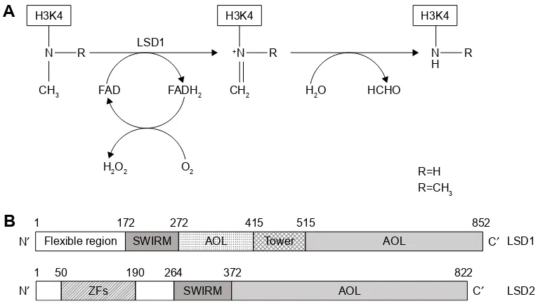
The molecular structure of LSD1 and LSD2 proteins has been determined. LSD2 is a homologue of LSD1 based on its sequence, and shares about 30% sequence similarity with LSD1. Several structural/functional domains have been characterized. Some of these domains are present in both LSD1 and LSD2 proteins: an AOL domain, essential for enzymatic activity (the AOL domain has two lobes, one forming a noncovalent FAD-binding site and the other a substrate-binding and -recognition site), and a SWIRM domain, typically observed in chromatin-associated proteins (the SWIRM domain is involved in protein–protein interac-tions; it is also involved in the association with the androgen receptor). Other domains are specific to LSD1, such as the TOWER domain, while others are specific to LSD2, such as the amino zinc-finger domain, whose function is unclear (Figure 1).64 As mentioned, the catalytic activity of both
LSD1 and LSD2 enzymes is in the AOL domain and requires the cofactor FAD.
The determination of the three-dimensional structure of LSD1 considerably helped in the understanding of the function of this protein at the molecular level. LSD1 forms a highly symmetric, packed domain structure from which a long helical tower domain protrudes. The SWIRM domain, which contributes to the stability of the protein, packs together with the AOL domain through numerous interac-tions. The active site cavity present at the level of the AOL domain is spacious and capable of accommodating several residues of the histone-tail substrate.65,66
The chemical demethylation reaction catalyzed by LSD1 is complex. During all the reactions catalyzed by LSD1, at each demethylation cycle a molecule of formaldheyde and H2O2 is produced and O2 is consumed. The chemical reac-tion involves the initial conversion of methylated lysine to an iminium cation by loss of a hydride anion captured by the oxidized FAD prosthetic groups (Figure 1). The imine cation is then hydrolyzed to produce a carbinol amine, decomposing to formaldehyde, and the demethylated residue. The reduced FAD produced during the initial step of the reaction is reoxi-dized by O2 to generate a molecule of H2O2 and regenerated oxidized FAD (Figure 1).67,68
The capacity of LSD1 to form molecular complexes with other nuclear proteins and transcriptional factors is an
OncoTargets and Therapy downloaded from https://www.dovepress.com/ by 118.70.13.36 on 25-Aug-2020
Dovepress
Castelli et al
essential property of its activity and the regulation thereof. In this context, it is important to point out that LSD1 was initially discovered as a molecular partner of HDAC2 in HeLa cells.69 Subsequently, many other studies have shown
that LSD1 participates in the formation of multisubunit com-plexes involving LSD1, CoREST (also known as RCOR1), HDAC1, HDAC2, ZNF217, PHF21A and HMG20B. This complex is commonly known as the CoREST transcription-repressor complex.70,71 The functional implications of all
these interactions are not completely defined. However, the LSD1–CoREST interactions are required to protect LSD1 from proteosomal degradation, while the association with HDAC1/HDAC2 is required for both demethylase and HDAC activity.72 The molecular protein complex formed by
LSD1 was covered in two recent reviews on LSD1.73,74
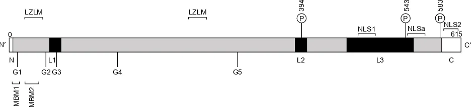
The LSD1 gene contains 19 exons, highly conserved among vertebrates. Through a process of alternative RNA splicing, two additional exons, E2a and E8a, can be included into mature mRNA and generate four possible LSD1 iso-forms, called LSD1, LSD1-2a, LSD1-8a, and LSD1-2a+8a.75
The inclusion of exon E2a can occur in all tissues, while LSD1 transcripts containing E8a are found only in brain and testis.73 While insertion of the 60-nucleotide-long E2a exon
does not modify the enzymatic activity of LSD1, insertion
of the 12-nucleotide-long exon E8a could modify LSD1 enzymatic activity (in fact, the LSD1-8a isoform plays a role in mediating H3K9 and H4K20 demethylation).
Role of LSD1 in normal hematopoietic
differentiation
Gene-knockout studies have contributed greatly to our understanding of the role of LSD1 in the control of normal hematopoiesis. Conditional knockout studies have shown that a deficit of LSD1 expression in the hematopoietic system determines an expansion of HSCs and hematopoietic progenitor cells (HPCs) not associated with a significant increase in marrow cellularity and only a slight increase in white blood cells.76 Analysis of bone marrow of
LSD1-deficient animals showed an inhibition of granulopoiesis and a stimulation of monocytopoiesis, thus suggesting that LSD1 plays an important role in lineage choice during granulo/monocytic differentiation.76 Analysis of
erythroid-cell lineage has shown that LSD1 knockdown results in anemia associated with perturbed terminal erythropoiesis and expansion of early erythroid progenitors.70 Other studies
have supported the role of LSD1 as an indispensable epi-genetic governor of hematopoietic differentiation.77 This
function is mainly exerted through the capacity of LSD1 to
1′ )OH[LEOHUHJLRQ &′
6:,50 $2/ 7RZHU $2/
&′
/6'
/6'
=)V 6:,50 $2/
1′
+. +. +.
1
$
%
5 1 5 1 5
+
&+
+2 2
&+ +2 +&+2
5 + 5 &+
)$' /6'
)$'+
Figure 1 Schematic representation of enzymatic activity and linear structure of human LSD1 and LSD2.
Notes: (A) Enzymatic reaction catalyzed by LSD1. LSD1 catalyzes the demethylation of Lys4 of histone H3 (H3K4) through a flavin-dependent oxidative reaction. LSD1 acts on both di- and monomethylated H3K4. The reaction involves the steps shown from the left to right: first, the histone substrate is bound by the enzyme and the methylated
Lys4 side chain is oxidized by the FAD prosthetic group, with consequent reduction of oxygen (O2) to hydrogen peroxide (H2O2); second, the resulting imine intermediate is
then hydrolyzed, thus generating the demethylated H3 tail and formaldehyde. (B) Overview of LSD1 and LSD2 structure. Human LSD1 is composed of 852 amino acids with
major domains: an N-terminal SWIRM (small α-helical domain), contributing to the steadiness of the molecule; a central protruding Tower domain; and a C-terminal amine
oxidase like (AOL) domain. Among these domains, the AOL and SWIRM domains pack together through various interactions (at the level of three-dimensional structure), determining the formation of a spherical structure. At the level of the N-terminal (1–172), there is an N-flexible region. Human LSD2 is composed of 822 amino acids and displays 31% of sequence similarity with LSD1. LSD2 shows three major domains, from the N- to the C-terminal: a ZFs area, a SWIRM, and an AOL domain. It is important
to note that LSD2 does not contain a Tower domain, the structure of which plays a crucial role in LSD1 for CoReST binding.
OncoTargets and Therapy downloaded from https://www.dovepress.com/ by 118.70.13.36 on 25-Aug-2020
Dovepress Histone methyltransferase and demethylase in AML
repress hematopoietic stem and progenitor gene-expression programs during hematopoietic cell differentiation.77
Defi-cient LSD1 expression causes a failure to silence HSC and HPC genes fully, compromising the different steps of the hematopoietic differentiation-cell program.77
The activity of LSD1 during hematopoietic differentia-tion and its regulatory effects on hematopoiesis are regulated by its interaction with CoREST/REST (Rcor) corepressors. Upadhyay et al showed that the three Rcor proteins (Rcor1, Rcor2, and Rcor3) regulate LSD1 activity and cellular differentiation in hematopoietic cells. All the three Rcor proteins interact with LSD1 and the transcription factor Gfi1b: while Rcor1 and Rcor2 facilitate LSD1-mediated nucleosome demethylation, Rcor3 inhibits demethylation.78 In line with
these observations, Rcor1 and Rcor2 favor differentiation, while Rcor3 inhibits differentiation.78 Rcor1 and Rcor3 levels
are differentially regulated in erythroid and megakaryocytic cells during terminal stages of maturation.78
The capacity of LSD1 to interact with some transcription factors acting as key master regulators of hematopoiesis is essential to explain its effects on hematopoietic differentia-tion. In hematopoietic cells, LSD1 and Rcor1 associate with Gfi1 and Gfi1b and repress most Gfi1b targets in erythroid cells.79 Gfi-transcription factors are master regulators of
hematopoietic cell differentiation: Gfi1 is essential for neutrophil differentiation, while Gfi1b is required for the development of erythroid and megakaryocytic lineages.80
Furthermore, LSD1–Rcor1 also associates with other transcription factors, such as Scl1/Tal1,81,82 Bcl11A,83 and
GATA2,84 in erythroid cells, inducing repression of their
target genes. The excessive and uncoordinated function of all these transcription factors induced by LSD1 deficiency in HPCs determines a derangement of erythroid and mega-karyocytic cell differentiation. The interaction between Gfi1 and LSD1 involves the SNAG domain of Gfi1: this domain needs to be methylated at the level of lysine 8 for optimal binding capacity to LSD1.85
Finally, recent studies provided evidence that LSD1 plays a relevant functional role at the level of events that deter-mine the initial specification of hematopoietic cell lineage during embryonic development. In vertebrates, the initial appearance of HSCs with long-term repopulation and mul-tilineage-differentiation capacities occurs at the level of the aorta–gonad–mesonephros region. This primitive population of HSCs is generated from a rare subpopulation of endothe-lial cells, known as hemogenic endothelia and is capable of transdifferentiation through a process of endothelial to hematopoietic transition. This unique transdifferentiation
process requires a molecular orchestration requiring from one side the repression of the endothelial differentiation program and from the other side the induction of the hematopoietic differentiation program. Gfi1 and Gfi1b proteins mark the hemogenic endothelia, and are strictly required for the hemogenic activity of the hemangioblasts: the Gfi proteins, through the recruitment of LSD1 protein, exert their repres-sive effects on the endothelial differentiation program.86
Furthermore, LSD1 activity in the hemangioblast is essential for the inhibition of the endothelial differentiation program through downregulation of the transcription factor Etv2, an essential regulator of vasculogenesis.87
Gfi1 and Gfi1B act as transcriptional repressors by recruiting histone-modifying enzymes to promoters and enhancers of target genes, and thus can be considered epi-genetic regulators that modify chromatin structure. Several rare hematological diseases are associated with acquired or inheritable mutations in the GFI1 and GFI1B genes; particularly, some patients with severe congenital neutro-penia carry mutations in GFI1 that determine the disruption of the C-terminal zinc-finger domains.88 Furthermore, recent
studies have suggested a possible role of GFi1 in human leukemias. Hones et al analyzed a large number of AML samples for GFi1 expression, showing that about 10% of these expressed low GFi1 levels.89 These leukemia types have
a poor outcome and frequently display an adverse cytogenetic FABM0 phenotype, NRAS mutations, elevated EVI1 expres-sion, and a leukemic stem-cell signature at the level of the gene-expression profile.83 In experimental mouse models, low
GFi1 expression accelerates leukemia development driven by oncofusion proteins, such as MLLAF9.89 Low GFi1 confers
sensitivity of leukemic cells to histone methyltransferase inhibitors, associated with resistance to HDAC inhibitors.89
Recently, Volpe et al analyzed AML with normal karyotype for GFi1 expression and observed that those displaying high expression of this transcription factor have frequent FLT3ITD
and NPM1 mutations, display an FLT3ITD signature, and
exhibit high expression of some leukemia-related genes, such as HOXA9, MEIS1, and PBX3.90 At variance with the
findings of Hönes et al89 obtained in the whole AML
popula-tion, in karyotype-normal AML, high GFi1 expression was associated with a worse outcome.90
Very interestingly, mice deleted for Rcor1 were shown to be markedly anemic, showing a block of erythroid pre-cursors at the transition from proerythroblasts to basophilic erythroblasts.91 Erythroid progenitors purified from
Rcor1-null bone marrow cultured in vitro form myeloid colonies, but fail to form erythroid colonies;91 mutant proerythroblasts
OncoTargets and Therapy downloaded from https://www.dovepress.com/ by 118.70.13.36 on 25-Aug-2020
Dovepress
Castelli et al
aberrantly express genes typically expressed in myeloid cells and in SCs/PCs.91 Evaluation of the myelomonocytic lineage
of Rcor1-/- animals showed absence of mature neutrophils associated with an increase in monocytes, a feature observed also in LSD1-/- mice.92
LSD1 in leukemia
LSD1 is not mutated in acute leukemias or in other blood neoplastic disease. However, LSD1 is overexpressed in many hematologic diseases, including AML, acute lymphoblastic leukemia (ALL), myeloproliferative neoplasms, chronic myelomonocytic leukemia, and MDS.93 In AMLs, LSD1
was overexpressed in about 60% of cases.93
Using a model of human MLLAF9 leukemia, Harris et al identified LSD1 as a key regulator of leukemic stem-cell potential, in that this demethylase acts at the level of the genomic loci bound by MLLAF9, favoring the effect of the MLL-fusion protein and thus preventing cell differentiation and apoptosis.94 In line with this finding, primary AML
cells bearing various MLL rearrangements were shown to be markedly inhibited by small-molecule LSD1 inhibitors.94
Interestingly, LSD1 was expressed in MLL-rearranged AML, as well as in other molecular subtypes of AML. In addition to MLL-rearranged AML, AML associated with PML-RARA and RUNX1–RUNX1T1 was shown to be sensitive to the LSD1 inhibitors.94 Consistent with these findings, LSD1
was found among the 5% most highly expressed genes in prospectively purified immunophenotypic leukemic SCs from a variety of distinct AML subtypes.95
According to these observations, elevated LSD1 expres-sion in AML and in other hematological neoplasia may contribute to leukemia development. To test this hypothesis, Wada et al evaluated the effect of LSD1 overexpression in HSCs and HPCs by generating transgenic mice that over-express LSD1 in HSCs/HPCs under control of the Sca1 promoter.96 First, these authors showed that among acute
leukemias LSD1 is expressed at the highest levels in T-cell ALL.88 The overexpression of the short LSD1 isoform, which
lacks E2a and E8a, induced a marked increase in the self-renewal activity of HSCs via upregulation of HOXA genes, but retained multidifferentiation capacities. Transgenic mice overexpressing LSD1 did not develop any hematological neoplasia; however, these mice developed high frequency T-cell ALL after γ-irradiation.96
Schenk et al explored the capacity of LSD1 inhibitors to confer to non-APL cells the capacity to differentiate in response to all-trans retinoic acid (ATRA). In fact, while APL cells have the remarkable property to differentiate
under treatment with ATRA, non-APL cells fail to undergo differentiation. Schenk et al showed that non-APL cell lines, as well as primary AML cells, undergo granulocytic differen-tiation when incubated with ATRA and an LSD1 inhibitor.97
Analysis of leukemic cells treated with LSD1 inhibitors showed that these drugs did not lead to a large-scale increase of H3K4me across the genome, but increased H3K4me and gene expression at the level of selected genes, particularly some genes involved in myeloid differentiation.97
LSD1 inhibitors
The development of specific LSD1 inhibitors has been the objective of many recent studies. These inhibitors represent a fundamental tool to improve our understanding of the role of LSD1 in normal and pathological conditions, and offer a unique strategy to develop preclinical studies to evaluate the therapeutic impact of LSD1 inhibition in animal models of various pathologic conditions and to translate these studies in the clinic.
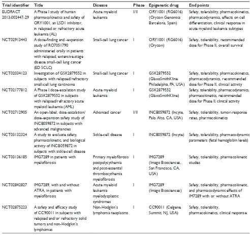
Some recent papers have analyzed in detail the structure and mechanism of action of the main LSD1 inhibitors that have been developed in the last 10 years.95 Here, we analyze
the main LSD1 inhibitors and particularly those under clinical development, with emphasis on studies focused on AML treatment (Table 1). According to Niwa and Umehara, LSD1 inhibitors can be subdivided into two main groups: irreversible covalent inhibitors and reversible noncovalent inhibitors.98
The irreversible covalent inhibitors can be subdivided into three subgroups: (±)-trans-2-phenylcyclopropylamine (2-PCPA), N-alkylated 2-PCPA derivatives, and inhibitors other than 2-PCPA derivatives.
Studies on the MAO inhibitor tranylcypromine have led to the discovery that this compound exhibited better LSD1-inhibitory activity than the other MAO inhibitors known at the time.99 It is important to briefly discuss the properties of
Tranylcypromine (TCP), the compound initially investigated for its capacity to inhibit LSD1. TCP was approved as a drug for the treatment of depression. In combination with ATRA, TCP is under exploration in three clinical trials in AML patients: TCP in combination with ATRA in AML patients who cannot undergo chemotherapy (NCT02261779), TCP at four doses (10, 20, 40, and 60 mg) in combination with ATRA in AML and MDS patients (NCT02273102), and TCP in combination with ATRA and the chemotherapy agent cytarabine (DRKS00006055) (Table 1).
The 2-PCPA scaffold represents the basic structure used for the development of the large majority of irreversible LSD1 inhibitors.100 The introduction of various chemical
OncoTargets and Therapy downloaded from https://www.dovepress.com/ by 118.70.13.36 on 25-Aug-2020
Dovepress Histone methyltransferase and demethylase in AML
modifications to this basic structure has led to the identi-fication of many new compounds, some exhibiting higher LSD1-inhibitory activity and a reduced MAO-inhibitory activity.98 Particularly, chemical substitutions on the phenyl
ring of tranylcypromine have considerably improved the potency and selectivity of these LSD1 inhibitors.99
Most LSD1 inhibitors under clinical development per-tain to the family of N-alkylated 2-PCPA derivatives. This category of compounds was discovered by Oryzon Genom-ics (Barcelona, Spain), showing that LSD1 inhibition is markedly improved by the development of N-alkylated 2-PCPA derivatives, exhibiting LSD1-inhibitory potency in
the nanomolar range. One of these compounds, ORY1001, displayed potent LSD1-inhibitory activity (IC50 of about 18 nM) and specificity (.1,000-fold selectivity over MAOs and LSD2).97 A cellular assay on the THP1 cell
line (MLL – AF9 cells) displayed pharmacologic activ-ity of ORY1001 (as evaluated by methylation and cell differentiation assay) at subnanomolar concentrations.100
Leukemic cells with MLL translocations were particularly sensitive to the inhibitory effects of this compound. Daily oral doses ,20 μg/kg have shown a potent antileukemic effect in mice transplanted with MV (4;11) cells.100 Finally,
preclinical pharmacologic and toxicologic studies showed Table 1 Main LSD1 inhibitors introduced into a plan of clinical development
Trial identifier Title Disease Phase Epigenetic drug End points
eUDRACT
2013-002447-29
A Phase i study of human pharmacokinetics and safety of ORY1001, an LSD1 inhibitor, in relapsed or refractory acute leukemia (AL)
Acute myeloid leukemia
i/ii ORY1001 (RG6016) (Oryzon Genomics Barcelona, Spain)
Safety, tolerability, pharmacokinetics, pharmacodynamics, effects on cell differentiation, clinical response in acute myeloid leukemia subtypes
NCT02913443 A dose-finding and -expansion
study of RO7051790 administered orally in patients
with relapsed, extensive-stage disease small-cell lung cancer
(eD SCLC)
Small-cell lung cancer i ORY1001 (RG6016) (Oryzon)
Safety, tolerability, recommended dose for Phase ii, overall survival
NCT02034123 investigation of GSK2879552 in subjects with relapsed/refractory
small-cell lung carcinoma
Small-cell lung cancer i GSK2879552 (GlaxoSmithKline Philadelphia, PA, USA)
Safety, tolerability, pharmacokinetics, pharmacodynamics, recommended dose for Phase ii, clinical activity
NCT02177812 A Phase I dose-escalation study
of GSK2879552 in subjects with relapsed/refractory acute myeloid leukemia (AML)
Acute myeloid leukemia
i GSK2879552
(GlaxoSmithKline)
Safety, tolerability, pharmacodynamics, pharmacokinetics, recommended dose for Phase ii, clinical activity
NCT02712905 An open-label, dose-escalation/ dose-expansion safety study of INCB059872 in subjects with
advanced malignancies
Advanced cancer i/ii INCB059872 (Incyte,
Palo Alto, CA, USA)
Safety, tolerability, tumor-response
rates, pharmacokinetics
NCT03132324 A study to evaluate safety, pharmacokinetic, and biological
activity of INCB059872 in subjects with sickle-cell disease
Sickle-cell disease i INCB059872 (Incyte) Safety, tolerability, pharmacodynamic parameters (fetal hemoglobin levels)
NCT03136185 iMG7289 in patients with
myelofibrosis
Primary myelofibrosis
postpolycythemia
and post-essential
thrombocythemia
myelofibrosis
i iMG7289
(imago Biosciences, San Francisco, CA, USA)
Safety, tolerability, pharmacokinetic studies
NCT02842827 iMG7289, with and without ATRA, in patients with
myelofibrosis
Acute myeloid leukemia myelodysplastic syndromes
i iMG7289
(imago Biosciences)
Safety, tolerability, pharmacokinetic, and pharmacodynamic effects of iM7289 with or without ATRA
NCT02875223 A safety and efficacy study
of CC90011 in subjects with relapsed and/or refractory solid
tumors and non-Hodgkin’s
lymphomas
Non-Hodgkin’s
lymphoma neoplasms
i CC90011 (Celgene,
Summit, NJ, USA)
Safety, tolerability,
pharmacokinetics, clinical response
Abbreviation: ATRA, all-trans retinoic acid.
OncoTargets and Therapy downloaded from https://www.dovepress.com/ by 118.70.13.36 on 25-Aug-2020
Dovepress
Castelli et al
good oral bioavailability and a good safety profile.101
Given this favorable pharmacologic profile, ORY1001 entered a plan of clinical development screening to inves-tigate its safety profile and possible antitumor effects in patients with non-small-cell lung cancer and AML (Table 1).
A preliminary report on the ongoing Phase I/II clinical study on AML patients (EUDRACT 2013-002447-29) was presented at the last meeting of the American Society of Hematology in December 2016. Patients with refractory/ relapsed AML were enrolled in this study, and the dose-escalation section allowed definition of a recommended dose of ORY1001 for the expanded phase, of 140 μg/m2/day.102
At this dose, the drug was relatively well tolerated, and the most frequent adverse events were asthenia, febrile neutro-penia, constipation, and peripheral edema.102 Importantly,
ORY1001 was evaluated in an extension cohort of 14 AML patients with relapsing AML subtypes predicted to be more sensitive according to preclinical studies (ie, ten AML MLL/ translocated and four acute erythroleukemia/M6). Objective responses were seen in 36% and blast-cell differentiation in blood observed in 64% of these patients.102 These
observa-tions further support additional studies exploring the anti-leukemic effects of ORY1001.
After the end of this Phase I study in AML, further devel-opment of ORY1001 will be carried out by Roche (Basel, Switzerland) and the compound name will be RG6016. RG6016 will be additionally evaluated in AML patients and in small-cell lung cancer patients in the context of a Phase I study (NCT02913443). Another LSD1 inhibitor, ORY2001, generated by Oryzon is under evaluation in a Phase I clinical study in neurodegenerative disease (Alzheimer’s disease, Parkinson’s disease, and Huntington’s disease). This study was preceded by one initiated in early 2016 to determine the safety, tolerability, and kinetics of ORY2001 in healthy volunteers.
Other N-alkylated compounds, RN1 and RN7, are cur-rently being evaluated in preclinical models. Both those compounds displayed a selective and potent LSD1 inhibitory capacity (IC50 10–30 nM); an important property of these compounds is in their brain penetration capacity, eliciting inhibitory effects on long-term, but not short-term memory formation.103 RN1 was evaluated in preclinical models for
its capacity to act as an agent stimulating fetal hemoglobin synthesis in adult erythroid cells and for its antileukemic properties. Concerning the first point, RN1 was evaluated in a mouse model of sickle-cell disease, showing that drug administration induced fetal hemoglobin synthesis in erythroid cells, reduced disease pathology,104 and reduced
oxidative-induced damage to erythroid cells.105 Preclinical
studies in baboons support the efficacy (evaluated by fetal hemoglobin increase) and safety of long-term administration of RN1 in baboons.106,107 These observations support further
development of the LSD1 inhibitor RN1 in the treatment of sickle-cell disease.106,107
Other studies on RN1 were focused on evaluating the antileukemic effects of this inhibitor. Studies performed on leukemic cell lines showed that leukemic cells with MLL translocations or bearing RUNX1–RUNX1T1 (AML1–ETO) translocations are particularly sensitive to cell death, cell dif-ferentiation, and inhibition of cell proliferation in vitro and in vivo induced by RN1.106 The in vitro assay indicated that
RN1 is a potent LSD1 inhibitor with an IC50 assayed on inhibi-tion of cell proliferainhibi-tion corresponding to 1–5 nM.108 These
observations suggest that both MLL-rearranged and RUNX1-rearranged AML may converge on similar downstream onco-genic pathways that are impacted by LSD1 inhibition.108
In 2015, the Cancer Epigenetic Department of Glaxo-SmithKline (Philadelphia, PA, USA) performed a screen-ing on a library of 2.5 million compounds, and discovered a series of small molecules with LSD1-inhibitory activity that led to the development of three potent LSD1 inhibitors: GSK2879552, GSKLSD1 and GSK2699537. All three mol-ecules are N-alkylated cyclopropylamine derivatives and act as potent irreversible LSD1 inhibitors. The pharmacokinetic and pharmacodynamic properties of GSK2879552 indicated that this compound was suitable for clinical development. GSK2879552 treatment of a large panel of tumor cell lines indicated that AML and small-cell lung cancer cells were uniquely sensitive.109 The antitumor effect against small-cell
lung cancer was characterized in detail, providing evidence that the sensitivity of these tumor cells to GSK2879552 correlated with DNA hypomethylation of a signature set of probes.109 GSK2879552 is under investigation in the context
of a Phase I study in a subset of cancer patients with relapsed/ refractory small-cell lung cancer (NCT02034123) (Table 1). Incyte (Palo Alto, CA, USA) recently developed a new LSD1 inhibitor, INCB059872, displaying properties suit-able for clinical development. This inhibitor was potent and selective for LSD1 and was orally bioavailable. Initial pharmacodynamic studies confirmed the capacity of this inhibitor, like other LSD1 inhibitors, to induce apoptosis and differentiation of leukemic cells displaying MLL rearrange-ments.108 Studies in xenograft mice transplanted with human
MLLAF9 leukemic cells showed prolonged in vivo effects of INCB059872, allowing its administration with an alternate-day regimen.110 Preclinical studies with INCB059872 showed
a synergistic interaction with ATRA111 and with a BET
inhibitor112 in inducing cell differentiation and inhibiting
OncoTargets and Therapy downloaded from https://www.dovepress.com/ by 118.70.13.36 on 25-Aug-2020
Dovepress Histone methyltransferase and demethylase in AML
the growth of non-APL AML cells. In another set of stud-ies, preliminary evidence was provided on possible synergy between INCB059872 and various signal-transduction inhibi-tors (such as PIM-kinase inhibiinhibi-tors, JAK1/2 inhibiinhibi-tors, or PI3Kδ-selective inhibitor) in some AML models.113 Other
studies were focused on exploring the possible antitumor effects of INCB059872 in some solid tumors. Interestingly, preclinical studies showed significant antitumor effects of this LSD1 inhibitor alone or in combination with chemotherapy in Ewing sarcoma cell lines.113 In vivo studies in mice
xeno-grafted with human Ewing sarcoma cell lines confirmed their sensitivity to the antitumor effects of INCB059872.114
Inter-estingly, using patient-derived xenograft models, evidence was provided that tumors with EWS–FLI translocations were particularly inhibited in their growth by the LSD1 inhibitor.115
Finally, other preclinical studies confirmed the observations made with ORY1001 and showed clear sensitivity of small-cell lung cancer to LSD1 inhibitors.116 On the basis of these
observations, Incyte has launched two clinical trials based on the administration of INCB059872: a Phase I/II study of dose escalation/dose expansion in patients with advanced cancer (NCT02712905), and a Phase I dose-escalation study in sickle-cell disease (NCT03132324) (Table 1).
Recently, new LSD1 inhibitors pertaining to this chemical subtype have been reported. These compounds are promising for their potency, specificity, and pharmacologic proper-ties. Takeda (Osaka, Japan) reported a new LSD1 inhibitor, T3775440, a tranylcypromine derivate characterized by high LSD1-inhibitory activity (EC50 2 nM). A screening of the effects of this compound against a large panel of leukemic cell lines showed a sensitivity of erythroid and megakaryocytic leukemic cell lines. The growth-inhibitory effect observed on these cells seems to be related to a trans-differentiating effect consisting of the induction of monocyte differentiation.117 In vivo experiments in normal mice showed
that T3775440, as well as other LSD1 inhibitors, exerted an inhibitory effect on erythroid and megakaryocytic progeni-tors, resulting in thrombocytopenia.117 This inhibitor effect
on erythromegakaryopoiesis could be related to the capac-ity of T3775440 to disrupt Gfi1b-containing transcriptional complexes.111 These observations support the hypothesis that
LSD1 inhibitors could be evaluated for the treatment of M6 and M7 AML. In line with this observation, as mentioned, M6 AML patients were shown to be frequent responders to treatment with the ORY1001 LSD1 inhibitor.102
Ogasawara et al developed novel LSD1 inhibitors, NCD25 and NCD38, which consist of two moieties: a tranylcypromine-based moiety with LSD1-inhibiting activity and a lysine that is designed to recognize, with high affinity, an LSD1
enzymatic pocket, thus allowing selective inhibitory activity restricted to LSD1 and not extended to other MAO enzy-mes.118 These two LSD1 inhibitors were recently
character-ized for their anticancer properties. Sugino et al explored the antileukemic activity of NCD25 and NCD38.119 NCD25
and NCD38 inhibited LSD1 with EC50 of about 500 nM. Through the study of a panel of sensitive leukemic cell lines and gene-expression studies in LSD1 inhibitor-treated cells, the conclusion was reached that these inhibitors derepress superenhancers of hematopoietic regulators (such as Gfi1, ERG, and CEBPA) that are abnormally silenced by LSD1 and via this mechanism inhibit leukemic programs and promote cell differentiation.119 Furthermore, NCD38 showed a marked
inhibitory effect on colony formation by primary leukemic cells derived from MLL-rearranged AML, erythroleukemia, and MDS.119 These findings provide a rationale for clinical
trials of LSD1 inhibitors in MDSs.119 Interestingly, NCD38
and NCL1, another lysine-specific LSD1 inhibitor,120 were
shown to inhibit glioma SCs.121
The use of noncovalent reversible LSD1 inhibitors could provide some advantages compared to irreversible inhibitors, particularly as regards a safer metabolic profile. Only a limited number of noncovalent LSD1 inhibitors have been devel-oped, and some are promising in preclinical assay in terms of development of antileukemic drugs.122 Several compounds
with these properties and submicromolar potency have been developed. Among these compounds, particularly interesting is the inhibitor SP2509, containing a benzohydrazide scaffold, and with a Ki on LSD1 activity of about 30 nM.123
The effect of SP2509 on leukemic cell lines was tested, providing evidence about effects comparable to those induced by irreversible LSD1 inhibitors (ie, inhibition of prolifera-tion, induction of apoptosis and cell differentiation).124 This
compound was tested also on primary AML cells, providing evidence that NPM1 and MLL-rearranged leukemic cells are particularly sensitive to the effects of SP2509.124 Interestingly,
cotreatment with an HDAC inhibitor (panobinostat) and SP2509 was synergistically inhibitory for AML blasts.124
Another reversible LSD1 inhibitor, JL1037, was recently identified through computer-aided drug-design technology.125
This compound displayed a reversible and selective LSD1-inhibitory activity with IC50 of 100 nM and antileukemic effects like those observed using other LSD1 inhibitors.125
Exploration of the inhibitory activity of JL1037 on leuke-mic cells revealed induction of apoptotic and autophagic responses.125
From high-throughput screening, a new series of com-pounds was identified, named 4H-thieno[3,2-b]pyrrole-5-carboxamide, with inhibitory activity against LSD1.126
OncoTargets and Therapy downloaded from https://www.dovepress.com/ by 118.70.13.36 on 25-Aug-2020