O R I G I N A L R E S E A R C H
Methyloleanolate Induces Apoptotic And
Autophagic Cell Death Via Reactive Oxygen
Species Generation And c-Jun N-terminal Kinase
Phosphorylation
This article was published in the following Dove Press journal:
OncoTargets and Therapy
Myoung Seok Jeong*
Ji Hoon Jung *
Hyemin Lee Chang Geun Kim Sung-Hoon Kim
College of Korean Medicine, Kyung Hee University, Dongdaemun-Gu, Seoul 02447, Republic of Korea
*These authors contributed equally to this work
Background: To develop a potent anticancer agent similar to oleanolate, the underlying mechanisms of its derivative, methyloleanolate, in the apoptosis and autophagy of A549 and H1299 cells were elucidated.
Purpose:The aim of the present study was to investigate the effect of methyloleanolate in inducing apoptotic and autophagic cell death in cancer cells.
Materials and methods: Flow cytometric analysis with Annexin V/PI staining, Western blot analysis, and immunofluorescence analysis were conducted in A549 and H1299 cells.
Results: Methyloleanolate increased the fraction of Annexin V/PI apoptotic cells and activated caspase-8, caspase-3, and death receptor 5 (DR5) more than oleanolate in A549 and H1299 cells pretreated with pancaspase inhibitor z-VAD-fmk and DR5 depletion. Also, methyloleanolate induced autophagic features of microtubule-associated protein light chain 3 3BII (LC3BII) conversion and puncta in A549 and H1299 cells, along with autophagosomes and vacuoles. Methyloleanolate blocked autophagy flux for impaired autophagy and chlor-oquine (CQ)-enhanced microtubule-associated protein LC3BII accumulation and cytotoxi-city in A549 and H1299 cells, although 3-methyladenine (3-MA) did not. Interestingly, LC3BII accumulation was detected only in methyloleanolate-treated autophagy-related gene 5 (ATG5)+/+ mouse embryonicfibroblast (MEF) cells but not in ATG5−/- MEF cells. Methyloleanolate reduced p-mTOR but activated p-c-Jun N-terminal kinases and reactive oxygen species production in A549 and H1299 cells. Conversely, N-acetyl-L-cysteine and SP600125 blocked apoptotic and autophagic cascades caused by methyloleanolate in A549 and H1299 cells.
Conclusion: Overall, the findings suggest that methyloleanolate induces apoptotic and autophagic cell death in non–small cell lung cancers via reactive oxygen species generation and c-Jun N-terminal kinase phosphorylation.
Keywords:methyloleanolate, apoptosis, autophagy, JNK, ROS
Introduction
Lung cancer causes 1.6 million deaths annually worldwide.1Eighty percent of lung
cancers are non–small cell lung cancers (NSCLCs), which have a poor prognosis.2
In particular, NSCLC is known to develop drug resistance by means of epidermal
growth factor receptor mutation and anaplastic lymphoma kinase translocation.3
Epidermal growth factor receptor inhibitor gefitinib4,5 and anaplastic lymphoma
kinase inhibitor crizotinib6have been applied to combat drug resistance. Recently,
Correspondence: Sung-Hoon Kim College of Korean Medicine, Kyung Hee University, 26, Kyungheedae-ro, Dongdaemun-gu, Seoul 02447, Republic of Korea
Tel +82-2-961-9233 Fax +82-2-961-9598 Email [email protected]
OncoTargets and Therapy
Dove
press
open access to scientific and medical research
Open Access Full Text Article
OncoTargets and Therapy downloaded from https://www.dovepress.com/ by 118.70.13.36 on 25-Aug-2020
natural compounds such as cucurbitacin7 1, fisetin,8
solamargine,9 and protocatechuic acid10 have been
reported to increase antitumor efficacy and reduce
che-moresistance in NSCLCs.
Programmed cell death is important in determining the fate of a cell under physiological and pathological
condi-tions. Programmed cell death is classified into three
cate-gories based on morphological changes:11,12apoptosis (type
I), autophagic cell death (type II), and necrosis (type III). Apoptosis is characterized by cell shrinkage, loss of adhe-sion to the extracellular matrix or neighbors, nuclear
con-densation, and fragmentation.13 Autophagy is a catabolic
process in which cells succumb to nutrient starvation,
damage, and various stresses.14 Misfolded proteins and
organelles are usually sequestered into autophagosomes and then degraded by lysozyme after the autophagosome
fuses with a lysosome.15 In contrast, excessive
accumula-tion of autophagic vacuoles in the cytoplasm can induce
autophagic cell death16–18 in a caspase-independent
pathway19different from apoptosis or necrosis.20,21
Oleanolic acid (3β-hydroxyolean-12-en-28-oic acid;
OA), a pentacyclic triterpenoid contained inSalvia
gluti-nosa, Phytolacca americana, Rosa woodsii, Oldenladia diffusa, Kochia scoparia, and other plants, is known to
have multiple biological activities, such as anti-infl
amma-tory, antiviral, antifungal, hepatoprotective, and antitumor
effects.22–24 The antitumor effect of oleanolic acid has
been considered to be limited in several cancers. The present study aimed to explore the underlying antitumor mechanism of methyloleanolate (MO), a derivative of OA,
in A549 and H1299 NSCLC cells and to determine MO’s
association with apoptotic and autophagic cell death sig-naling pathways.
Materials And Methods
Reagents And Antibodies
OA and MO (Figure 1) were isolated fromSalvia glutinosa25
by Dr Namin Baek, a pharmacognosist and professor at
Kyung Hee University, and purified up to 98% by means of
high-performance liquid chromatography. Dulbecco’s
Modified Eagle’s Medium, Roswell Park Memorial
Institute (RPMI) 1640 medium, and fetal bovine serum (FBS) were purchased from Welgene (Gyeongsan, Korea). 3-Methyladenine (3-MA), N-Acetyl-L-cysteine (NAC),
2′,7′-dichlorofluorescin diacetate (DCF-DA) and β-actin
were purchased from Sigma-Aldrich (St Louis, MO, USA). Lipofectamine 2000 reagent was purchased from Invitrogen
(Carlsbad, CA, USA). Antibodies for poly (ADP-ribose) polymerase (PARP), caspase-3, caspase-8, FasL, DR4, Bid, mammalian or mechanistic target of rapamycin (mTOR), p-mTOR (Ser2448), SAPK/JNK, p-SAPK/JNK (Thr183/ Tyr185), ATG5, ATG7 p62 sequestosome (p62/SQSTM1), and beclin 1 were purchased from Cell Signaling Technology (Beverly, MA, USA). Antibodies for LCB3B and DR5 were purchased from Novous (San Diego, CA, USA) and Santa Cruz Biotechnology (Santa Cruz, CA, USA), respectively. SP600125 and z-VAD-fmk were obtained from Calbiochem (San Diego, CA, USA).
Cell Cultures
Non–small cell lung cancer (NSCLC) cell lines A549
(ATCC® ccl-185™) and H1299 (ATCC® CRL-5803™)
and normal lung fibroblast HEL299 cells (ATCC®
CCL-137™) were purchased from American Type Culture
Collection (Manassas, VA, USA). ATG5+/+ and ATG5−
/-MEF cells were obtained from Professor Guillermina Lozano (The University of Texas MD Anderson Cancer
Center, USA) and were approved (19–084) by the Living
Modified Organism (LMO) committee of Kyung Hee
University. The cells were maintained in RPMI 1640 sup-plemented with 10% FBS (Gibco, Carlsbad, CA, USA), 1% penicillin/streptomycin, and 2 µM L-glutamine
(WelGene, Deagu, South Korea) at 37 °C in 5% CO2.
The cells were incubated in the growth medium under a
humidified, 5% CO2atmosphere at 37 °C.
Cell Viability Assay
The cytotoxic effects of OA and MO were evaluated by means of the 3-(4,5-dimethylthiazol-2-yl)-2,5-diphenyltetra-zolium bromide (MTT) assay. In brief, A549 and H1299 cells
(1 × 10⁴cells per well) were seeded onto 96-well culture
plates and exposed to various concentrations of OA or MO for 24 or 48 hrs. The cells were incubated with MTT (1 mg/mL) for 2 hrs and then treated with MTT lysis solution for 1 hr. Optical density was measured with a microplate reader (Molecular Devices Co, San Jose, USA) at 570 nm. Cell viability was calculated as the number of viable cells in the groups treated with OA or MO as a percentage of the number of viable cells in the untreated control.
Apoptosis Detection By Annexin V/PI
Double Staining
A549 and H1299 cells were exposed to OA (50 or 100μMÞ
or MO (20 or 40 μMÞ for 12 hrs, washed with
OncoTargets and Therapy downloaded from https://www.dovepress.com/ by 118.70.13.36 on 25-Aug-2020
H3C
H
H H3C
H O
C H3
H3C H O H
O C H3
C H3
C H3
H3C
H
H H3C
H O
C H3
H3C H
O
O C H3
C H3
C H3
C H3
OA MO
Con OA 50 μM OA 100 μM MO 20 μM MO 40 μM
A549
H1299
0.79% 1.96%
0.46% 1.23%
1.32% 1.32%
3.34% 1.06%
8.41% 15.02%
0.97% 2.14%
0.76% 2.07%
1..04% 2.49%
0.47% 1.75%
2.80% 12.22%
PI
Annexin V
A
B
0 5 10 15 20
25
A549
H1299
Con OA
50 100OA MO20 MO40 Con OA50 100OA MO20 MO40
%
o
f ap
op
to
tic
cel
ls
C
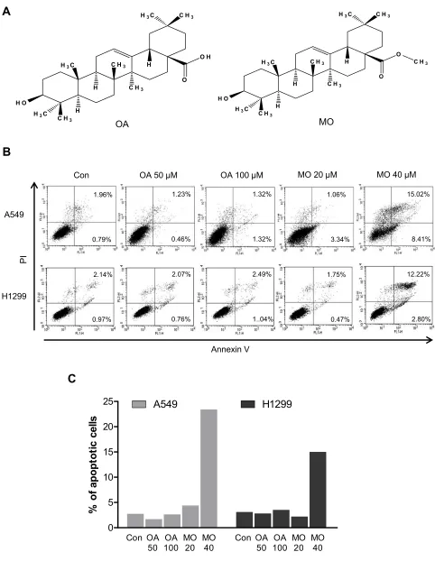
Figure 1Methyloleanolate (MO) increased apoptosis (indicated by Annexin V/PI staining) more than oleanolate (OA) did in A549 and H1299 non–small cell lung cancer cells. (A) Chemical structure of OA and MO. (B) Apoptotic fraction of OA- or MO-treated A549 and H1299 cells, determined by FACSCalibur with Annexin V/PI staining. A549 and H1299 cells were exposed to OA (50, 100μMÞor MO (20, 40μMÞfor 12 hrs and apoptotic cells were analyzed by FACSCalibur with Annexin V/PI staining. N = 6 per group. Two-way ANOVA with a post-hoc Tukey’s test was used for all the statistical analyses. (C) The effect of OA and MO on the fraction of apoptotic A549 and H1299 cells.
OncoTargets and Therapy downloaded from https://www.dovepress.com/ by 118.70.13.36 on 25-Aug-2020
phosphate-buffered saline (PBS), and harvested by trypsini-zation. The cells were gently resuspended in a binding buffer (10 mM 4-(2-hydroxyethyl)-1-piperazineethanesulfonic acid
(HEPES), pH 7.4, 140 mM NaCl, 2.5 mM CaCl2) and
incubated with Annexin V-FITC and propidium iodide for 15 mins at room temperature in the dark with the Annexin V-Apoptosis Detection kit (Biovision, Mipitas, USA). Apoptotic cells were analyzed by FACSCalibur (Becton
Dickinson, San Jose, USA) and were defined as the number
of cells positively stained by Annexin V/PI as apoptotic portion.
Western Blot Analysis
A549 and H1299 cells were seeded onto 60-mm dishes at
5 × 10⁵cells per dish in RPMI 1640 with 10% FBS and
exposed to OA (50 or 100μMÞor MO (20 or 40μMÞfor
12 hrs. The cells were then washed with cold PBS and harvested by centrifugation. The pellets were lysed in radioimmunoprecipitation assay (RIPA) lysis buffer con-taining RIPA buffer (50 mM Tris-HCl, 150 mM NaCl, 2 mM EDTA, and 1% TritonX-100) containing protease inhibitors (Roche, Basel, Switzerland). Cell lysates were centrifuged at 13,000 rpm for 15 mins at 4 °C, and the
supernatant was collected. Protein content was quantified
with the DC Protein Assay Kit (Bio-Rad) according to the
manufacturer’s instructions. An equal amount of protein
(30 μg) was subjected to 10% to 15% sodium dodecyl
sulfate and polyacrylamide gel SDS-PAGE) and
trans-ferred onto 0.45μm nitrocellulose membranes. The
mem-branes were blocked with 5% skim milk or 3% Bovine Serum Albumin (BSA) in PBS with 0.1% Tween-20 and incubated with primary antibody at 4 °C. The membranes were washed and incubated with secondary antibodies. Proteins were detected using the enhanced
chemilumines-cence (ECL) system (Amersham, Buckinghamshire,
England) and analyzed by densitometry with Image J (Bethesda, USA) software.
Immuno
fl
uorescence Analysis For LC3
Puncta
A549 and H1299 cells were seeded at 5 × 10⁴cells onto
glass discs, and one disc was placed in each well of 24-well plates and exposed to OA or MO for 12 hrs. The
cells were fixed with 4% paraformaldehyde in PBS for
15 mins at room temperature and the discs were treated with 0.01% Triton X-100 for 10 mins and blocked with 2% BSA for 30 mins at room temperature. The cells were
incubated with anti-LC3 antibody at 4 °C overnight and cultured with Alexa Fluor 488-conjugated secondary anti-body for 1 hr. Nuclear staining was then performed with
DAPI (Sigma) and fluorescence images of the cells were
observed under a Zeiss LSM 710 microscope (Boston,
USA). Also, the number of green fluorescent protein
(GFP)-LC3 puncta per cell was counted from randomly selected photographs.
Autophagic Flux Assay With Tandem
Sensor Red And Green Fluorescent
Protein LC3 Constructs
To investigate whether autophagy flux was impaired by
MO, an autophagy flux assay was conducted. A549 and
H1299 cells were transfected with tandem sensor LC3
con-jugated to red fluorescent protein and green fluorescent
protein (RFP-GFP-LC3) constructs (a gift from Professor Hongbo Hu from the Department of Nutrition and Health, College of Food Science and Nutritional Engineering, China Agricultural University) by a transient transfection
method using Lipofectamine 2000 (Thermo Fisher
Scientific, New York, USA). The cells were then exposed
to 40 µM MO for 12 hrs. Then, the merged red or green channel signals were observed in MO-treated A549 and H1299 cells under the Zeiss LSM 710 confocal microscope.
RNA Interference
A549 and H1299 cells were transfected with scrambled siRNA or DR5 siRNA plasmids with Lipofectamine 2000 and InterferinTM transfection reagent (Polyplus-transfec-tion Inc, New York, NY, USA). The mixtures of DR5 siRNA (40 nM) and transfection reagent were incubated for 10 mins and then the cells were incubated at 37 °C for
48 hrs before exposure to MO (40μMÞfor 12 hrs.
Measurement Of Reactive Oxygen
Species Production
A549 and H1299 cells were seeded at 3 × 10⁵per well in
6-well plates in a phenol red–free RPMI 1640 with 10%
FBS. The cells were pretreated with or without NAC for
1 hr and then exposed to MO (10, 20, 30, or 40μMÞ for
12 hrs. After incubation, fluorescent 2ʹ,7ʹ –dichlorofl
uor-escin diacetate (DCF-DA) was added at 37 °C for 30 mins before cells were harvested. Reactive oxygen species
(ROS) production was analyzed by means of flow
cyto-metry and confocal microscopy.
OncoTargets and Therapy downloaded from https://www.dovepress.com/ by 118.70.13.36 on 25-Aug-2020
Statistical Analysis
Values were presented as the mean plus or minus standard
deviation. Statistical significance was evaluated by
Student’s t-test using SigmaPlot software (Systat
Software Inc, San Jose, USA) or ANOVA followed by
Tukey post-hoc test when the F statistic was significant.
P < 0.05 was considered statistically significant. At least
three independent experiments were performed in dupli-cate for each assay.
Results
Effect Of MO On The Fraction Of
Apoptotic Cells In A549 And H1299
Non
–
Small Cell Lung Cancer Cells
To compare the apoptotic effect of MO and OA (Figure 1A), a flow cytometry assay with Annexin V/PI staining was conducted on A549 and H1299 NSCLC cells. MO increased the fraction of apoptotic A549 and H1299
cells at concentrations of 20 and 40 μM, while OA only
weakly increased the fraction of apoptotic cells even at 50
and 100μM (Figure 1B andC). But MO and OA showed
weak cytotoxicity in normal lungfibroblast HEL 299 cells
(Supplementary Figure 1).
Effect Of MO On Extrinsic Apoptosis
Through DR5 Activation In A549 And
H1299 Cells
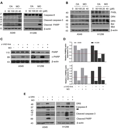
To determine whether the cytotoxic effect of MO is due to apoptosis induction, Western blotting was performed on A549 and H1299 cells. MO markedly activated caspase-8 and caspase-3 and cleaved PARP in MO-treated A549 and
H1299 cells (Figure 2A), while DR5 was upregulated and
no effect was seen on FasL, DR4, and tBid (Figure 2B).
However, pretreatment with pancaspase inhibitor
z-VAD-fmk or knockdown of DR5 reduced cytotoxicity and cleavage of PARP and caspase-3 in MO-treated A549
and H1299 cells (Figure 2CandD).
Effect Of MO On Autophagy In A549
And H1299 Cells
Based onfindings that OA can induce protective autophagy
in A549 cells26at 100 µg/mL, the effect of MO on autophagy
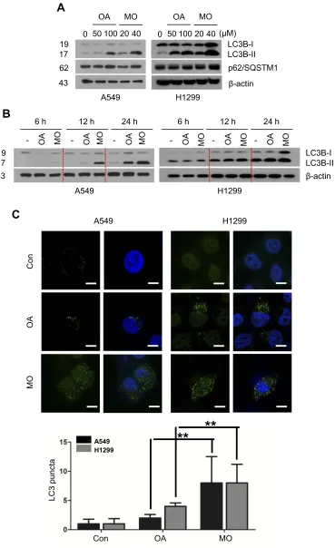
was evaluated in A549 and H1299 cells. MO increased LC3B-II accumulation in a concentration- and
time-depen-dent manner without significant effect on p62 in A549 and
H1299 cells more than OA did (Figure 3A and B). MO
consistently enhanced the formation of GFP-LC3 puncta and autophagic vacuoles more than OA did in A549 and
H1299 cells (Figure 3C,Supplementary Figure 2).
Effect Of MO On Incomplete Autophagy
Flux In A549 And H1299 Cells
To evaluate whether the elevation of LC3 lipidation induced by MO was due to fusion with autolysosomes or
increased degradation, an autophagy flux assay was
con-ducted in A549 and H1299 cells transfected with
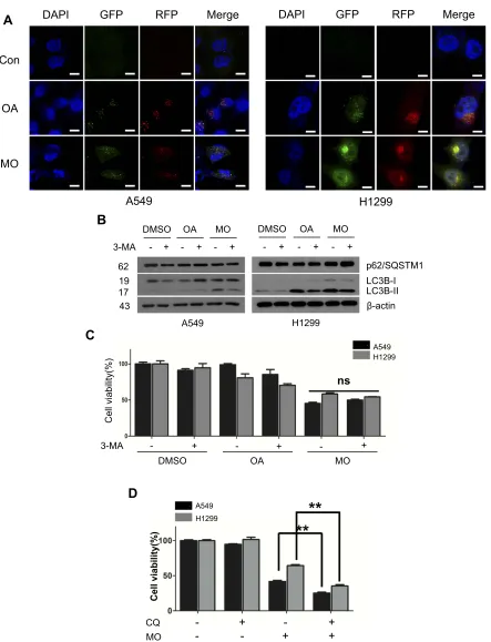
RFP-GFP-LC3 constructs. As shown inFigure 4A,
immu-nofluorescence revealed the merged yellow color in
MO-treated A549 and H1299 cells, whereas more red puncta were observed in OA-treated A549 and H1299 cells. Of note, an early stage autophagy inhibitor, 3-MA,
did not significantly affect the cytotoxicity and weakly
attenuated the expression of LC3BII in MO-treated A549
and H1299 cells (Figure 4B and C), while a late-stage
autophagy inhibitor, chloroquine (CQ), enhanced the
cyto-toxicity of MO in A549 and H1299 cells (Figure 4D).
Effect Of MO On ATG5-Dependent
Autophagy In A549 And H1299 Cells
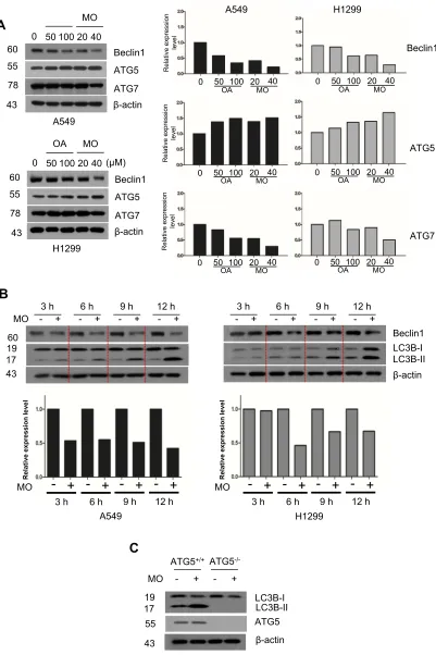
To elucidate the mechanism of MO-induced autophagy, the effect of MO on beclin 1, ATG5, and ATG7 was examined in A549 and H1299 cells. Interestingly, MO attenuated the expression of beclin 1 and ATG7 but increased the expression of ATG5 in A549 and H1299cells more than OA did (Figure 5A). Furthermore, MO
reduced the expression of beclin 1 in a time-dependent
fashion in A549 and H1299 cells (Figure 5B). To confirm
the role of ATG5 in MO-induced autophagy, Western
blotting was performed in ATG5+/+ and ATG5−/- MEF
cells. As shown inFigure 5C, MO induced LC3II
conver-sion and upregulation of ATG5 inATG5+/+MEF cells but
it did not inATG5−/-MEF cells.
Effect Of ROS Generation On
MO-Induced Autophagy In A549 And
H1299 Cells
Emerging evidence shows that ROS play a critical role in
apoptotic and autophagic cell death.12,27–29 To confirm
whether MO is associated with ROS generation, ROS
pro-duction was measured by flow cytometric analysis with
DCFDA staining in A549 and H1299 cells. MO signifi
-cantly induced ROS production in a concentration- and
time-dependent manner (Figure 6A and B). Conversely,
OncoTargets and Therapy downloaded from https://www.dovepress.com/ by 118.70.13.36 on 25-Aug-2020
ROS inhibitor NAC suppressed the number of greenfl
uor-escent ROS particles observed by immunofluorescence after
DCFDA staining (Figure 6C). ROS inhibitor NAC blocked
phosphorylation of c-Jun N-terminal kinases (JNK),
activation of DR5, LC3BII conversion, and PARP cleavage
induced by MO in A549 and H1299 cells (Figure 6D).
NAC consistently suppressed cytotoxicity and LC3 puncta
by MO in A549 and H1299 cells (Figure 6EandF).
Figure 2Methyloleanolate (MO) induced cell death by activation of caspase-3 and caspase-8 and death receptor 5 (DR5) more than oleanolate (OA) did in A549 and H1299 non–small cell lung cancer cells. (A) Effect of OA or MO on caspase-8, cleaved caspase-3, and PARP in A549 and H1299 cells. The cells were treated with OA (50, 100μMÞ
or MO (20, 40μMÞfor 12 hrs and subjected to Western blotting with antibodies of caspase-8, caspase-3, cleaved PARP, andβactin. (B) Effect of OA or MO on FASL, DR4, DR5, and Bid in A549 and H1299 cells. (C) Effect of pancaspase inhibitor z-VAD-fmk on PARP cleavage in MO-treated A549 and H1299 cells. (D) Effect of pancaspase inhibitor z-VAD-fmk on the viability of A549 and H1299 cells in the presence or absence of MO or doxorubicin (Dox). (E) Effect of DR5 depletion on DR5, caspase-8, caspase-3, and PARP in MO-treated A549 and H1299 cells. Cells were transfected with control or DR5 siRNA plasmids with or without MO (40μM) for 12 hrs and subjected to Western blotting with antibodies of DR5, caspase-8, caspase-3, PARP, andβactin.
OncoTargets and Therapy downloaded from https://www.dovepress.com/ by 118.70.13.36 on 25-Aug-2020
0 50 10020 40(μM)
OA MO
LC3B-I LC3B-II 0 50 100 20 40
OA MO
p62/SQSTM1 β-actin
A549 H1299
β-actin LC3B-I LC3B-II - OA MO
6 h
- OA MO 12 h
- OA MO 24 h
- OA MO 6 h
- OA MO 12 h
- OA MO 24 h
A549 H1299
Con
OA
MO
A549 H1299
**
**
A549 H1299
LC3 punct
a
Con OA MO
A
B
C
62 19 17
43
19 17 43
Figure 3Methyloleanolate (MO) induced 1A/1B-light chain 3BII (LC3BII) conversion and puncta in A549 and H1299 cells better than oleanolate (OA) did. (A) Effect of OA and MO on LC3BII and p62/SQSTM1 in A549 and H1299 cells. Cells were incubated with OA (50, 100μMÞor MO (20, 40μMÞfor 12 hrs, and Western blotting was performed. (B) Time-dependent effect of OA (100μMÞor MO (40μMÞon LC3BII accumulation for 6 hrs, 12 hrs, and 24 hrs in A549 and H1299 cells. (C) Effect of OA and MO on LC3 puncta in A549 and H1299 cells. Immunofluorescence shows that OA (100μM) or MO (40μM) formed LC3 puncta in A549 and H1299 cells. Thefluorescence images were taken by confocal microscopy. LC3 puncta were counted as means ±SD from three independent experiments. **P< 0.01 between OA- and MO-treated groups. Bar: 10μm.
OncoTargets and Therapy downloaded from https://www.dovepress.com/ by 118.70.13.36 on 25-Aug-2020
DAPI GFP RFP Merge
Con
OA
MO
A549
DAPI GFP RFP Merge
H1299
3-MA - + DMSO
- + OA
- + MO
p62/SQSTM1 LC3B-I LC3B-II β-actin - +
DMSO - +
OA
- + MO
A549 H1299
Cell viability
(%)
3-MA - +
DMSO
- +
OA
- +
MO
A549 H1299
ns
- - + +
CQ MO
- + - +
A549
H1299
**
**
Cell v
iability
(%
)
A
B
C
D
62 19 17 43
Figure 4Methyloleanolate (MO)-impaired autophagyflux in A549 and H1299 cells. (A) Effect of oleanolate (OA) and MO on autophagyflux in A549 and H1299 cells. A549 and H1299 cells were transfected with LC3 conjugated to redfluorescent protein and greenfluorescent protein constructs and exposed to OA (100μM) or MO (40μM) for 12 hrs. Autophagyflux was evaluated with confocal microscopy. (B) Effect of 3-methyladenine (3-MA) on 1A/1B-light chain 3BII (LC3BII) and p62/SQSTM1 in OA- or MO-treated A549 and H1299 cells. The cells were pretreated with 3-MA for 1 hr and incubated with OA (100μM) or MO (40μM) for 12 hrs. Then western botting was conducted and antibodies of LC3BII and p62/SQSTM1 were used for detection. (C) Effect of 3-MA on the viability of OA- or MO-treated A549 and H1299 cells. The cells were pretreated with 3-MA for 1 hr, exposed to OA (100μM) or MO (40μM) for 12 hrs, and an MTT assay was performed. (D) Effect of chloroquine (CQ) on the viability of MO-treated A549 and H1299 cells. The cells were pretreated with CQ for 1 hr and exposed to MO (40μM) for 12 hrs and a 3-(4,5-dimethylthiazol-2-yl)-2,5-diphenyltetrazolium bromide assay was performed. Data are shown as means ±SD from three independent experiments. **P< 0.01 between CQ- and MO-treated groups. N = 6 per group.
Abbreviation:NS, not significant.
OncoTargets and Therapy downloaded from https://www.dovepress.com/ by 118.70.13.36 on 25-Aug-2020
0 50 10020 40(μM) OA MO Beclin1 ATG5 ATG7 β-actin 0 50 100 20 40
MO A549 H1299 3 h - + 6 h - + 9 h - + 12 h - + β-actin Beclin1 LC3B-I LC3B-II MO 3 h - + 6 h - + 9 h - + 12 h - + A549 H1299 3 h MORelativ
- +
e expression lev el 6 h
- +
9 h- +
12 h- +
3 h MORelativ- +
e expression lev el 6 h
- +
9 h- +
12 h- +
ATG5+/+ ATG5
-/-MO - + - +
LC3B-I LC3B-II ATG5 β-actin Relativ e ex pression lev el Relativ e ex pression lev el Relativ e ex pression lev el
0 50 100 20 40
OA MO 0 50 100 20 40OA MO
0 50 100 20 40
OA MO 0 50 100 20 40OA MO
0 50 100 20 40
OA MO 0 50 100 20 40OA MO
Beclin1 ATG5 ATG7 A549 H1299 Beclin1 ATG5 ATG7 β-actin
A
B
C
60 55 43 78 60 55 43 78 60 55 43 43 19 17 19 17Figure 5Autophagy-related gene 5 (ATG5) plays a critical role in methyloleanolate (MO)-induced autophagy in A549 and H1299 cells. (A) Concentration-dependent effect of oleanolate (OA) or MO on beclin 1, ATG5 and ATG7 in A549 and H1299 cells. Cells were incubated with OA (50, 100 µM) or MO (20, 40 µM) for 12 hrs and subjected to Western blotting and antibodies of beclin 1, ATG5, and ATG7 were used for detection. (B) Time-dependent effect of MO (40μM) on beclin 1 and 1A/1B-light chain 3BII (LC3BII) in A549 and H1299 cells. (C) Effect of endogenous ATG5 on beclin 1, LC3BII, and ATG5 inATG5+/+
orATG5−
/-MEF cells.ATG5+/+ orATG5−
/-MEF cells were treated with MO (40μM) for 12 hrs and Western blotting was performed with LC3BII and ATG5 antibodies.
OncoTargets and Therapy downloaded from https://www.dovepress.com/ by 118.70.13.36 on 25-Aug-2020
Control 10 µM 20 µM 30 µM 40 µM
Control 6 hr 12 hr
Con
(-) NA
C
MO
(+) NA
C
NAC - + - +
DMSO MO
p-JNK
JNK
LC3B-I LC3B-II Cleaved PARP
β-actin DR5
- + - +
DMSO MO
A549 H1299
Con MO
(-) NA
C
(+)
NA
C
H1299
LC3
punta
te
cells (fold)
Con NAC MO NAC + MO
**
NAC
MO
-+
-+
+
+
A549 H1299
Cell
vi
a
b
il
it
y(
%)
**
**
A
B
C
D
E
F
54 46 54
43 48 19 17 89 46
Figure 6Reactive oxygen species (ROS) mediate cytotoxicity and cell death induced by methyloleanolate (MO) in H1299 cells. (A) Effect of MO on ROS production in H1299 cells. ROS level was measured as DCFDA byflow cytometry in H1299 cells treated with various concentrations of MO (10, 20, 30, 40μM) for 12 hrs. (B) Time-dependent effect of MO on ROS production in H1299 cells. The cells were exposed to 40μM MO for 6 and 12 hrs, and ROS production was analyzed byflow cytometry after DCFDA staining for 30 mins. (C) ROS were generated by MO (40μM) with or without N-acetyl-L-cysteine (NAC) by using DCFDA, and the greenfluorescence images were analyzed byfluorescence microscopy. (D) Effect of NAC on pJNK DR5, LC3BII, and c-PARP in A549 and H1299 cells. Cells were treated with MO (40μM) for 12 hrs, after pretreatment with 4 mM NAC, and subjected to Western blotting with p-JNK, JNK, DR5, LC3BII, and cleaved PARP antibodies. (E) Effect of NAC on LC3 puncta in MO-treated H1299 cells. H1299 cells were exposed to MO (40μM) for 12 hrs and LC3B puncta were detected by confocal microscopy. **P<0.01 versus control. Bar: 10μm. (F) Effect of NAC on the viability of MO-treated A549 and H1299 cells. Cells were exposed to MO (40μM) for 12 hrs and a 3-(4,5-dimethylthiazol-2-yl)-2,5-diphenyltetrazolium bromide assay was performed. Results represent means ±SD from three independent experiments. **P< 0.01 versus control. N = 6 per group.
OncoTargets and Therapy downloaded from https://www.dovepress.com/ by 118.70.13.36 on 25-Aug-2020
Effect Of JNK On MO-Induced
Autophagy In A549 And H1299 Cells
It is well documented that JNK and the mammalian or mechanistic target of rapamycin (mTOR) play crucial rolesin autophagy induction.30–32 Here, Western blots revealed
that MO weakly attenuated the expression of mTOR and remarkably activated the phosphorylation of JNK in a time- and concentration-dependent fashion more than OA
did in A549 and H1299 cells (Figure 7A). Conversely,
JNK inhibitor SP600125 blocked phosphorylation of JNK, LC3BII accumulation, DR5 activation, and PARP cleavage induced by MO in A549 and H1299 cells (Figure 7B). SP600125 consistently reduced the number of LC3 puncta and cytotoxicity by MO in H1299 cells (Figure 7CandD).
Discussion
Lung cancer has a high death rate because it is difficult to
detect at an early stage and thus has often metastasized to
other organ tissues before it is diagnosed and treated.33In
the current study, the underlying antitumor mechanism of MO was comparatively assessed in A549 and H1299 NSCLCs with OA. There have been no previous reports on the antitumor effect of MO, although there have been
reports on its antidiabetic activity.34 The results of this
study showed that MO increased the fraction of Annexin
V/PI–positive apoptotic cells and activated caspases (8 and
3) and DR5 in A549 and H1299 cells, and did not affect Bid. This implies that MO has an extrinsic apoptotic effect in NSCLCs. Pancaspase inhibitor z-VAD-fmk consistently reduced the cytotoxicity and cleavage of PARP and cas-pase-3. DR5 knockdown also attenuated the cleavage of PARP and caspase-8 and caspase-3 in MO-treated A549 and H1299 cells, which indicates that the cytotoxicity of MO mainly occurs via an extrinsic apoptosis signaling pathway.
Interestingly, MO induced autophagic features in A549 and H1299 cells, including LC3BII accumulation and puncta, along with autophagosomes and vacuoles (observed by transmission electron microscopy). These results may indicate that MO causes autophagic cell death, because excessive accumulation of autophagic vacuoles in the cyto-plasm may indicate hyperactivation of autophagy which can
lead to autophagic cell death.16–18 However, the effects of
different concentrations of MO may be complex. Research
has shown that low concentrations offlavonoids are
protec-tive in rat H4IIE cells whereas high concentrations cause
DNA damage and apoptosis.35In addition, low
concentra-tions of nonsteroidal anti-inflammatory drugs can induce
autophagy at early stages, whereas higher concentrations
of nonsteroidal anti-inflammatory drugs can inhibit
autop-hagy and induce apoptosis at later stages.36In light of these
results, further study on the effect of MO on autophagy and apoptosis at low and high concentrations is warranted.
Autophagy is a catabolic process consisting of four distinct steps, initiation, nucleation, elongation, and degra-dation, which are regulated by autophagy-related genes
(ATGs).37 Among the ATG proteins, autophagy-related
gene 5 (ATG5) is one of the key regulators of autophagic
cell death,38while ATG6 and beclin 1 have multiple
func-tions are not autophagy-specific39and ATG7 is an
impor-tant protein in the elongation of the autophagosome
membranes.40 The results reported here show that MO
attenuated the expression of mTOR, but activated ATG5
in A549 and H1299 cells. However, depletion or over-expression of ATG6 or beclin 1 did not affect LC3BII accumulation in A549 and H1299 cells (data not shown).
In contrast, MO-induced LC3BII accumulation inATG5+/+
MEF cells but not inATG5−/-MEF cells, which indicates
the pivotal role of ATG5 in the MO-induced autophagic effect in A549 and H1299 cells.
Emerging evidence reveals that autophagy has dual effects: it can have a cytoprotective effect on healthy cells
and can cause autophagic cell death in cancer cells.41–44To
confirm the exact autophagic mechanism of MO, an
autop-hagy flux assay and an autophagy inhibitor study were
carried out in the current study. Immunofluorescence
revealed a yellowish color in MO-treated A549 and H1299 cells transfected with RFP-GFP-LC3 constructs, which indicates incomplete autophagy because the green
fluorescent proteinfluorescence was not reduced under the
acidic lysosomal environment. Furthermore, early-stage
autophagy inhibitor 3-MA did not significantly affect the
cytotoxicity and protein expression of LC3BII and p62/ SQSTM-1, whereas late-stage autophagy inhibitor CQ enhanced the cytotoxicity of MO in A549 and H1299 cells. This implies that the cytotoxicity of MO is mainly caused by autophagic cell death via autophagosome accu-mulation, which occurs despite the impaired autophagy process in A549 and H1299 cells.
There is accumulating evidence that excessive ROS
generation can be toxic and induce cell death.45 Herein,
MO significantly induced ROS production in a
concentra-tion- and time-dependent fashion and ROS inhibitor NAC
blocked cytotoxicity, PARP cleavage, and LC3BII
OncoTargets and Therapy downloaded from https://www.dovepress.com/ by 118.70.13.36 on 25-Aug-2020
p-mTOR 0 50 100 20 40
OA MO 6 h
0 50 100 20 40 OA MO
12 h
mTOR
p-JNK
JNK
β-actin (μM) 0 50 100 20 40
OA MO 6 h
0 50 10020 40 OA MO
12 h
A549 H1299
p-JNK
SP600125 - + - +
JNK
β-actin DMSO MO
LC3B-I LC3B-II
Cleaved PARP DR5
- + - + DMSO MO
A549 H1299
MO
(-) SP
60
01
25
(+)
SP60012
5
H1299
LC3 punta
te
d cells
(fold)
MO
SP600125 SP600125 + MO
**
SP600125
MO
-
-
+
-
+
-
+
+
Cell viability(%)
A549 H1299
*
*
Con
Con
A
B
C
D
54 46 54 46 289 289
43
54 46 54 46 19 17 48 89
43
Figure 7Pivotal role of c-Jun N-terminal kinases (JNK) in cytotoxicity, and apoptotic and autophagic cell death in MO-treated A549 and H1299 cells. (A) Effect of oleanolate (OA) and methyloleanolate (MO) on p-mTOR and p-JNK in A549 and in H1299 cells. Cell were exposed to OA (50, 100μMÞor MO (20, 40μMÞfor 12 hrs and subjected to Western blotting with antibodies of p-mTOR, mTOR, p-JNK, JNK, andβactin. (B) Effect of JNK inhibitor SP600125 on p-JNK LC3BII, DR5, and c-PARP in A549 and in H1299 cells. Cell were exposed to dimethyl sulfoxide (DMSO) or MO (40μM) for 12 hrs and subjected to Western blotting with antibodies of p-mTOR, mTOR, p-JNK, JNK, LC3BII, DR5, c-PARP, andβactin in the presence or absence of SP600125. (C) Effect of SP600125 on LC3 puncta in H1299 cells. H1299 cells were exposed to MO (40μM) with or without SP600125 for 12 hrs and were stained by LC3 antibody. Immunofluorescence images for LC3 puncta were detected by confocal microscopy. LC3 puncta were counted as means ±SD from three independent experiments. **P< 0.01, versus control. Bar: 10μm. (D) Effect of SP600125 on the viability of MO-treated H1299 cells. Cell viability was determined in MO- (40μM) treated H1299 cells with or without SP690125 by 3-(4,5-dimethylthiazol-2-yl)-2,5-diphenyltetra-zolium bromide assay. Results represent means ±SD from three independent experiments. *P< 0.05, versus control. N = 6 per group.
OncoTargets and Therapy downloaded from https://www.dovepress.com/ by 118.70.13.36 on 25-Aug-2020
accumulation with puncta and activation of p-JNK and DR5 induced by MO in A549 and H1299 cells. This demonstrates the crucial role of ROS in activating p-JNK in MO-induced apoptotic and autophagic cell death in
NSCLCs. Similarly, Song et al46revealed that OA
deriva-tive SZC017 induced ROS-dependent apoptosis in A549 cells through inactivation of Akt and JAK2/STAT3 signaling.
It is well documented that transient activated JNK is associated with cell survival, sustained JNK activation
induces apoptosis,47 and chemoresistance is acquired
through autophagy mediated by JNK and mTOR
pathways.45,48,49 Our study showed that MO-induced
phosphorylation of JNK in A549 and H1299 cells. Conversely, JNK inhibitor SP600125 blocked cytotoxicity, PARP cleavage, LC3BII accumulation with puncta and activation of p-JNK and DR5 induced by MO in A549 and H1299 cells, which indicates a critical role of p-JNK in MO-induced apoptotic and autophagic cell death in
NSCLCs. Similarly, Liu et al50 demonstrated OA-induced
apoptosis in A549 cells via activation of ROS/ASK1/p38 MAPK pathways, but not JNK signaling. Further in vitro, in vivo, and clinical studies are required to elucidate the detailed mechanisms, pharmacokinetics, and toxicology of MO, especially given that MO showed better anticancer potential in NSCLC cells than OA did.
Conclusion
MO increased the fraction of Annexin V/PI apoptotic
cells, activated caspase-3, caspase-8, DR5, ATG5,
p-JNK, and ROS production, and induced autophagic fea-tures of LC3BII conversion. NAC and JNK inhibitor SP600125 blocked the apoptotic and autophagic cell death by MO in A549 and H1299 cells. Overall, the
findings reported here suggest that MO induces apoptotic
and autophagic cell death in NSCLC cells more effectively than OA does through ROS generation and JNK phosphorylation.
Acknowledgments
We thank the Kim lab members for active discussion and suggestions. The work reported here was supported by the National Research Foundation of Korea Grant funded by the Korean Government (no. 2017R1A2A1A17069297).
Author Contributions
All authors contributed to data analysis, drafting or
revis-ing the article, gave final approval of the version to be
published, and agree to be accountable for all aspects of the work.
Disclosure
The authors report no conflicts of interest in this work.
References
1. Torre LA, Bray F, Siegel RL, Ferlay J, Lortet-Tieulent J, Jemal A.
Global cancer statistics, 2012.CA.2015;65(2):87–108. doi:10.3322/
caac.21262
2. Zhou C, Yao LD. Strategies to improve outcomes of patients with
EGRF-mutant non-small cell lung cancer: review of the literature.J
Thorac Oncol.2016;11(2):174–186. doi:10.1016/j.jtho.2015.10.002 3. Shi SB, Wang M, Tian J, Li R, Chang CX, Qi JL. MicroRNA 25,
microRNA 145, and microRNA 210 as biomarkers for predicting the
efficacy of maintenance treatment with pemetrexed in lung
adeno-carcinoma patients who are negative for epidermal growth factor receptor mutations or anaplastic lymphoma kinase translocations.
Transl Res.2016;170:1–7. doi:10.1016/j.trsl.2015.11.006
4. Liu Z, Yao L, Tan B, Li L, Chen B. Detection of microRNA-200b
may predict the inhibitory effect of gefitinib on non-small cell lung
cancer and its potential mechanism.Oncol Lett. 2016;12(6):5349–
5355. doi:10.3892/ol.2016.5365
5. Paez JG, Janne PA, Lee JC, et al. EGFR mutations in lung cancer:
correlation with clinical response to gefitinib therapy. Science.
2004;304(5676):1497–1500. doi:10.1126/science.1099314
6. You L, Shou J, Deng D, et al. Crizotinib induces autophagy through inhibition of the STAT3 pathway in multiple lung cancer cell lines.
Oncotarget.2015;6(37):40268–40282. doi:10.18632/oncotarget.5592 7. Hsu HS, Huang PI, Chang YL, et al. Cucurbitacin I inhibits tumori-genic ability and enhances radiochemosensitivity in nonsmall cell
lung cancer-derived CD133-positive cells. Cancer. 2011;117
(13):2970–2985. doi:10.1002/cncr.25869
8. Kang KA, Piao MJ, Hyun JW. Fisetin induces apoptosis in human
nonsmall lung cancer cells via a mitochondria-mediated pathway.In
Vitro Cell Dev Biol Anim.2015;51(3):300–309. doi:10.1007/s11626-014-9830-6
9. Liang CH, Shiu LY, Chang LC, Sheu HM, Tsai EM, Kuo KW. Solamargine enhances HER2 expression and increases the suscept-ibility of human lung cancer H661 and H69 cells to trastuzumab and
epirubicin. Chem Res Toxicol. 2008;21(2):393–399. doi:10.1021/
tx700310x
10. Tsao SM, Hsia TC, Yin MC. Protocatechuic acid inhibits lung cancer cells by modulating FAK, MAPK, and NF-kappaB pathways.
Nutr Cancer. 2014;66(8):1331–1341. doi:10.1080/01635581.2014. 956259
11. Clarke PG. Developmental cell death: morphological diversity and
multiple mechanisms. Anat Embryol (Berl). 1990;181(3):195–213.
doi:10.1007/bf00174615
12. Baehrecke EH. How death shapes life during development.Nat Rev
Mol Cell Biol.2002;3(10):779–787. doi:10.1038/nrm931
13. Nishida K, Yamaguchi O, Otsu K. Crosstalk between autophagy and
apoptosis in heart disease. Circ Res. 2008;103(4):343–351.
doi:10.1161/CIRCRESAHA.108.175448
14. Towers CG, Thorburn A. Therapeutic targeting of autophagy.
EBioMedicine.2016. doi:10.1016/j.ebiom.2016.10.034
15. Song S, Tan J, Miao Y, Li M, Zhang Q. Crosstalk of autophagy and apoptosis: involvement of the dual role of autophagy under ER stress.
J Cell Physiol.2017. doi:10.1002/jcp.25785
16. Bursch W. The autophagosomal-lysosomal compartment in
pro-grammed cell death. Cell Death Differ. 2001;8(6):569–581.
doi:10.1038/sj.cdd.4400852
OncoTargets and Therapy downloaded from https://www.dovepress.com/ by 118.70.13.36 on 25-Aug-2020
17. Lockshin RA, Zakeri Z. Programmed cell death and apoptosis:
ori-gins of the theory. Nat Rev Mol Cell Biol. 2001;2(7):545–550.
doi:10.1038/35080097
18. Edinger AL, Thompson CB. Death by design: apoptosis, necrosis and
autophagy.Curr Opin Cell Biol.2004;16(6):663–669. doi:10.1016/j.
ceb.2004.09.011
19. de Bruin EC, Medema JP. Apoptosis and non-apoptotic deaths in
cancer development and treatment response. Cancer Treat Rev.
2008;34(8):737–749. doi:10.1016/j.ctrv.2008.07.001
20. Bialik S, Zalckvar E, Ber Y, Rubinstein AD, Kimchi A. Systems
biology analysis of programmed cell death. Trends Biochem Sci.
2010;35(10):556–564. doi:10.1016/j.tibs.2010.04.008
21. Eisenberg-Lerner A, Bialik S, Simon HU, Kimchi A. Life and death
partners: apoptosis, autophagy and the cross-talk between them.Cell
Death Differ.2009;16(7):966–975. doi:10.1038/cdd.2009.33
22. Sheng H, Sun H. Synthesis, biology and clinical significance of
pentacyclic triterpenes: a multi-target approach to prevention and
treatment of metabolic and vascular diseases. Natural Product
Reports.2011;28(3):543–593. doi:10.1039/c0np00059k
23. Liby KT, Sporn MB. Synthetic oleanane triterpenoids: multifunc-tional drugs with a broad range of applications for prevention and
treatment of chronic disease.Pharmacol Rev.2012;64(4):972–1003.
doi:10.1124/pr.111.004846
24. Pollier J, Goossens A. Oleanolic acid.Phytochemistry.2012;77:10–
15. doi:10.1016/j.phytochem.2011.12.022
25. Topcu G, Tan N, Kokdil G, Ulubelen A. Terpenoids from Salvia
glutinosa.Phytochemistry.1997;45(6):1293–1294.
doi:10.1016/s0031-9422(97)00042-3
26. Liu J, Zheng L, Zhong J, Wu N, Liu G, Lin X. Oleanolic acid induces protective autophagy in cancer cells through the JNK and mTOR
path-ways.Oncol Rep.2014;32(2):567–572. doi:10.3892/or.2014.3239
27. Zhang L, Wang K, Lei Y, Li Q, Nice EC, Huang C. Redox signaling: potential arbitrator of autophagy and apoptosis in therapeutic
response. Free Radic Biol Med. 2015;89:452–465. doi:10.1016/j.
freeradbiomed.2015.08.030
28. Poillet-Perez L, Despouy G, Delage-Mourroux R, Boyer-Guittaut M. Interplay between ROS and autophagy in cancer cells, from tumor
initiation to cancer therapy.Redox Biol.2015;4:184–192. doi:10.10
16/j.redox.2014.12.003
29. Kaminskyy VO, Zhivotovsky B. Free radicals in cross talk between
autophagy and apoptosis.Antioxid Redox Signal.2014;21(1):86–102.
doi:10.1089/ars.2013.5746
30. Zhou YY, Li Y, Jiang WQ, Zhou LF. MAPK/JNK signalling: a
potential autophagy regulation pathway. Biosci Rep. 2015;35:3.
doi:10.1042/BSR20150111
31. He W, Wang Q, Srinivasan B, et al. A JNK-mediated autophagy path-way that triggers c-IAP degradation and necroptosis for anticancer
chemotherapy.Oncogene.2014;33(23):3004–3013. doi:10.1038/onc.20
13.256
32. Kim YC, Guan KL. mTOR: a pharmacologic target for autophagy
regulation.J Clin Invest.2015;125(1):25–32. doi:10.1172/JCI73939
33. Simmons CP, Koinis F, Fallon MT, et al. Prognosis in advanced lung
cancer–A prospective study examining key clinicopathological factors.
Lung Cancer.2015;88(3):304–309. doi:10.1016/j.lungcan.2015.03.020 34. Yoshikawa M, Matsuda H. Antidiabetogenic activity of oleanolic
acid glycosides from medicinal foodstuffs. Biofactors. 2000;13(1–
4):231–237.
35. Watjen W, Michels G, Steffan B, et al. Low concentrations offl
avo-noids are protective in rat H4IIE cells whereas high concentrations
cause DNA damage and apoptosis. J Nutr. 2005;135(3):525–531.
doi:10.1093/jn/135.3.525
36. Yu C, Li WB, Liu JB, Lu JW, Feng JF. Autophagy: novel applications
of nonsteroidal anti-inflammatory drugs for primary cancer.Cancer
Med.2018;7(2):471–484. doi:10.1002/cam4.1287
37. Kohli L, Roth KA. Autophagy: cerebral home cooking.Am J Pathol.
2010;176(3):1065–1071. doi:10.2353/ajpath.2010.090850
38. YousefiS, Simon HU. Apoptosis regulation by autophagy gene 5.
Crit Rev Oncol Hematol. 2007;63(3):241–244. doi:10.1016/j. critrevonc.2007.06.005
39. Wirawan E, Lippens S, Vanden Berghe T, et al. Beclin1: a role in
membrane dynamics and beyond.Autophagy.2012;8(1):6–17. doi:10.
4161/auto.8.1.16645
40. Gomez-Puerto MC, Folkerts H, Wierenga AT, et al. Autophagy proteins ATG5 and ATG7 are essential for the maintenance of
human CD34(+) hematopoietic stem-progenitor cells. Stem Cells.
2016;34(6):1651–1663. doi:10.1002/stem.2347
41. Abedin MJ, Wang D, McDonnell MA, Lehmann U, Kelekar A. Autophagy delays apoptotic death in breast cancer cells following
DNA damage.Cell Death Differ.2007;14(3):500–510. doi:10.1038/
sj.cdd.4402039
42. Claerhout S, Verschooten L, Van Kelst S, et al. Concomitant
inhibi-tion of AKT and autophagy is required for efficient cisplatin-induced
apoptosis of metastatic skin carcinoma. Int J Cancer. 2010;127
(12):2790–2803. doi:10.1002/ijc.25300
43. Ren JH, He WS, Nong L, et al. Acquired cisplatin resistance in human
lung adenocarcinoma cells is associated with enhanced autophagy.Cancer
Biother Radiopharm.2010;25(1):75–80. doi:10.1089/cbr.2009.0701
44. O’Donovan TR, O’Sullivan GC, McKenna SL. Induction of
autop-hagy by drug-resistant esophageal cancer cells promotes their survi-val and recovery following treatment with chemotherapeutics.
Autophagy.2011;7(5):509–524. doi:10.4161/auto.7.6.15066 45. Aoki H, Takada Y, Kondo S, Sawaya R, Aggarwal BB, Kondo Y.
Evidence that curcumin suppresses the growth of malignant gliomas in vitro and in vivo through induction of autophagy: role of Akt and
extracellular signal-regulated kinase signaling pathways. Mol
Pharmacol.2007;72(1):29–39. doi:10.1124/mol.106.033167 46. Song Y, Kong L, Sun B, et al. Induction of autophagy by an oleanolic
acid derivative, SZC017, promotes ROS-dependent apoptosis through Akt and JAK2/STAT3 signaling pathway in human lung cancer cells.
Cell Biol Int.2017;41(12):1367–1378. doi:10.1002/cbin.10868 47. Kyriakis JM, Banerjee P, Nikolakaki E, et al. The stress-activated
protein kinase subfamily of c-Jun kinases. Nature. 1994;369
(6476):156–160. doi:10.1038/369156a0
48. Xavier CP, Lima CF, Pedro DF, Wilson JM, Kristiansen K, Pereira-Wilson C. Ursolic acid induces cell death and modulates autophagy through JNK
pathway in apoptosis-resistant colorectal cancer cells.J Nutr Biochem.
2013;24(4):706–712. doi:10.1016/j.jnutbio.2012.04.004
49. Nakano H, Nakajima A, Sakon-Komazawa S, Piao JH, Xue X, Okumura K. Reactive oxygen species mediate crosstalk between
NF-kappaB and JNK. Cell Death Differ. 2006;13(5):730–737.
doi:10.1038/sj.cdd.4401830
50. Liu J, Wu N, Ma LN, et al. p38 MAPK signaling mediates mitochondrial
apoptosis in cancer cells induced by oleanolic acid.Asian Pac J Cancer
Prev.2014;15(11):4519–4525. doi:10.7314/apjcp.2014.15.11.4519
OncoTargets and Therapy downloaded from https://www.dovepress.com/ by 118.70.13.36 on 25-Aug-2020
OncoTargets and Therapy
Dove
press
Publish your work in this journal
OncoTargets and Therapy is an international, peer-reviewed, open access journal focusing on the pathological basis of all cancers, potential targets for therapy and treatment protocols employed to improve the management of cancer patients. The journal also focuses on the impact of management programs and new therapeutic
agents and protocols on patient perspectives such as quality of life, adherence and satisfaction. The manuscript management system is completely online and includes a very quick and fair peer-review system, which is all easy to use. Visit http://www.dovepress.com/ testimonials.php to read real quotes from published authors.
Submit your manuscript here:https://www.dovepress.com/oncotargets-and-therapy-journal
OncoTargets and Therapy downloaded from https://www.dovepress.com/ by 118.70.13.36 on 25-Aug-2020