http://www.sciencepublishinggroup.com/j/jps doi: 10.11648/j.jps.20180601.11
ISSN: 2331-0723 (Print); ISSN: 2331-0731 (Online)
In vitro
Plantlet Regeneration from Nodal Explant and
Callus Induction of
Vernonia amygdalina
Delile
Chen Mei-Yin, Hamsawi Sani
Department of Plant Science and Environmental Ecology, Faculty of Science and Resource Technology, University Malaysia Sarawak, Kota Samarahan, Malaysia
Email address:
myinchen@gmail.com (Chen Mei-Yin), awi@unimas.my (H. Sani)
To cite this article:
Chen Mei-Yin, Hamsawi Sani. In vitro Plantlet Regeneration from Nodal Explant and Callus Induction of Vernonia amygdalina Delile. Journal of Plant Sciences. Vol. 6, No. 1, 2018, pp. 1-6. doi: 10.11648/j.jps.20180601.11
Received: December 7, 2016; Accepted: October 16, 2017; Published: December 20, 2017
Abstract:
Vernonia amygdalina is a vegetable and medicinal plant used to treat various ailments such as diabetes, malaria, gastrointestinal disorders, and parasitic infections. The present study investigated the effect of supplementing different concentrations of 6-benzylaminopurine (BAP) and 1-naphthaleneacetic acid (NAA) either alone or in combination to Murashige and Skoog (MS) medium on in vitro plantlet regeneration of V. amygdalina from nodal explants. Control treatment without plant growth regulators was ideal for in vitro plantlet regeneration of V. amygdalina. In vitro plantlets regenerated from nodal explants supplemented with BAP and/or NAA showed growth abnormalities including chlorosis, basal callus, and excessive adventitious rooting. Callus cultures were induced from leaf explants on MS medium supplemented with different concentrations of BAP, NAA, and 2,4-dichlorophenoxyacetic acid (2,4-D) either alone or in combination. Maximum callus induction frequency (100%) was recorded in leaf explants cultured on MS medium supplemented with 0.5 – 2.0 mg/L 2,4-D. Fresh weight of calli increased up to 11-fold when treated with 0.5 mg/L 2,4-D after 8 weeks of culture.Keywords:
Vernonia amygdalina, Plantlet Regeneration, Callus1. Introduction
Vernonia amygdalina Delile, commonly known as bitter leaf, is an ethnomedicinal plant found in tropical Africa [14]. In Nigeria, the dark green leaves are consumed as a vegetable in soup preparations [23]. V. amygdalina is one of the most frequently used species in the Vernonia genus along with V. cinerea, V. colorata, V. guineensis, and V. kotschyana [31]. In regions where shortage of plant proteins is severe, consuming the plant can supplement the nutritional values of low-nitrogen foods [1]. Wild chimpanzees have been observed consuming the pith to possibly treat parasites although it is a non-dietary food of the animal [16, 21, 24]. Leaf curl virus is the major disease of bitter leaf which threatens the availability of high quality leaves. Northern Nigeria in particular had recorded many pests attacking the plant which includes thrips, aphids, ants, and white fly as well as bitter leaf weevil (Lixus camerunus) that tunnels into the stems and branches, causing vulnerability to breaking [14].
A number of medicinally-important plant species of the Asteraceae family have been micropropagated using tissue
culture techniques [2]. Micropropagation can produce virus-free plantlets and inter-specific crossing between Vernonia amygdalina with V. colorata or V. hymenolepsis could potentially produce high-yielding hybrids [25]. Plant cell culture is an alternative approach towards the production of bioactive plant secondary metabolites with commercial and medicinal value [17, 26, 29]. Studies have shown that root cultures of V. amygdalina have antioxidant and antileukemic activities [20]. Alkaloids have been isolated from callus, cell suspension, and roots cultures of V. cinerea [22]. At present, large-scale culture of plant cells in bioreactors is limited to expensive end-products. However, with proper screening, selection, and medium optimization, the production of plant secondary metabolites can be scaled up to 30-fold [32].
2. Materials and Methods
2.1. Plant Material
V. amygdalina plants were collected from the greenhouse of University Malaysia Sarawak (UNIMAS). The plant species was identified and confirmed by the Herbarium of Faculty of Resource Science and Technology, UNIMAS.
2.2. Plantlet Regeneration
V. amygdalina was cultured using modified methods [19]. Nodal explants (1cm segments) were surface sterilized with 70% (v/v) ethanol for 1 min followed by 20% (v/v) Clorox® (bleach) for 15 min then rinsed three times with sterilized distilled water. The nodal explants were cultured on MS medium containing 3% sucrose, 3 g/L Gelrite and supplemented with different concentrations of BAP and NAA at 25 ± 1°C under light conditions (24 hr photoperiod) with cool white fluorescent light at a photon flux density of 32.5 µmol m-2 s-1. Plant growth regulator-free medium was used as the control treatment. After 4 weeks, the number of shoots and roots, shoot length, and number of leaves proliferated from the shoots were recorded.
2.3. Callus Induction
Leaf explants (1cm segments) were sliced from 4-week-old in vitro plantlets of V. amygdalina and cultured on MS medium supplemented with 30 g/L sucrose, 3 g/L Gelrite, and different concentrations (0.5 – 4.0 mg/L) of BAP, NAA, and 2,4-D alone or in combination at 25 ± 1°C under dark conditions. Plant growth regulator-free medium was used as the control treatment. After 8 weeks, callus induction frequency (%) was determined based on percentage of leaf explants with callus.
2.4. Determination of Callus Biomass
Friable calli with initial inoculum size of 1 g was sub-cultured on MS medium supplemented with different concentrations (0.1 - 4.0 mg/L) of 2,4-D and cultured at 25 ± 1°C under dark conditions. After 8 weeks, fresh and dry weights of six randomly harvested samples of callus were measured.
2.5. Statistical Analysis
Analysis of significant statistical difference was performed using one-way analysis of variance (ANOVA) on SPSS version 20. Post-hoc test of Tukey’s HSD was used to determine significant differences among the parameters for plantlet regeneration and callus induction frequency while Duncan’s test was used for callus biomass determination.
3. Results
3.1. Plant Regeneration
After 8 weeks of culture, multiple shoots were regenerated from nodal explants treated with different concentrations (0.5 – 2.0 mg/L) BAP and NAA alone or in combination. Meanwhile, only one shoot was produced in each nodal explant in the control treatment (Table 1).
Mean number of shoots was significantly higher in nodal explants treated with 2.0 mg/L BAP + 2.0 mg/L NAA (2.73) as compared to control, NAA alone, 0.5 mg/L BAP + 1.0 - 2.0 mg/L NAA, and 1.0 mg/L BAP + 0.5 – 2.0 mg/L NAA (1.00 – 1.67). Significantly higher mean of shoot length was recorded in nodal explants treated with 0.5 mg/L NAA (6.55 cm) as compared to treatments of 2.0 mg/L BAP/NAA, 0.5 mg/L BAP + 2.0 mg/L NAA, and combinations of 1.0 – 2.0 mg/L BAP with 0.5 – 2.0 mg/L NAA (1.31 – 2.57 cm). The results showed that supplementation of BAP alone produced fewer roots in the nodal explants. Besides that, mean number of roots was significantly highest in nodal explants treated with 0.5 mg/L NAA (20.93).
Meanwhile, significantly higher mean number of leaves proliferated from the shoots was recorded in nodal explants treated with 1.0 mg/L BAP (10.20) than treatment of 2.0 mg/L NAA (3.47). Observations on plantlet quality showed that nodal explants treated with BAP/NAA alone and 0.5 mg/L BAP + 0.5 – 2.0 mg/L NAA produced adventitious roots on the stem. Observations recorded that nodal explants treated with 2.0 mg/L NAA and 2.0 mg/L BAP + 0.5 – 2.0 mg/L NAA had hyperhydricity, a condition in which the leaves appeared translucent or ‘glassy’ and were malformed (Figure 1). Only nodal explants in the control treatment had normal plantlet regeneration without basal callus.
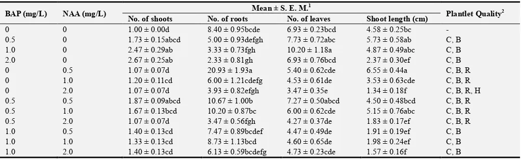
Table 1. Mean number of shoots, roots, and leaves, and shoot length regenerated from nodal explants.
BAP (mg/L) NAA (mg/L) Mean ± S. E. M.
1
Plantlet Quality2
No. of shoots No. of roots No. of leaves Shoot length (cm)
0 0 1.00 ± 0.00d 8.40 ± 0.95bcde 6.93 ± 0.23bcd 4.58 ± 0.25bc -
0.5 0 1.73 ± 0.15abcd 5.00 ± 0.93defgh 7.73 ± 0.72abc 5.73 ± 0.58ab C, B
1.0 0 2.47 ± 0.29ab 3.33 ± 0.73fgh 10.20 ± 1.18a 4.87 ± 0.49abc C, B
2.0 0 2.67 ± 0.25ab 2.33 ± 0.81gh 6.93 ± 0.76bcd 2.37 ± 0.30ef C, B
0 0.5 1.07 ± 0.07d 20.93 ± 1.93a 5.40 ± 0.62cde 6.55 ± 0.44a C, B, R
0 1.0 1.20 ± 0.11cd 6.00 ± 1.21cdefg 4.53 ± 0.61de 3.53 ± 0.63cde C, B, R
0 2.0 1.07 ± 0.07d 3.93 ± 0.82efgh 3.47 ± 0.35e 1.34 ± 0.18f C, B, R, H
0.5 0.5 1.87 ± 0.09abcd 10.67 ± 1.00b 7.27 ± 0.50abcd 4.50 ± 0.48bcd C, B, R
0.5 1.0 1.67 ± 0.13bcd 10.20 ± 0.87bc 6.00 ± 0.62cde 5.15 ± 0.76abc C, B, R
0.5 2.0 1.07 ± 0.07d 3.47 ± 0.56fgh 4.27 ± 0.37de 1.83 ± 0.17ef C, B, R
1.0 0.5 1.40 ± 0.13cd 7.47 ± 0.89bcdef 4.47 ± 0.49de 1.91 ± 0.19ef C, B
1.0 1.0 1.33 ± 0.13cd 8.73 ± 1.13bcd 4.60 ± 0.65de 1.98 ± 0.24ef C, B
BAP (mg/L) NAA (mg/L) Mean ± S. E. M. 1
Plantlet Quality2
No. of shoots No. of roots No. of leaves Shoot length (cm)
2.0 0.5 2.00 ± 0.24abcd 1.60 ± 0.67gh 5.07 ± 0.46cde 1.31 ± 0.14f C, B, H
2.0 1.0 2.13 ± 0.38abc 2.00 ± 0.49gh 6.33 ± 0.72bcde 1.45 ± 0.12f C, B, H
2.0 2.0 2.73 ± 0.47a 0.87 ± 0.24h 9.07 ± 0.73ab 2.57 ± 0.38def C, B, H
1
Mean ± S.E.M. followed by the same letter along each column are not significantly different. 2C= Chlorosis, B= Basal callus, R = Adventitious roots on stem, H = Hyperhydricity.
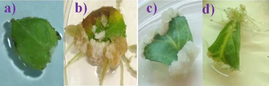
Figure 1. Effect of BAP and/or NAA on plantlet regeneration after 8 weeks of culture: a) Basal callus and chlorosis in 0.5 mg/L BAP b) Adventitious roots produced on stem in 0.5 mg/L NAA c) Hyperhydricity of leaf in 2.0 mg/L NAA d) Basal callus and growth of small leaves in 2.0 mg/L BAP + 2.0 mg/L NAA f) Normal rooting in control treatment.
3.2. Callus Induction
3.2.1. BAP, NAA, and 2,4-D Alone
Leaves excised from regenerated plantlets were used as explants for callus induction. Calli formed were friable and brownish white. The highest callus induction frequency (100%) was recorded when treated with 0.5 – 2.0 mg/L 2,4-D (Table 2). Meanwhile, callus was absent in the control treatment and when treated with 0.5 – 2.0 mg/L BAP, and 0.5 – 1.0 mg/L NAA. Observations showed that leaf explants treated with BAP and NAA alone had chlorosis and/or necrosis within 4 to 8 weeks of culture. Instead, leaf explants treated with 2,4-D remained green throughout the study. Adventitious roots were produced from the leaf midribs in the control treatment whereas calli and rooting from the point of excision were recorded when treated with 2.0 – 4.0 mg/L NAA (Figure 2).
Table 2. Callus induction frequency of leaf explants treated with BAP, NAA, and 2,4-D alone after 8 weeks.
Plant growth regulator (mg/L) Callus induction frequency (%)
Control - 0c
BAP
0.5 0c
1.0 0c
2.0 0c
4.0 0c
NAA
0.5 0c
1.0 0c
2.0 14c
4.0 4c
2,4-D
0.5 100a
1.0 100a
2.0 100a
4.0 52b
3.2.2. BAP + NAA
Leaf explants were treated with different concentrations of BAP in combination with NAA. After 8 weeks, treatment of 2.0 mg/L BAP + 2.0 mg/L NAA produced significantly higher callus induction frequency (72%) as compared to 0.5 mg/L BAP + 1.0 mg/L NAA and 4.0 mg/L BAP + 1.0 – 2.0 mg/L NAA (2 – 6%) (Table 3). All leaf explants treated with BAP + NAA recorded chlorosis and/or necrosis after 4 to 8 weeks of culture. In addition, several leaf explants treated with BAP + NAA exhibited hyperhydricity and callus browning (Figure 3).
Table 3. Callus induction frequency of leaf explants treated with BAP + NAA after 8 weeks.
Plant growth regulator (mg/L) Callus induction
frequency (%)
BAP NAA
0.5 0.5 20ab
0.5 1.0 4b
0.5 2.0 42ab
0.5 4.0 24ab
1.0 0.5 50ab
1.0 1.0 24ab
1.0 2.0 32ab
1.0 4.0 34ab
2.0 0.5 8ab
2.0 1.0 8ab
2.0 2.0 72a
2.0 4.0 52ab
4.0 0.5 30ab
4.0 1.0 6b
4.0 2.0 2b
4.0 4.0 24ab
3.2.3. BAP + 2,4-D
Leaf explants were treated with different concentrations of BAP in combination with 2,4-D. After 8 weeks, results showed that treatment of 0.5 mg/L BAP + 0.5 mg/L 2,4-D and 2.0 mg/L BAP + 4.0 mg/L 2,4-D produced significantly higher callus induction frequency (92 – 100%) when compared to 0.5 mg/L BAP + 4.0 mg/L 2,4-D, 1.0 – 2.0 mg/L BAP + 0.5 mg/L 2,4-D, and 4.0 mg/L BAP + 0.5 – 4.0 mg/L 2,4-D (0 – 12%) (Table 4). Observations showed that all leaf explants treated with BAP + 2,4-D recorded chlorosis and/or necrosis after 4 to 8 weeks of culture (Figure 3).
Table 4. Callus induction frequency of leaf explants treated with BAP + 2,4-D after 8 weeks.
Plant growth regulator (mg/L) Callus induction
frequency (%)
BAP 2,4-D
0.5 0.5 92a
0.5 1.0 20bc
0.5 2.0 28bc
0.5 4.0 12c
1.0 0.5 0c
1.0 1.0 24bc
1.0 2.0 16bc
1.0 4.0 50abc
2.0 0.5 8c
2.0 1.0 50abc
2.0 2.0 74ab
2.0 4.0 100a
Plant growth regulator (mg/L) Callus induction
frequency (%)
BAP 2,4-D
4.0 0.5 12c
4.0 1.0 4c
4.0 2.0 0c
4.0 4.0 12c
Figure 3. Effect of BAP in combination with NAA or 2,4-D on callus induction after 8 weeks of culture: a) 0.5 mg/L BAP + 1.0 mg/L NAA b) 1.0 mg/L BAP + 2.0 mg/L 2,4-D.
3.3. Determination of Callus Biomass
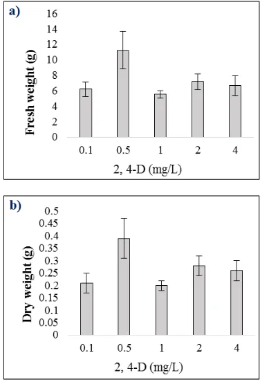
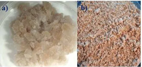
Calli were sub-cultured on fresh MS medium supplemented with different concentrations of 2,4-D to determine the effect on callus biomass. Results of the callus biomass after 8 weeks as presented in Figure 4 indicated that higher means of fresh (11.27 g) and dry (0.39 g) weights were recorded when treated with 0.5 mg/L 2,4-D. Observations showed that fresh calli sub-cultured onto fresh MS medium supplemented with different concentrations of 2,4-D were light brown in colour and remained friable after 8 weeks of culture while dried calli were a darker brown and powdery (Figure 5).
Figure 5. Brown calli proliferated after 8 weeks of culture: a) fresh b) dry.
4. Discussion
The findings of this present study indicated that in vitro
plantlets of V. amygdalina can be successfully regenerated from nodal explants and influenced by plant growth regulators. We tested the effects of BAP and NAA based on an earlier study on shoot regeneration of V. amygdalina [19]. Addition of BAP and/or NAA to MS medium in this present study produced growth abnormalities in the plantlets including chlorosis, basal callus that interfered with normal root growth, excess production of adventitious roots on the stems, and hyperhydricity in several plantlets that caused malformation and stunted growth of leaves. Studies have shown that output of endogenous cytokinin stimulates bud growth [27] whereas endogenous auxin is capable of down-regulating the synthesis of cytokinin in the nodes of stems [30]. Hence, further addition of exogenous growth regulators can bypass the actions of endogenous plant growth regulators and/or alter regulatory processes that control hormone levels after wounding [11].
Callus exhibits a sink effect that traps plant growth regulators [3]. A number of reports indicated that the occurrence of basal callus which inhibits growth was caused by plant growth regulators such as the treatment of
Neotchihatchewia isatidea with BAP [15], treatment of
Celosia argentea [5], and Wattakaka volubilis with NAA [33] as well as when BAP and/or NAA was supplemented to Aster scaber [8]. Plant growth regulators in addition to other properties of the culture medium and atmospheric conditions of the culture vessels have been associated with hyperhydricity [10, 13]. Nodal explants of V. amygdalina
exhibited the disorder when treated with NAA alone or in combination with BAP. The effect of cytokinin on induction of hyperhydricity in in vitro cultureshave been reported such as the case of carnation [28], aloe [4], and agave [9]. Other reports suggest that gelling agent in nutrient medium triggers hyperhydricity [6, 18].
Results from this present study indicated that control treatment without plant growth regulators is the ideal and cost-efficient culture condition for the regeneration of in vitro V. amygdalina plantlets from nodal explants. Observations showed that the control treatment produced plantlets with large, green leaves and profuse proliferation of roots due to the absence of basal callus.
Callus induction frequency in leaf explants of V. amygdalina
in this present study was influenced by plant growth regulators. Significant difference in callus induction was observed with the use of the auxins NAA and 2,4-D alone or in combination with the cytokinin BAP. Leaf explants in MS medium containing 0.5 - 2.0 mg/L 2,4-D showed 100% callus induction frequency and less chlorosis. The supplementation of a higher concentration of 2,4-D produced less callus instead. Compared to Vernonia cinerea, maximum cell suspension biomass was obtained when treated with 0.1 mg/L BA + 1.0 mg/L NAA [22] which indicated that response of callus induction towards plant growth regulators is species-dependent.
Results in this present study showed that supplementation of 0.5 mg/L 2,4-D into MS medium increased the highest fresh callus biomass of V. amygdalina by 11-fold to 11.27g after 8 weeks of culture. In comparison with another earlier study, maximum fresh callus biomass of 3.56g was obtained with treatment of 1.5 mg/L 2,4-D [12].
5. Conclusion
MS medium without plant growth regulator was the optimum culture condition for in vitro regeneration of V. amygdalina from nodal explants. The optimum plant growth regulator for callus induction and further callus proliferation was 2,4-D. The established regeneration and callus induction protocol provides a baseline for further studies on the breeding of the multipurpose plant as well as utilization of cell cultures for potential production of bioactive secondary metabolites.
References
[1] Aletor, O., Oshodi, A. A., and Ipinmoroti, K. (2002) Chemical composition of common leafy vegetables and functional properties of their leaf protein concentrates. Food Chemistry, 78 (1), 63-68.
[2] Amin, S., Kaloo, Z. A., Singh, S., and Altaf, T, (2013) Micropropagation of medicinally important plant species of family Asteraceae – A review. International Journal of Recent Scientific Research, 4 (8), 1296-1303.
[3] Bairu, M. W., and Kane, M. E. (2011) Physiological and developmental problems encountered by in vitro cultured plants. Plant Growth Regulators, 63, 101-103.
[4] Bairu, M. W., Stirk, W. A., Dolezal, K., and Van Staden, J. (2007) Optimizing the micropropagation protocol for the endangered Aloe polyphylla: can meta-topolin and its derivatives served as replacement for benzyladenine and zeatin. Plant Cell Tissue and Organ Culture, 90, 15-23.
[5] Bakar, D. A., Ahmed, B. A., and Taha, R. M. (2014) In vitro callus induction and plant regeneration of Celosia argentea – An important medicinal plant. Brazilian Archives of Biology and Technology, 57 (6), 860-866.
[7] Bidarigh, S., and Azarpour, E. (2013) Evaluation of the effect of MS medium levels on rooting in micro cuttings of tea (Camellia sinensis L.) under in-vitro culture condition. Asian Research Publishing Network (ARPN) Journal of Agricultural and Biological Science, 8 (1), 24-28.
[8] Boo, K. H., Cao, D. V., Pamplona, R. S., Lee, D., Riu, K. Z., and Lee, D. S. (2015) In vitro plant regeneration of Aster scaber via somatic embryogenesis. Bioscience, Biotechnology, and Biochemistry, 79 (5), 725-731.
[9] Caraballo, M. G., Oramas, G. G., García, S. A., Cruz, E. A., Bravo, K. Q., Caligari, P. D. S., and Garcíia-González, R. (2010) Management of auxin-cytokinin interactions to improve micropropagation protocol of henequen (Agave fourcroydes Lem.). Chilean Journal of Agricultural Research, 70 (4), 545-551.
[10] Chakrabarty, D., Park, S. Y., Ali, M. B., Shin, K. S., and Paek, K. Y. (2005) Hyperhydricity in apple: ultrastructural and physiological aspects. Tree Physiology, 26, 377-388.
[11] Chen, X., Qu, Y., Sheng, L., Liu, J., Huang, H., and Xu, L. (2014) A simple method suitable to study de novo organogenesis. Frontiers in Plant Science, 5, 208.
[12] Daffalla, H. M., Abdellatef, E., and Khalafalla, M. (2014) Callogenesis and rhizogenesis in Vernonia amygdalina leaf: step for secondary metabolites production. Proceedings of 5th International Workshop Biotechnology and its Role in Economic Development in the Arab World.
[13] Debergh, P. C. (1983) Effects of agar brand and concentrations on the tissue culture medium. Physiologia Plantarum, 59, 270-276.
[14] Fomum, F. U. (2004) Vernonia amgydalina Delile. In Grubben, G. J. H. and Denton, O. A. (Eds.), Plant Resources of Tropical Africa 2: Vegetables. Netherlands: PROTA Foundation/Backhuys Publishers/CTA, pp 543-546.
[15] Gümüşçü, A., Çöçü, S., Uranbey, S., Ipek, A., Çalıskan, M., and Arslan, N. (2008). In vitro micro-propagation of endangered ornamental plant-Neotchihatchewia isatidea (Boiss.) Rauschert. African Journal of Biotechnology, 7 (3), 234-238.
[16] Huffman, M. A., and Seifu, M. (1989) Observations on the illness and consumption of a possibly medicinal plant Vernonia amgydalina (Del.), by a wild chimpanzee in the Mahale Mountains National Park, Tanzania. Primates, 30 (1), 51-63.
[17] Hussain, M. S., Fareed, S., Ansari, S., Rahman, M. A., Ahmad, I. Z., and Saeed, M. (2012) Current approaches toward production of secondary plant metabolites. Journal of Pharmacy and BioAllied Sciences, 4 (1), 10-20.
[18] Ivanova, M., Novák, O., Strnad, M., and Van Staden, J. (2006) Endogenous cytokinins in shoots of Aloe polyphylla cultured in vitro in relation to hyperhydricity, exogenous cytokinins and gelling agents. Plant Growth Regulation, 50 (2-3), 219-230.
[19] Khalafalla, M. M., Abdellatef, E., Daffalla, H. M., Nassrallah, A. A., Aboul-Enein, K. M., Lightfoot, D. A., Cocchetto, A., and El-Shemy, H. A. (2009) Antileukemic activity from root cultures from Vernonia amygdalina. Journal of Medicinal Plants Research, 556-562.
[20] Khalafalla, M. M., Elgaali, E. I., and Ahmed, M. M. (2007) In vitro multiple shoot regeneration from nodal explants of Vernonia amygdalina – An important medicinal plant. Proceedings from African Crop Science Conference.
[21] Koshimizu, K., Ohigashi, H., and Huffman, M. A. (1994) Use of Vernonia amygdalina by wild chimpanzee: Possible roles of its bitter and related constituents. Physiology and Behaviour, 56 (6), 1209-1216.
[22] Maheshwari, P., Songara, B., Kumar, S., Jain, P., Srivastava, K., and Kumar, A. (2007) Alkaloid production in Vernonia cinerea: Callus, cells suspension and root cultures. Biotechnology Journal, 2(8), 1026-1032.
[23] Oboh, G. (2006) Nutritive value and haemolytic properties (in vitro) of the leaves of Vernonia amygdalina on human erythrocyte. Nutrition and health, 18 (2), 151-160.
[24] Ohigashi, H., Huffman, M. A., Izutsu, D., Koshimizu, K., Kawanaka, M. Sugiyama, H., Kirby, G. C., Warhurst, D. C., Allen, D., Wright, C. W., David Phillipson, J., Timon-David, P., Delmas, F., Elias, R., and Balansard, G. (1994) Toward the chemical ecology of medicinal plant use in chimpanzees: The case of Vernonia amgydalina, a plant used by wild chimpanzees possibly for parasite-related diseases. Journal of Chemical Ecology, 20 (3), 541-553.
[25] Opabode, J. T., and Adebooye, O. C. (2005) Application of biotechnology for the improvement of Nigerian indigenous leaf vegetables. African Journal of Biotechnology, 4 (3), 138-142.
[26] Rao, S. R., and Ravishankar, G. A. (2002) Plant cell cultures: Chemical factories of secondary metabolites. Biotechnology Advances, 20 (2), 101-153.
[27] Shimizu-Sato, S., Tanaka, M., and Mori, H. (2009) Auxin-cytokinin interactions in the control of shoot branching. Plant Molecular Biology, 69, 429-435.
[28] Simona, L., Cerasela, P., Alexandru, L., and Maria, B. (2012) Influence of growth regulators on morphogenetic processes under in vitro condition. Journal of Horticulture, Forestry and Biotechnology, 16 (2), 197-202.
[29] Smetanska, I. (2008) Production of secondary metabolites using plant cell cultures. Advances in Biochemical Engineering and Biotechnology, 111, 187-228.
[30] Tanaka, M., Takei, K., Kojima, M., Sakakibara, H., and Mori, H. (2006) Auxin controls local cytokinin biosynthesis in the nodal stem in apical dominance. The Plant Journal, 45 (6), 1028-1036.
[31] Toyang, N. J., and Verpoorte, R. (2013) A review of the medicinal potentials of plants of the genus Vernonia (Asteraceae). Journal of Ethnopharmacology, 146 (3), 681-723.
[32] Verpoorte, R., Contin, A., and Memelink, J. (2002) Biotechnology for the production of plant secondary metabolites. Phytochemistry Reviews, 1 (1), 13-25.