© 2018 IJSRST | Volume 4 | Issue 2 | Print ISSN: 2395-6011 | Online ISSN: 2395-602X Themed Section: Science and Technology
Brain Tumor Classification Using Pattern Recognition Techniques: the
Comprehensive Review
Rahul B. Mapari*1, Dr. Anilkumar N. Holambe2
*1Maharashtra Institute of Technology, Aurangabad, Maharashtra, India
2TPCT College of Engineering, Osmanabad, Maharashtra, India
ABSTRACT
A Brain tumor is very serious disease causing deaths of many individuals. The detection and classification system must be available so that it can be diagnosed at early stages. Detection of the brain tumor and its type in its early stage is essential for right treatment. So classification of brain tumor is very important. Tumor classification has been one of the most challenging tasks in clinical diagnosis. Different image processing techniques such as image segmentation, image enhancement and feature extraction are used for detection of the brain tumor in the MRI images of the cancer affected patients. Medical Image Processing is the fast growing and challenging field now days. Image processing and neural network techniques are used to improve the performance of detecting and classifying brain tumor in MRI images. The objective of this review paper is to presents a comprehensive overview for MRI brain tumor segmentation methods. In this paper, various segmentation techniques have been discussed. Comparative analysis of existing techniques has been done in brief.
Keywords: Brain Tumor, Segmentation, Classification, Feature Extraction, Discrete Wavelet Transform, Discrete Cosine Transform, SVM, Probabilistic Neural Network, Artificial Neural Network, MRI.
I.
INTRODUCTION
Abnormal or uncontrollable growth of tissues within the brain is known as a brain tumor. Brain tumor is a group of tissue that is prearranged by a slow addition of irregular cells. It occurs when cell get abnormal formation within the brain. Brain tumor is one of the most common and deadly diseases in the world. The seriousness of brain tumor is very big among all the variety of cancers, so to save a life immediate detection and proper treatment to be done.
Each year over 190,000 people in the United States and 10,000 people in Canada are diagnosed with various types of brain tumor. Brain tumors are the second leading cause of cancer based deaths among children in the age group between 0 and 19 years. And also they are the second leading cause of cancer
based deaths in young men up to age 39 years and the fifth leading cause of cancer based deaths among young women between ages 20 and 39 years. In India, totally 80,271 people are affected by various types of brain tumor.
Brain tumor is classified into three types: Normal, Benign and Malignant. A benign brain tumor grows slowly, has distinct boundaries and spreads rarely. While a malignant brain tumor grows quickly and spreads to nearby brain areas. These have irregular boundaries but don’t spread to organs outside the brain that’s why they are also known as brain cancers [25].
Interpretation of images is based on organized and explicit classification of brain MRI.
The brain related disease can be identified with imaging techniques like MRI. Magnetic Resonance Imaging (MRI) is a radiology technique, widely used by physicians and radiologists to visualize internal organ of human body in detail. It is also called as Nuclear Magnetic Resonance Imaging (NMRI) or Magnetic Resonance Tomography (MRT). MRI inherits the properties of nuclear magnetic resonance to generate digital image of atoms inside human body. MRI is a safe diagnostic and painless procedure to scan internal organ and structure of human body through strong magnet and radio waves. For the past 40 decades, it is used as a significant tool in scientific research and medicine diagnose. MRI is the most suitable choice for investigating brain tumor as it is more sensitive than CT scan in identifying small tumors and it gives better visualization [4].
The first step to minimize the death rate of brain tumor is to develop an effective diagnosing method. Magnetic Resonance Imaging (MRI) is one of the best imaging system to diagnose brain tumor, even though it is crucial for the clinicians to diagnose the early stage of brain tumor. So, image segmentation is the only possible tool for early detection of brain tumor. Manual segmentation by an expert will consume more time and it is very difficult to do proper segmentation. Hence, computer aided automatic classification techniques are preferred in detection and classification of brain tumors.
Accurate detection of the type of brain abnormality is highly essential for treatment planning in order to minimize diagnostic errors. The accuracy can be improved by using computer aided diagnosis (CAD) systems. Image processing methods are very useful to detect and classify the brain tumors [26].
II.
LITERATURE REVIEW
[2] Noramalina Abdullah, Lee Wee Chuen, Umi Kalthum Ngah, Khairul Azman Ahmad, in this article the classifications of brain tumors is done by using support vector machine (SVM). The accuracy in classification data with and without the implementation of principal component analysis (PCA) is compared. It is observed that using PCA method, the number of feature vector has been reduced from 17689 to 200 and increase the percentage of accuracy.
[15] Vijay Wasule, Poonam Sonar, proposed method use LCM technique to extract the texture features from images and stored as a feature vector. The extracted features are classified using supervised SVM and KNN algorithm. The accuracy of the proposed system is 96% and 86% for SVM and KNN respectively for clinical database and 85% and 72.50% for SVM and KNN respectively for Brats database.
[3] Sandeep Chaplot, L.M. Patnaik, N.R. Jagannathan, In this paper, a new technique is proposed which use wavelets as input to neural network self-organizing maps. Then SVM is used for classification of MRI images of human brain. The proposed method classifies MR brain images as either normal or abnormal. The proposed approach is evaluated using a dataset of 52 MR brain images. 94% accuracy was achieved using the neural network self-organizing maps (SOM) and 98% from support vector machine. It is observed that the classification rate is high for a support vector machine classifier compared to self-organizing map-based approach.
reference MR image. These distances are further fed to k-Means classifier to classify the MR images as normal and abnormal images.
[1] D.Shridhar, Murali Krishna, this paper proposed a technique for brain tumor classification using Probabilistic Neural Network with Discrete Cosine Transform. The process involved two stages, a) Feature extraction and reducing dimensionality using the Discrete Cosine Transform and b) classification of tumors using Probabilistic Neural Network (PNN). The dataset has 20 Brain Tumor images for evaluation. The proposed method is efficient and gives better recognition rate as compared to previous classifiers. The main advantage of this method is its high speed computing ability and low computational requirements.
[4] Y. Zhang, L. Wu, In this paper, a technique to classify a given MR brain image as normal or abnormal is proposed. In this method wavelet transform is used to extract features from images, followed by applying principle component analysis (PCA) to reduce the dimensions of features. The images with reduced dimensions are submitted to a kernel support vector machine. The strategy of K-fold stratified cross validation is used to enhance generalization of Kernel support vector machine. The technique was evaluated with four kernels, and it is observed that the GRB kernel achieves the highest classification accuracy than others.
[5] El-Sayed A. El-Dahshan, Heba M. Mohsen , Kenneth Revett , Abdel-Badeeh M. Salem, proposed technique is using the feedback pulse-coupled neural network for segmentation of images, the DWT for features extraction, the PCA for reducing the dimensionality of the wavelet coefficients, and the feed forward back-propagation neural network to classify the brain MRI images into normal or abnormal types. The classification accuracy on both training and test images is very high ie around 99%.
[7] Pauline John, the proposed method classified brain tumors into normal, benign and malignant brain tumor in three stages, (1) wavelet decomposition, (2) textural feature extraction and (3) classification. The Discrete Wavelet Transform with Daubechies wavelet (db4) is used for decomposing the MR image into different levels of approximate and detailed coefficients and then the gray level co-occurrence matrix is formed, from which the texture statistics such as entropy, correlation, energy, homogeneity, and contrast are obtained. The results of co-occurrence matrices are then fed into a PNN for further classification and tumor detection. The accuracy of proposed method is very high.
[8] El-Sayed A. El-Dahshan, Abdel-Badeeh M. Salem, And Tamer H. Younis, the proposed method has three stages, feature extraction, dimensionality reduction, and classification. In the first stage, features related with MRI images are extracted using discrete wavelet transformation. In the second stage, the features of MRI images are reduced using PCA and in last step, two classifiers based on supervised machine learning have been developed. The first classifier based on feed forward back-propagation artificial neural network (FP-ANN) and the second classifier based on k-nearest neighbor (k-NN). The classifiers have been used to classify subjects as normal or abnormal MRI human images. These evaluation of method shows that the proposed hybrid technique is effective compared with others.
accuracy, sensitivity, and specificity, assessed by leave-one-out cross-validation on 102 brain tumors, are respectively 87%, 89%, and 79% for discrimination of metastases from gliomas, and 87%, 83%, and 96% for discrimination of high grade from low grade neoplasms. Multi-class classification is also performed via a one-versus-all voting scheme.
[12] R.Lavanyadevi, M.Machakowsalya, J.Nivethitha, A.Niranjil Kumar, in this paper the neural network is used to classify brain tumor as benign, malignant or normal. Feature extraction is done by using the Gray Level Co-Occurrence Matrix (GLCM). Image recognition and image compression is done by using the Principal Component Analysis (PCA) method and also large dimensionality of the data is reduced. Classification is done by using probabilistic neural network (PNN). Segmentation process is done by using K-means clustering algorithm and also detects the brain tumor spread region. It is found that PNN is fastest technique and also provide the good classification accuracy.
[9] Yudong Zhang, Zhengchao Dong , Lenan Wu, Shuihua Wanga, in this paper, they have present a method which use neural network to classify a brain image as normal or abnormal. This method first employs wavelet transform to extract features from images, and then applies the technique of principle component analysis (PCA) to reduce the dimensions of features. The reduced features are sent to a back propagation (BP) NN, with which scaled conjugate gradient (SCG) is adopted to find the optimal weights of the NN. The classification accuracies on both training and test images are 100%, and the computation time is also very low.
[13] Mahmoud Khaled Abd-Ellah, Ali Ismail Awad, Ashraf A. M. Khalaf, and Hesham F. A. Hamed, in this
paper, a two-stage method is proposed for detection and classification of brain tumors. In the first stage, the system classifies brain tumor MRI into normal and abnormal images. In the second stage, the type of tumor is classified as benign or malignant. MRI images are segmented by K-means clustering, feature extraction using discrete wavelet transform (DWT), feature reduction by principal component analysis (PCA). The two-stage classification has been conducted using a support vector machine (SVM). Performance evaluation of the proposed CAD has achieved good results.
[14] S.K. Shil, Et.al, in this scheme, K- means clustering is used for segmentation. Discrete cosine transform and Principal of component analysis is used for feature extraction and reduced features are submitted to support vector machine for classification. Performance evaluation is done using specificity, sensitivity and accuracy parameters.
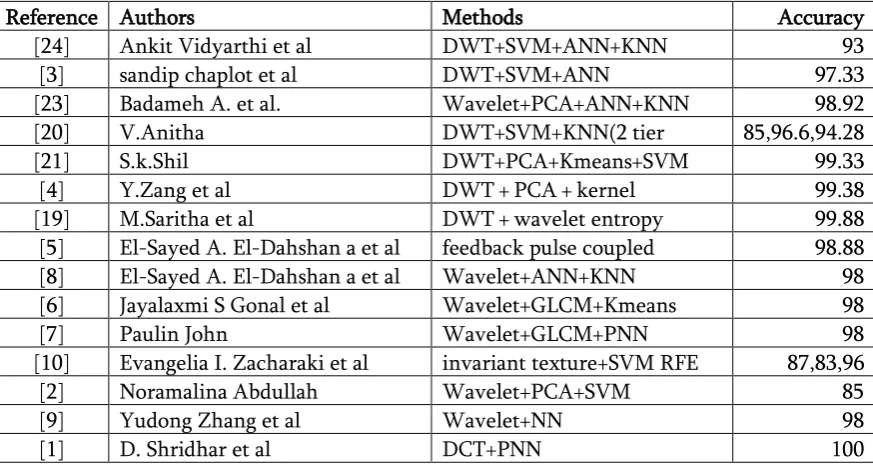
Table 1.Comparative Analysis Of Existing Brain Tumor Classification Techniques
Reference Authors Methods Accuracy
[24] Ankit Vidyarthi et al DWT+SVM+ANN+KNN 93
[3] sandip chaplot et al DWT+SVM+ANN 97.33
[23] Badameh A. et al. Wavelet+PCA+ANN+KNN 98.92
[20] V.Anitha DWT+SVM+KNN(2 tier
classifier)
85,96.6,94.28
[21] S.k.Shil DWT+PCA+Kmeans+SVM 99.33
[4] Y.Zang et al DWT + PCA + kernel SVM + Gaussian radial basis
99.38 [19] M.Saritha et al DWT + wavelet entropy
(WE) + spider web plots +PNN
99.88 [5] El-Sayed A. El-Dahshan a et al feedback pulse coupled
NN + DWT + PCA + FP-ANN
98.88
[8] El-Sayed A. El-Dahshan a et al Wavelet+ANN+KNN 98
[6] Jayalaxmi S Gonal et al Wavelet+GLCM+Kmeans 98
[7] Paulin John Wavelet+GLCM+PNN 98
[10] Evangelia I. Zacharaki et al invariant texture+SVM RFE 87,83,96
[2] Noramalina Abdullah Wavelet+PCA+SVM 85
[9] Yudong Zhang et al Wavelet+NN 98
[1] D. Shridhar et al DCT+PNN 100
III. METHODOLOGIES
Following methodologies are used in the process of brain tumor classification:
A. Feature Extraction using Discrete Wavelet Transform
Wavelet transform is an effective tool for feature extraction from MR brain images, because it allows analysis of images at various levels of resolution due to its multi-resolution analytic property. However, this technique requires large storage and is computationally expensive [24].
The wavelet used to extract the wavelet coefficient from MR images. Wavelets are localized basis
functions, which are scaled and shifted versions of some fixed mother wavelets. The main advantage of wavelets is that they provide localized frequency information about a function of a signal, which is particularly beneficial for classification [16][19].
The discrete wavelet transform (DWT) is a powerful implementation of the WT using the dyadic scales and positions. Formally DWT is defined as, Suppose x(t) is a square-integrable function, then the continuous WT of x(t) relative to a given wavelet ᴪ(t) is defined as
B. Feature dimensionality reduction using Principal Component Analysis
Excessive features increase computation times and storage memory. Furthermore, they sometimes make classification more complicated, which is called the curse of dimensionality. It is required to reduce the number of features. PCA is an efficient tool to reduce the dimension of a data set consisting of a large number of interrelated variables while retaining most of the variations. It is achieved by transforming the data set to a new set of ordered variables according to their variances or importance. This technique has three effects: it orthogonalizes the components of the input vectors so that uncorrelated with each other, it orders the resulting orthogonal components so that those with the largest variation come first, and eliminates those components contributing the least to the variation in the data set. It should be noted that the input vectors be normalized to have zero mean and unity variance before performing PCA [17][18].
extracted from the image but it will not change, improve or anything to do with Original image. In PCA, the input feature space is transformed into a lower-dimensional feature space using the largest eigenvectors of the correlation matrix. PCA is the most widely used subspace projection technique. When a set of data is given, PCA finds the linear lower-dimensional representation of the data such that the variance of the data is preserved. Using a system of feature reduction based on PCA limits the feature vectors to the component selected by the PCA which leads to an efficient classification algorithm. So, the main idea behind using PCA is to reduce the dimensionality of the wavelet coefficients which results in a more efficient and accurate classifier. C. Classification:
i) Artificial Neural Network
An ANN is a mathematical model consisting of a number of highly interconnected processing elements organized into layers, geometry and functionality of which have been resembled to that of the human brain. The ANN may be regarded as possessing learning capabilities in as much as it has a natural propensity for storing experimental knowledge and making it available for later use. The neural network which was employed as the classifier required in this study had three layers. The first layer consisted of 7 input elements in accordance with the 7 feature vectors that selected from the wavelet coefficients by the PCA. The number of neurons in the hidden layer was four. The single neuron in the output layer was used to represent normal and abnormal human brain [23].
The most frequently used training algorithm in classification problems is the back-propagation (BP) algorithm, which is used in this work also. The details of back-propagation (BP) algorithm are well documented in the literature. The neural network has been trained to adjust the connection weights and biases in order to produce the desired mapping. At the training stage, the feature vectors are applied as input
to the network and the network adjusts its variable parameters, the weights and biases, to capture the relationship between the input patterns and outputs [19][20].
ii) K Nearest Neighbor based classifier:
One of the simplest classification techniques is the k- Nearest Neighbor classifier. Classification of an input feature vector X is done by determining the k closest training vectors according to a suitable distance metric. The vector X is then assigned to that class to which the majority of those k nearest neighbors belong to. The k-NN algorithm is based on a distance function and a voting function in k nearest neighbors, the metric employed is the Euclidean distance. The k-nearest neighbor classifier is a conventional nonparametric supervised classifier that is said to yield good performance for optimal values of k. Like most guided learning algorithms, k-NN algorithm consists of a training phase and a testing phase. In the training phase, data points are given in a n-dimensional space. These training data points have labels associated with them that designate their class. In the testing phase, unlabeled data are given and the algorithm generates the list of the k nearest (already classified) data points to the unlabeled point. The algorithm then returns the class of the majority of that list [15][20].
KNN algorithm
1: Determine a suitable distance metric.
2: In the training phase: Stores all the training data set P in pairs( according to the selected features) P = (yi, ci), i=1. . .n , where yi is a training pattern in the training data set, ci is its corresponding class and n is the amount of training patterns.
3: During the test phase: Computes the distances between the new feature vector and all the stored features (training data).
satisfactory, the k value can be tuned until a reasonable level of correctness is achieved.
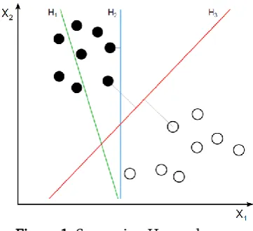
iii) Support Vector Machine
Support vector machine is a supervised method used to find pattern and perform classification and regression analysis. Given a set of training data marked with class, the SVM classifier builds a model that assigns new unseen data into a category. This can be used for multiclass classification using kernel tricks.[21]
The key concept of SVM is the use of hyper planes to define decision boundaries separating between data points of different classes. SVMs are to handle simple, linear, classification tasks, as well as more complex i.e. nonlinear, classification problems. Both separable and non separable problems are handled by SVMs in the linear and nonlinear case. In this research there are two classified data of normal and abnormal (Tumor) brain images. The hyper planes for SVMs are used to separate these classified data as normal data and tumor data. SVMs are capable to handle very large feature spaces and have good generalization properties compared to conventional classifiers because in training the SVM classifier the so-called structural misclassification risk is to be minimized, whereas traditional classifiers are usually trained so that empirical risk is minimized. It performs binary classification tasks. In our study, the two classes are normal or abnormal brain. The kernel function used in our SVM classifier is radial basis function (RBF) kernel. Two major RBF parameters applied in SVM, namely C and y , are obtained by GA-SVM model. SVM-based classification takes N samples, trains the classifier on N-l samples, and then uses the remaining sample for testing. Classification is continued in this manner using a different test sample in every iteration. Final performance is reported as an average of results for all N trials. Thus, the normal and the abnormal region are classified [22].
Figure 1. Separating Hyperplanes
IV. CONCLUSION
In this paper we have completed a detailed review of existing techniques for detection and segmentation of brain tumors in MRI images. Various image processing methods are involved for pre-processing, feature extraction and segmentation of images in entire process. We have also discussed their advantages and disadvantages as comparative analysis.
There are many techniques which can detect the tumor efficiently and accurately. This work will be extended in near future for the development of new algorithm for brain tumor classification and detection, which will provide more accurate, efficient and reliable result than the existing methods for early detection and classification of brain tumors to save the lives of patients suffering from brain tumor.
V.
REFERENCES
[1]. D.Shridhar, Murali Krishna, "Brain Tumor Classification U sing Discrete Cosine Transform and Probabilistic Neural Network", 2013 Internationa Conference on Signal Processing, Image Processing and Pattern Recognition [ICSIPR], 978-1-4673-4862-1/13/$31.00 2013 IEEE.
Principal Component Analysis", 2011 IEEE International Conference on Control System, Computing and Engineering, 978-1-4577-1642-3/11/$26.00 2011 IEEE.
[3]. Sandeep Chaplot, L.M. Patnaik, N.R. Jagannathan, "Classification of magnetic resonance brain images using wavelets as input to support vector machine and neural network", Biomedical Signal Processing and Control 1, pp. 86-92,2006 Elsevier.
[4]. Y. Zhang, L. Wu, "An Mr Brain Images Classifier Via Principal Component Analysis And Kernel Support Vector Machine", Progress In Electromagnetics Research, Vol. 130, pp.369-388, 2012.
[5]. El-Sayed A. El-Dahshan, Heba M. Mohsen , Kenneth Revett , Abdel-Badeeh M. Salem, "Computer-aided diagnosis of human brain tumor through MRI: A survey and a new algorithm", Expert Systems with Applications 41,pp.5526-5545, 2014 Elsevier.
[6]. Jayalaxmi S. Gonal, Vinayadatt V. Kohir, "Classification of Brain MR Images using Wavelets Texture Features and k-Means Classifier", International Conference on Electrical, Electronics, Signals, Communication and Optimization (EESCO), 978-1-4799-7678-2/15/$31.00 2015 IEEE.
[7]. Pauline John, "A Classifier to Detect Tumor Disease in MRI Brain Images", 2012 IEEE/ACM International Conference on Advances in Social Network Analysis and Mining, 978-0-7695-4799-2/12 $26.00 2012 IEEE.
[8]. El-Sayed A. El-Dahshan, Abdel-Badeeh M. Salem, And Tamer H. Younis, "A Hybrid Technique For Automatic Mri Brain Images Classification", Studia Univ. Babes-Bolyai, Informatica, Volume LIV, Number 1, 2009. [9]. Yudong Zhang, Zhengchao Dong , Lenan Wu,
Shuihua Wanga, "A hybrid method for MRI brain image classification", Expert Systems with Applications, (2011), pp. 10049-10053, Elsevier.
[10]. Evangelia I. Zacharaki, Sumei Wang, Sanjeev Chawla, Dong Soo Yoo, Ronald Wolf, Elias R. Melhem, Christos Davatzikos, "MRI-Based Classification Of Brain Tumor Type And Grade Using SVM-RFE", 2009 IEEE International Symposium on Biomedical Imaging: From Nano to Macro, 978-1-4244-3932- 4/09/$25.00 2009 IEEE
[11]. Praveen, Amritpal Singh, "Detection of Brain Tumor in MRI Images, using Combination of Fuzzy C-Means and SVM", 2015 2nd International Conference on Signal Processing and Integrated Networks (SPIN), 978-1-4799-5991-4/15/$31.00 2015 IEEE.
[12]. R.Lavanyadevi, M.Machakowsalya, J.Nivethitha, A.Niranjil Kumar, "Brain Tumor Classification and Segmentationin MRI Images using PNN", 2017 International Conference on Electrical, Instrumentation and Communication Engineering (ICEICE2017).
[13]. Mahmoud Khaled Abd-Ellah, Ali Ismail Awad, Ashraf A. M. Khalaf, and Hesham F. A. Hamed, "Design and Implementation of a Computer-Aided Diagnosis System for Brain Tumor Classification", 2016 28th International Conference on Microelectronics (ICM) 978-1-5090-5721-4/16/$31.00 2016 IEEE.
[14]. S.K. Shil, F.P.Polly, M.A.Hossain, M.S.Ifthekhar, M.N.Uddin,Y.M.Jang, "An improved brain tumor detection and classification mechanism", 2017 International Conference on Information and Communication Technology Convergence (ICTC), 978-1-5090-4032-2/17/$31.00 2017 IEEE.
[15]. Vijay Wasule, Poonam Sonar, "Classification of Brain MRI Using SVM and KNN Classifier", 2017 IEEE 3rd International Conference on Sensing, Signal Processing and Security (ICSSS), 978-1-5090-4929-5 2017 IEEE.
Wavelet Transform for Image Classification", The 5th Student Conference on Research and Development -SCOReD 2007,11-12 December 2007, Malaysia, 1-4244-1470- 9/07/$25.OO D2007 IEEE.
[17]. Y.D. Zhang, L.N. Wu, and G. Wei, "A New Classifier For Polarimetric Sar Images", Progress In Electromagnetics Research, PIER 94, 83-104, 2009.
[18]. Pasi Luukka , "Classification Based on Fuzzy Robust PCA Algorithms and Similarity Classifier", Expert Systems with Applications, Volume 36, Issue 4, May 2009, Pages 7463-7468, Elsevier.
[19]. M. Saritha, K. Paul Joseph, Abraham T. Mathew, "Classification of MRI brain images using combined wavelet entropy based spider web plots and probabilistic neural network", Pattern Recognition Letters Volume 34, Issue 16, 1 December 2013, pp. 2151-2156.
[20]. V. Anitha, S. Murugavalli, Brain tumour classification using two-tier classifier with adaptive segmentation technique", IET Comput. Vis., pp. 1-9, The Institution of Engineering and Technology 2015.
[21]. Isabelle Guyon, Jason Weston, Stephen Barnhill, M.D., Vladimir Vapnik, "Gene Selection for Cancer Classification using Support Vector Machines", Machine Learning, ISSN: 0885-6125 (Print) 1573-0565 (Online). January 2002, Volume 46, Issue 1-3, pp 389-422.
[22]. Omkishor Sahu, Vijay Anand, Vivek Kanhangad, Ram Bilas Pachori, "Classification of Magnetic Resonance Brain Images using Bi-dimensional Empirical Mode Decomposition and Autoregressive Model", Biomed Eng Lett (2015) 5:311-320, Springer.
[23]. Amer Al-Badarneh, Hassan Najadat, Ali M. Alraziqi, "A Classifier to Detect Tumor Disease in MRI Brain Images", 2012 IEEE/ACM International Conference on Advances in Social
Networks Analysis and Mining, 978-0-7695-4799-2/12 $26.00 2012 IEEE.
[24]. Ankit Vidyarthi, Namita Mittal, "Comparative Study for Brain Tumor classification on MR/CT images",
https://www.researchgate.net/publication/25953 1035_Comparative_Study_for_Brain_Tumor_Cl assification_on_MRCT_Images.
[25]. Mohammed Sabbih Hamoud Al-Tamimi, Ghazali Sulong, "Tumor Brain Detection Through MR Images: A Review Of Literature", Journal of Theoretical and Applied Information Technology, Vol. 62 No.2 ISSN: 1992-8645, 2014, pp. 387-403.