University of Pennsylvania
ScholarlyCommons
Publicly Accessible Penn Dissertations
1-1-2014
T-Lineage Specification and Commitment
Requires Constraint of Myeloid Gene Expression
Programs by Hes1
Maria Elena De Obaldia
University of Pennsylvania, [email protected]
Follow this and additional works at:
http://repository.upenn.edu/edissertations
Part of the
Allergy and Immunology Commons,
Immunology and Infectious Disease Commons,
and the
Medical Immunology Commons
This paper is posted at ScholarlyCommons.http://repository.upenn.edu/edissertations/1255 For more information, please [email protected].
Recommended Citation
De Obaldia, Maria Elena, "T-Lineage Specification and Commitment Requires Constraint of Myeloid Gene Expression Programs by Hes1" (2014).Publicly Accessible Penn Dissertations. 1255.
T-Lineage Specification and Commitment Requires Constraint of
Myeloid Gene Expression Programs by Hes1
Abstract
Early thymic progenitors (ETPs) are not committed to the T cell lineage; however, whether ETPs realize alternative (non T cell) lineage potentialsin vivois not well understood and indeed controversial. Notch signaling induces T cell lineage gene expression and discourages alternative fate outcomes; however, the mechanisms by which this occurs remain unclear. The work described here provides insight into two related questions in the field of early T cell development: first, we address whether ETPs adopt alternative fates in the thymus (Chapter 2) and second, we investigate the mechanisms used to constrain alternative gene expression programs as progenitors commit to the T cell lineage (Chapter 3 and 4). We found that ETPs do in fact access myeloid developmental fatesin vivo, since the majority of thymic granulocytes appear to derive from ETPs. Next, we identified the Notch target and transcriptional repressor Hes1 as an important mechanism that constrains myeloid gene expression programs in T cell progenitors. Hes1 deficiency in hematopoietic progenitors severely compromises T cell development; however, this defect can be completely rescued by deletion of the myeloid regulator C/EBPα. Thus, our findings indicate that ETPs are bona fide myelo-lymphoid progenitors and establishes the critical importance of constraining myeloid developmental programs early in T cell development.
Degree Type Dissertation
Degree Name
Doctor of Philosophy (PhD)
Graduate Group Immunology
First Advisor Avinash Bhandoola
Keywords
Hematopoiesis, Hes1, Notch signaling, T cell development, T lineage commitment
Subject Categories
Allergy and Immunology | Immunology and Infectious Disease | Medical Immunology
T-LINEAGE SPECIFICATION AND COMMITMENT REQUIRES CONSTRAINT OF MYELOID
GENE EXPRESSION PROGRAMS BY HES1
Maria Elena De Obaldia
A DISSERTATION
in
Immunology
Presented to the Faculties of the University of Pennsylvania
in
Partial Fulfillment of the Requirements for the
Degree of Doctor of Philosophy
2014
Supervisor of Dissertation
_________________________________
Avinash Bhandoola, MBBS, PhD
Professor of Pathology and Laboratory Medicine
Graduate Group Chairperson
_________________________________
Avinash Bhandoola, MBBS, PhD, Professor of Pathology and Laboratory Medicine
Dissertation Committee
David Allman, PhD, Associate Professor of Pathology and Laboratory Medicine Terri M. Laufer, MD, Associate Professor of Medicine
Warren S. Pear, MD, PhD, Gaylord P. and Mary Louise Harnwell Professor of Pathology and Laboratory Medicine
T-LINEAGE SPECIFICATION AND COMMITMENT REQUIRES CONSTRAINT OF MYELOID
GENE EXPRESSION PROGRAMS BY HES1
COPYRIGHT
2014
Maria Elena De Obaldia
This work is licensed under the Creative Commons Attribution- NonCommercial-ShareAlike 3.0 License
To view a copy of this license, visit
iii
DEDICATION
This thesis is dedicated to my family, for always being there. You’ve continuously gone above and beyond to help me pursue my goals, and I
could not have gotten here without you.
To my dad, who taught me the importance of always putting forth my best effort, in tasks both big and small.
To my mom, who personifies generosity and true grit, for providing infinite love and encouragement.
To Lisa and Cristy, for being wonderful sisters and always cheering me on.
To my extended family in the US and Panama: to all my loving and supportive cousins, aunts and uncles, especially Rosalie Gabriel (POTDFC) and Susie Spohn (VPOTDFC).
In remembrance of my grandmother, Elena de De Obaldia and to my grandfather, Jose A. De Obaldia, who is my biggest fan. To my late grandparents: Edward W. Petrillo
iv
ACKNOWLEDGMENTS
I have a lot of people to thank for a wonderful time at Penn:
To the Penn IGG faculty, administrators, and students for making this a fantastic learning environment.
To the members of the Bhandoola lab, past and present. It has been great to be among so many smart people who are also very committed to their work. In particular, Jeremiah
Bell, a former postdoc in the Bhandoola lab, for patiently teaching me during my rotation, and for only telling me years later how frustratingly slow I was in the lab at first. To Dan Zlotoff, for helping me learn the bread-and-butter of the Bhandoola lab, like flow cytometry and making chimeras. To Qi Yang, for teaching me a lot along the way and for many helpful discussions. To Xinxin Zhang and Christelle Harly, for helping
me get through the home stretch of the Hes1 resubmission. I especially want to thank Shirley Zhang, my partner in crime in the lab, for always being there. We did this
together. Sometimes I don’t know how we did it, but we did!
A big thank you is due to my mentor, Avinash Bhandoola, for providing a highly intellectual environment in which to do my PhD. You have taught me so much and I will always be grateful for that. Thank you for getting into the nitty gritty of the science with me since day one. You have been a wonderful mentor, and I am so happy that we had the
chance to work together these past few years. I will send you data (plural) in the future. And I will remember to ask myself whether I have a “good insight into an important question” and “do you feel lucky?” Please keep in touch and advise me as to when I can
finally “write my memoirs.”
To my thesis committee: Dave Allman, Terri Laufer, Nancy Speck, Warren Pear, and previously, Steve Reiner, for providing expert insights and suggestions during my
v
To Warren Pear, for his unflagging enthusiasm, encouragement, and for training grant funding.
To Steve Reiner, former Chairman of IGG, for making it an easy decision to attend Penn for grad school, and for continued guidance along the way.
To Susan Ross, for being a wonderful mentor, and for giving me the chance to work in her lab as a high school student, which opened up a lot of doors for me.
To Ellen Rothenberg (Caltech), for many stimulating discussions and for her insights on the Hes1 project. I have been very lucky to have her as a colleague.
To Joyce Bischoff (Harvard Medical School/Children’s Hospital Boston) for being an excellent mentor to me during my undergraduate thesis work.
To all my classmates, especially Julie Horowitz, Will Bailis, Martin Naradikian, Greg Sonnenberg, Lisa Korn, Aisling O’Hara-Hall, Sheila Rao, Rebecca May, Natalie Steinel,
and Carolyn Gray for being wonderful colleagues and friends.
To my friends from Moorestown, Harvard, and Philly: Sophia Dwosh, Anjum Unwala, Stephanie Weaver, Tu Dan, Meaghan Beattie, Amanda Shanks, Lauren Gibilisco, Sierra
Vance, Yin Miao, Michelle Oboite, Sam Teller, and Nathan Sharp for always cheering me on.
To the many mice involved in this work, particularly the critical male for the Chapter 4 experiments: eartag #632 (though, in a perfect world they would have reproduced in
closer approximation of Mendelian ratios…).
Last, but not least, the 38th and Spruce Wawa, for being open 24 hours, without which
vi
PEER-REVIEWED PUBLICATIONS
1. De Obaldia ME, Bell JJ, Wang X, Harly C, Yashiro-Ohtani Y, DeLong JH, Zlotoff DA, Sultana DA, Bhandoola A. (2013) T cell development requires
constraint of C/EBPα by Notch target gene and transcriptional repressor Hes1.
Nature Immunology,14(12),1277-84.
2. De Obaldia ME, Bell JJ, Bhandoola A. (2013) Early thymic progenitors are the major granulocyte precursors in the adult mouse thymus. Blood, 121(1) 64-71.
3. Yang Q, Monticelli LA, Saenz SA, Chi AW, Sonnenberg GF, Tang J, De Obaldia
ME, Bailis W, Bryson JL, Toscano K, Huang J, Haczku A, Pear WS, Artis D,
Bhandoola A. (2013) T cell factor 1 is required for group 2 innate lymphoid cell generation. Immunity, 18;38(4):694-704.
4. Chen MJ, Li Y, De Obaldia ME, Yang Q, Yzaguirre AD, Yamada-Inagawa T,
Vink CS, Bhandoola A, Dzierzak E, Speck NA. (2011) Erythroid/myeloid
progenitors and hematopoietic stem cells originate from distinct populations of endothelial cells. Cell Stem Cell, 2;9(6):541-52.
5. Cai X, Gaudet JJ, Mangan JK, Chen MJ, De Obaldia ME, Oo Z, Ernst P, Speck
NA. (2011) Runx1 loss minimally impacts long-term hematopoietic stem cells. PLoS One, 6(12):e28430.
6. Zlotoff DA, Zhang SL, De Obaldia ME, Hess PR, Todd SP, Logan TD,
Bhandoola A. (2011) Delivery of progenitors to the thymus limits T-lineage reconstitution after bone marrow transplantation. Blood, 118(7):1962-70.
7. Sultana DA, Bell JJ, Zlotoff DA, De Obaldia ME, Bhandoola A. (2010) Eliciting
vii
ATTRIBUTIONS
This thesis contains data from publications 1,2,and 3 listed on the previous page. Unpublished data figures are highlighted in gray in the List of Figures.
Jeremiah Bell started the Hes1 project and initially observed the expanded myeloid
output of Hes1-deficient CCR9+ multipotent progenitors in T-inductive culture
conditions. Jeremiah generated the data in Figure 2-6, Figure 3-6, and Figure 4-1 A,C.
Dan Zlotoff bred the CCR7–/–CCR9–/– mice used in Figure 2-8 and Figure 2-10, and
observed the ETP defect in CCR7–/–CCR9–/– competitive chimeras.
Xinxin Wang provided excellent assistance with many of the experiments required for the revision of publication 1.
Christelle Harly provided expert assistance to generate the data in Figure 3-16.
Yumi Ohtani expertly performed ChIP in DN3 thymocytes in Figure 4-4.
Anthony W.S. Chi provided expertise in designing luciferase reporter constructs in Figure
4-5.
Jonathan DeLong assisted with performing the 293T transfection and luciferase reporter
assays in Figure 4-5, when he was a rotation student in the lab.
viii ABSTRACT
T-LINEAGE SPECIFICATION AND COMMITMENT REQUIRES CONSTRAINT OF
MYELOID GENE EXPRESSION PROGRAMS BY HES1
Maria Elena De Obaldia
Avinash Bhandoola
Early thymic progenitors (ETPs) are not committed to the T cell lineage; however,
whether ETPs realize alternative (non T cell) lineage potentials in vivo is not well
understood and indeed controversial. Notch signaling induces T cell lineage gene
expression and discourages alternative fate outcomes; however, the mechanisms by
which this occurs remain unclear. The work described here provides insight into two
related questions in the field of early T cell development: first, we address whether ETPs
adopt alternative fates in the thymus (Chapter 2) and second, we investigate the
mechanisms used to constrain alternative gene expression programs as progenitors
commit to the T cell lineage (Chapter 3 and 4). We found that ETPs do in fact access
myeloid developmental fates in vivo, since the majority of thymic granulocytes appear to
derive from ETPs. Next, we identified the Notch target and transcriptional repressor
Hes1 as an important mechanism that constrains myeloid gene expression programs in T
cell progenitors. Hes1 deficiency in hematopoietic progenitors severely compromises T
cell development; however, this defect can be completely rescued by deletion of the
myeloid regulator C/EBPα. Thus, our findings indicate that ETPs are bona fide
myelo-lymphoid progenitors and establishes the critical importance of constraining myeloid
ix
TABLE OF CONTENTS
DEDICATION ... iii
ACKNOWLEDGMENTS ... iv
PEER-REVIEWED PUBLICATIONS ... vi
ATTRIBUTIONS ... vii
ABSTRACT ... viii
TABLE OF CONTENTS ... ix
LIST OF FIGURES ... xi
CHAPTER 1: INTRODUCTION ... 1
1.1
ESTABLISHMENT OF CELL IDENTITY ... 1
1.2 HEMATOPOIESIS OVERVIEW ... 5
1.3 GENERATION OF LYMPHOID PROGENITORS ... 8
1.4 T LYMPHOPOIESIS OVERVIEW ... 10
1.5 T CELL LINEAGE COMMITMENT ... 14
1.6 TRANSCRIPTION FACTOR NETWORKS IN HEMATOPOIESIS ... 21
1.7 FIGURES ... 25
CHAPTER 2: ETP GENERATE T CELL AND MYELOID PROGENY IN VIVO ... 29
2.1 ABSTRACT ... 29
2.2 INTRODUCTION ... 30
2.3 RESULTS ... 32
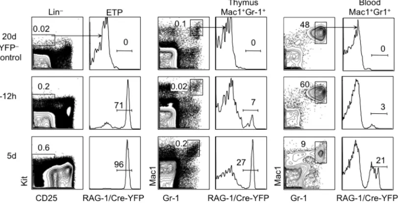
2.3.1 Thymic granulocytes have a developmental history of RAG-‐1 expression ... 32
2.3.2 ETPs can generate granulocytes in vivo ... 35
2.3.3 Thymic granulocyte development is impaired in the absence of thymic settling by T cell progenitors ... 36
2.3.4 Thymic ETP and granulocyte development depend on IL-‐7Rα and the common γ-‐chain ... 38
2.4 DISCUSSION ... 40
2.5 FIGURES ... 44
CHAPTER 3: NOTCH-MEDIATED INHIBITION OF MYELOID DEVELOPMENT REQUIRES HES1 ... 57
3.1 ABSTRACT ... 57
3.2 INTRODUCTION ... 59
3.3 RESULTS ... 62
3.3.1 Hes1 is essential for T cell lymphopoiesis ... 62
3.3.2 Notch constrains myeloid potential through Hes1 in multipotent progenitors ... 64
3.3.3 Lymphoid and multipotent progenitors respond differently to the absence of Hes1 ... 67
3.3.4 Hes1-‐deficient lymphoid progenitors generate expanded myeloid progeny in the presence of myeloid cytokines ... 70
3.3.5 Characterization of Hes1/TCF-‐1 doubly-‐deficient progenitors ... 72
3.4 DISCUSSION ... 74
3.5 FIGURES ... 80
CHAPTER 4: THE ESSENTIAL FUNCTION OF HES1 IS TO CONSTRAIN C/EBPα -DEPENDENT MYELOID PROGRAMS ... 102
4.1 ABSTRACT ... 102
x
4.3 RESULTS ... 108
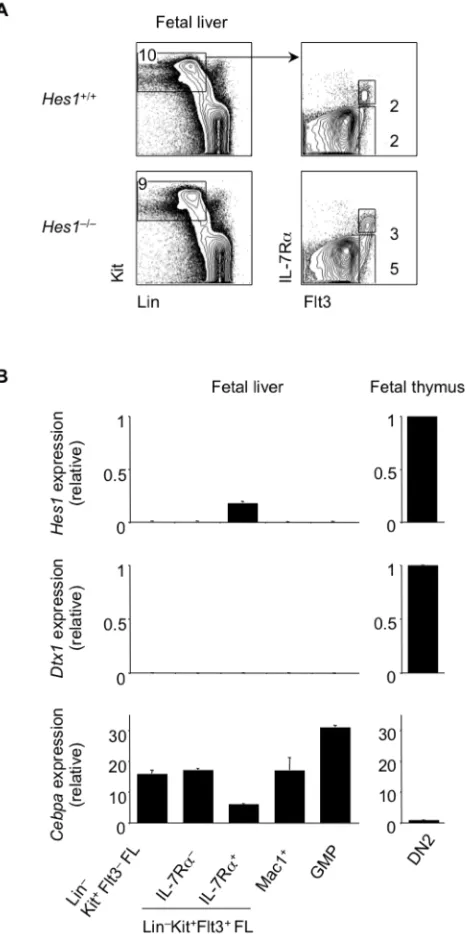
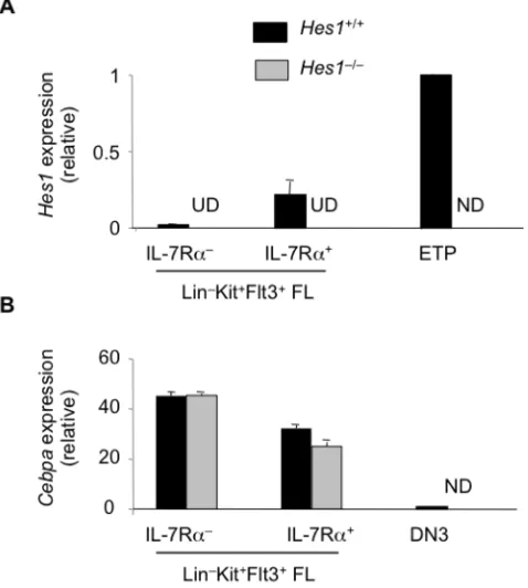
4.3.1 Hes1 represses Cebpa expression ... 108
4.3.2 Deletion of C/EBPαrestores T cell lymphopoiesis in the absence of Hes1 in vitro and in vivo ... 110
4.3.3 Deletion of C/EBPαin Hes1-‐deficient FL restores T cell lymphopoiesis from both multipotent and lymphoid progenitors ... 113
4.4 DISCUSSION ... 114
4.5 FIGURES ... 117
CHAPTER 5: DISCUSSION/FUTURE DIRECTIONS ... 129
5.1 SUMMARY OF FINDINGS ... 129
5.2 KEY IMPLICATIONS OF THIS WORK ... 130
5.2.1 Many physiological TSPs likely express IL-‐7R at functional levels and retain the ability to access myeloid gene expression programs ... 131
5.2.2 Thymic granulocytes are generated predominantly from Hes1-‐dependent ETP/DN2 thymocytes, unlike many other alternative lineage cells in the thymus. ... 133
5.2.3 Hes1-‐dependent ETPs (those which generate thymic granulocytes in vivo) are responsible for most T cell development. ... 135
5.2.4 Distinct mechanisms inhibit myeloid programs in periphery (Hes1-‐independent) versus thymus (Hes1-‐dependent). ... 136
5.2.5 Lineage-‐specific mechanisms to inhibit myeloid potential likely exist, since ILC2s, which use many elements of the core T cell program (TCF-‐1, GATA-‐3) develop independently of Hes1. ... 137
5.2.6 Multiple mechanisms exist to constrain different lineage potentials in uncommitted, intrathymic T cell progenitors. Protracted access to myeloid gene expression programs may reflect an association between these programs and stem/progenitor programs that drive robust T cell progenitor expansion. ... 138
5.2.7 Failure to inhibit myeloid programs in the absence of Hes1 causes defective in vivo T cell development from physiological TSPs. Cebpa deletion rescues Hes1 T cell developmental defect from both lymphoid progenitors and multipotent progenitors in vitro. ... 142
5.3 FUTURE DIRECTIONS ... 142
5.3.1 Further elucidation of Hes1 mechanism of repression ... 142
5.3.2 How is myeloid silencing maintained in committed T-‐lineage cells? ... 143
5.3.3 Clarifying the role of Hes1 in T-‐ALL ... 145
CHAPTER 6: METHODS ... 146
6.1 Mice ... 146
6.2 Flow cytometry and cell sorting ... 147
6.3 Intravenous transfers and intrathymic transfers ... 148
6.4 Stromal cell co-‐culture ... 149
6.5 Annexin Staining ... 150
6.6 Retroviral transduction ... 150
6.7 Hes1 mutant constructs ... 150
6.8 Chromatin Immunoprecipitation (ChIP) Assay ... 151
6.9 Luciferase assay ... 152
6.10 Wright-‐Giemsa staining ... 153
6.11 Quantitative PCR (qPCR) ... 153
6.12 Statistical analysis ... 153
xi
LIST OF FIGURES
Figure 1-1 Overview of hematopoiesis ... 10
Figure 1-2 Overview of T lymphopoiesis ... 11
Figure 1-3 Notch signaling overview ... 12
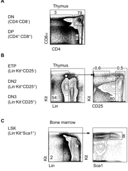
Figure 1-4 Gating of thymus and BM progenitors ... 13
Figure 2-1 Thymus Mac1+Gr-1+ cells are confirmed to be C/EBPα-dependent myeloid cells ... 26
Figure 2-2 Adult thymic granulocytes have a history of RAG-1 expression, ... 27
Figure 2-3 Neonatal thymic granulocytes mostly lack a history of RAG-1 expression, in contrast to ETPs ... 28
Figure 2-4 ETPs generate thymic granulocytes with a history of RAG-1 expression ... 29
Figure 2-5 Myeloid potential of ETPs is short-lived ... 30
Figure 2-6 Some BM progenitors will generate myeloid cells in thymus ... 31
Figure 2-7 Schematic of bone marrow chimera generation ... 32
Figure 2-8 Abrogation of thymic settling diminishes thymic granulocytes ... 33
Figure 2-9 Gating strategy: granulocytes, macrophages, and B cells in the thymus, spleen, or BM ... 34
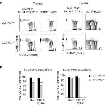
Figure 2-10 Thymic macrophages and B cells develop independently of thymus settling by T cell progenitors ... 35
Figure 2-11 Thymic granulocytes are IL-7Rα-dependent ... 36
Figure 2-12 Thymic granulocytes are CD132 (γc)-dependent ... 37
Figure 2-13 Model: ETPs generate most adult thymic granulocytes ... 38
Figure 3-1 Hes1 expression is upregulated in the thymus ... 58
Figure 3-2 Hes1 is required for T cell development in vivo ... 59
Figure 3-3 Notch fails to inhibit myelopoiesis from FL MPPs in the absence of Hes1 .... 60
Figure 3-4 Hes1-deficient MPPs show decreased T cell and increased myeloid progenitor frequency ... 61
Figure 3-5 Notch fails to downregulate myeloid genes in MPPs lacking Hes1 ... 62
Figure 3-6 Following intrathymic injection, Hes1-deficient MPPs generate expanded myeloid progeny in vivo ... 63
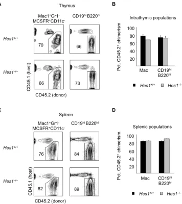
Figure 3-7 Thymus granulocytes and DCs are not expanded in Hes1-deficient FL chimeras ... 64
Figure 3-8 Thymus macrophages and B cells are not expanded in Hes1-deficient FL chimeras ... 65
Figure 3-9 CCR9+ FL MPPs are heterogeneous for IL-7Rα expression ... 66
Figure 3-10 Characterization of FL lymphoid progenitors ... 67
Figure 3-11 Hes1-deficient FL lymphoid progenitors effectively downregulate Cebpa expression ... 68
Figure 3-12 Hes1-deficient FL progenitors generate B cells and myeloid cells normally 69 Figure 3-13 Hes1-deficient MPPs (but not lymphoid progenitors) generate expanded myeloid progeny in T-inductive conditions ... 70
Figure 3-14 Notch continues to inhibit B cell potential in the absence of Hes1 and TCF-1 ... 71
Figure 3-15 Hes1 is not necessary for lung ILC2 development ... 72
xii
Figure 3-17 Hes1-deficient lymphoid progenitors generate expanded myeloid progeny
when OP9-Delta cultures are supplemented with myeloid cytokines ... 74
Figure 3-18 Hes1-deficient MPPs fail to downregulate Cebpa expression in the presence of Notch ... 75
Figure 3-19 Hes1/TCF-1 deficient lymphoid progenitors generate expanded myeloid progeny in T-inductive conditions ... 76
Figure 3-20 Early T cell development is abrogated in Hes1/TCF-1 double-deficient FL chimeras ... 77
Figure 3-21 Thymic myeloid cells are not expanded in Hes1/TCF-1 double-deficient FL chimeras ... 78
Figure 3-22 Hes1 defect precedes TCF-1 defect in intrathymic T cell development ... 79
Figure 4-1 Hes1 inhibits myeloid Cebpa mRNA and protein expression ... 93
Figure 4-2 Potential modes of Hes1-mediated gene repression ... 94
Figure 4-3 Repression of Cebpa requires DNA binding and Groucho interaction domains of Hes1 ... 95
Figure 4-4 Hes1 binds mouse Cebpa promoter in DN3 thymocytes ... 96
Figure 4-5 Hes1 inhibits mouse Cebpa promoter activity ... 97
Figure 4-6 C/EBPα is necessary for myeloid development ... 98
Figure 4-7 Cebpa deletion restores T lineage competence to Hes1-deficient progenitors in vitro ... 99
Figure 4-8 Cebpa deletion restores T cell progenitor frequency in Hes1-deficient progenitors ... 100
Figure 4-9 Cebpa deletion restores in vivo T cell development from Hes1-deficient progenitors ... 101
Figure 4-10 Cebpa deletion restores all stages of in vivo T cell development from Hes1-deficient progenitors ... 102
Figure 4-11 Cebpa deletion restores T cell development from both lymphoid and multipotent progenitors ... 103
Figure 4-12 Model: Hes1 constrains myeloid gene expression programs during early T cell development ... 104
1
CHAPTER 1: INTRODUCTION
1.1 Establishment of cell identity
Cellular differentiation is loosely defined as the process by which a less
specialized cell type becomes a more specialized cell type. As a cell differentiates, it takes
on new properties, and may change its size, shape, metabolic requirements, and
responsiveness to external stimuli. These phenotypic changes are driven by changes in
gene expression. Generally, cellular differentiation is thought to proceed in one
direction, since gene regulatory mechanisms exist that simultaneously allow new gene
expression and disallow past gene expression. As stem cells differentiate, they begin to
express genes characteristic of a particular terminally differentiated cell type and
progressively lose the ability to access gene expression programs associated with
alternative cell fates. These processes are referred to as lineage specification and
commitment, respectively. Lineage specification entails the positive regulation of genes
associated with the fate being adopted. It is clear that activation of lineage-specific gene
expression is not itself sufficient to explain acquisition of a particular cellular identity.
In order to “choose” one identity, multipotent progenitors, must also “renounce” the
ability to develop into all other alternative cell types by the process of lineage
commitment.
A remarkable exception to the unidirectionality of differentiation is the
phenomenon of “reprogramming” by which introduction of exogenous transcription
2
adopt another fate, possibly via an induced pluripotent stem cell (iPS cell) or
dedifferentiated progenitor intermediate state. The 2012 Nobel Prize in Medicine was
awarded to Shinya Yamanaka and Sir John Gurdon for their work on reprogramming.
Gurdon first showed in 1962 that somatic cells transferred into enucleated frog eggs
could be reprogrammed to an embryonic state1. Yamanaka’s group found that four
transcription factors (Oct4, Sox2, Klf4, and c-Myc) could recapitulate this
reprogramming process to generate iPS cells2. The identification of these “Yamanaka”
factors provided support to the idea that just a handful of transcriptional regulators
could exert powerful, deterministic effects on cell fate. This belief is at the heart of the
concept of “master regulators” of cell identity, which has become widespread in recent
years. So-called “master regulators” are transcription factors that are necessary and
sufficient for the acquisition of lineage identity. Thus, “master regulators” often have the
ability to both activate lineage-specific gene expression and to repress expression of
other factors promoting an alternative lineage. The identification and characterization of
master regulators has traditionally been achieved using gene-specific knockout strategies
and ectopic expression assays.
Genome-wide studies of gene expression, transcription factor binding, and
chromatin modifications are leading to a more complete picture of how lineage-specific
gene expression is achieved. Metazoan gene regulation is thought to depend upon the
interaction of promoters with enhancers, gene regulatory elements that may be located
at a distance from the promoters they regulate. It is thought that enhancers are
especially critical for the activation of lineage-specific gene expression3. Next-generation
sequencing has revealed that many enhancer elements are marked with a permissive
3
trimethylation (H3K4me3)4–6. Enhancer elements may also be identified by evolutionary
conservation and an abundance of DNAse hypersensitivity sites7.
Enhancers may display either a poised or transcriptionally competent
configuration after receiving appropriate signals. Poised enhancers are marked by
repressive (H3K27me3) and activating (H3K4me3) chromatin modifications78. Poised
enhancers are transcriptionally inactive, but can be rapidly activated when
differentiation signals are received. The activity of specific enhancers can be determined
by examining combination of the H3K4me1 mark with histone acetyltransferase (HAT)
p300 binding or deposition of H3K27ac acetylation marks9–13. Moreover, other factors,
such as the HATs CBP or SAGAs may regulate activity of enhancer landscapes
independently of p30014,15. Since H3K4me1 modifications are more broadly distributed
than p300 binding, it is possible that their deposition precedes that of p30016.
“Pioneering factors” may deposit H3K4me1 marks on poised chromatin to begin to setup
an enhancer, where downstream regulators can then bind16,17.
Like gene activation, the process of gene repression relies upon the use of
cis-regulatory elements and sequence-specific DNA binding factors; however, less is known
about the mechanisms used to achieve target gene repression compared to activation18.
For example, it is not known how many different types of factors are needed to
accomplish transcriptional repression; whereas, the transcriptional activation machinery
is relatively well characterized. Repressive regulatory elements can be identified by loss
of histone acetylation, reduced H3k4 methylation, and reduced DNase accessibility due
to compaction of the nucleosomal array19–21. Because of compaction, repressed elements
4
complicate systematic analysis of repression18. The H3K27me3 chromatin modification
is found to mark some repressed regions, following deposition by the Polycomb
repressive complex 2 (PRC2); however, it appears that this mark only is placed after
inhibition of transcription has already occurred22. This temporal issue makes it difficult
to identify the factors that initially caused repression. It is possible that additional
chromatin modifications which mark repressed regions remain to be identified. Whereas
eukaryotic transcriptional activators generally interacting with the core transcriptional
machinery or through recruitment of chromatin modifying co-activators, it appears that
repressors largely rely on indirect interactions with co-repressor proteins, and rarely
modify activity of the core transcriptional machinery23. Since lineage commitment is
thought to rely heavily on the repression of alternative lineage gene expression, a better
understanding of repressive mechanisms is needed in order to elucidate lineage
5 1.2 Hematopoiesis overview
Hematopoiesis is an excellent model system for the study of cell fate decisions,
since in many cases progenitor intermediates have been defined by cell surface markers,
allowing for isolation by fluorescence activated cell sorting (FACS) and the use of
single-cell assays. Models of hematopoietic differentiation may reflect either developmental
potential (“what cells can do”) or physiological cell fate (“what cells actually do”,
presumably in vivo). In any case, the complement of developmental potentials possessed
by a population is presumably a broader category that must encompass the physiological
cell fates realized by that progenitor cell type. Identification of progenitor-progeny
relationships in vivo has relied on adoptive transfer of progenitors into irradiated
animals. Because the cytokine milieu may be altered in the irradiated host, it is necessary
to validate adoptive transfer studies using genetically based fate-mapping approaches in
the steady state.
Single cell analyses of developmental potential are often more informative than
bulk assays, due to heterogeneity within prospectively isolated populations based on
currently available cell surface markers. The development of single cells can be more
easily studied and quantified in vitro than in vivo, due to technical limitations. While in
vitro developmental assays are commonly thought to be more permissive than in vivo
assays, in that they may allow for detection of potentials that are not necessarily realized
in vivo; however, the opposite may also be true, since in vitro assays are optimized to
support development of particular cell types. Thus, particular assays may underrepresent
6
detect the full complement of developmental potentials possessed by single cells of a
particular population.
All cellular components of the blood derive from hematopoietic stem cells (HSCs)
through a complex process of differentiation. Hematopoiesis proceeds in a hierarchical
fashion, with primitive HSCs at the apex of the hierarchy (Figure 1-1). Functionally, HSCs
were identified as single cells able to rescue the entire hematopoietic compartment of a
lethally irradiated mouse24–27. HSCs reside in the fetal liver (FL) during gestation and in
the bone marrow (BM) of the adult, and have the unique properties of being pluripotent
and capable of self-renewal. HSCs are enriched within the BM Lin–Sca1+Kit+ (LSK)
compartment, since they express high levels of stem cell antigen 1 (Sca1) and the growth
receptor tyrosine kinase Kit. HSCs may also be purified using SLAM markers CD150 and
CD48 as CD150+CD48- cells28–31. HSCs give rise to downstream progenitors, such as
multipotent progenitors (MPPs), which have lost self-renewal capacity and upregulated
expression of FMS-related tyrosine kinase 3 (Flt3). As progenitors undergo
differentiation, the complement of developmental potentials they possess becomes
“restricted” in a stepwise fashion, until progenitors are generated that are “committed”
to generate only one terminally differentiated cell lineage. Lineage commitment is
induced by both cell intrinsic mechanisms, such as transcription factors and regulatory
RNAs, as well as extrinsic mechanisms like cytokines, cell-cell interactions, and other
environmental cues. Since the loss of a given developmental potential may theoretically
occur in several ways, it follows that cells may commit to a fate via distinct pathways.
Evidence for such flexibility exists; however, "major" pathways can usually be identified.
7
Historically, mature blood cells have been broadly classified into several major
groups: megakaryocyte/erythroid, myeloid, and lymphoid lineages. Lymphocytes were
initially distinguished from myeloid cells based on their relative lack of cytoplasm and
their enrichment in lymph, lymph nodes, spleen, and thymus. Myeloid cells were
identified as phagocytic cells with abundant cytoplasm that were enriched in the BM. In
most cases, hematopoietic cells develop in the FL of embryos and the BM of the adult,
with the exception of T cells, which complete their maturation in a specialized organ, the
thymus. The dichotomy between myeloid and lymphoid lineages gave rise to what is
commonly known as the “classical model” of hematopoiesis in which there is an early,
obligate bifurcation between myeloid and lymphoid lineages. The identification of
common lymphoid progenitors (CLP)32, and later common myeloid progenitors
(CMP)33,34, in the BM by Irv Weissman’s group represented a major conceptual advance
in the field of hematopoiesis that provided experimental support for the classical model.
This discovery of lineage-restricted progenitors downstream of HSCs indicated that
developmental potentials are lost in a stepwise fashion.
While the classical model has been instrumental for developing our
understanding of hematopoiesis, its central tenet, the idea that lymphoid and myeloid
lineages strictly separate upstream of the CLP and CMP, has been challenged. Instead, a
“revised model” of hematopoiesis suggests that there are multiple alternative
developmental pathways by which early hematopoietic progenitors undergo
lymphoid-myeloid branching35. Sten Eirik Jacobsen’s group identified lymphoid-primed
multipotent progenitors (LMPPs) as Flt3hi MPPs, cells which had lost
megakaryocyte/erythroid (MegE) potential, but retained myeloid and lymphoid
8
with or without MegE potential, CMPs or LMPPs, respectively. LMPPs are thought to be
the upstream precursors of CLPs, cells which maintain Flt3 expression, downregulate
Sca1 and Kit (expressed at high levels on HSCs and LMPPs), and upregulate the
lymphoid cytokine receptor IL-7Rα. The process by which multipotent progenitors,
such as LMPPs differentiate to form lymphoid progenitors is not well understood, and is
discussed in more detail below.
1.3 Generation of lymphoid progenitors
How some descendants of HSCs “decide” to become lymphoid progenitors is not
well understood, but may involve signals from the BM niche, cytokines, and
transcription factors such as E2A, Ikaros, Bcl11a, PU.1, STAT5a/b, HOXA9, Satb1, and
others37–44. Some HSCs may be lymphoid-biased (reviewed in 45). Lymphoid priming in
HSCs may be important for downstream lymphoid development46.One critical aspect of
generating CLPs is the attenuation of myeloid potential. It is likely that E-proteins play a
role in initiating this process in the bone marrow37,47,48. As lymphoid progenitors
subsequently commit to a specific lymphoid lineage, there are likely to be lineage-specific
mechanisms to repress and eventually silence myeloid fates. In this thesis, we investigate
one such lineage-specific mechanism, showing that Hes1 constrains myeloid gene
expression programs during T cell development in the thymus. Interestingly, Hes1
appears not to be necessary to attenuate myeloid potential during the generation of BM
9
E2A is important for the initiation of lymphoid gene expression and the
generation of BM LMPPs37. Interestingly, E2A protein is not increased in LMPPs
compared to HSCs37. It may be that the decreased expression of E protein dimerization
partners (such as TAL1) in LMPPs results in increased availability of E proteins to
function in homodimeric complexes that activate lymphoid gene expression49. While
E2A is not required for extinction of MegE potential, it does appear to impact upon
myeloid versus lineage decisions, since residual LMPPs remaining in E2A-deficient mice
demonstrate enhanced myeloid cloning efficiency and deregulation of many lymphoid
genes: Rag1, Il7ra, Dntt, Igh-6, Notch1, CCR9, and Ets137. E2A may promote lymphoid
gene expression by collaborating with other transcription factors which are expressed in
LSKs and MPPs, such as PU.1, Ikaros, and Gfi-1, and which do not depend on E2A for
their expression49. DNA sequences flanking the transcriptional start sites of
E2A-dependent genes were enriched for Ikaros and PU.1 binding sites, providing evidence for
cooperation of these factors with E2A37. The cytokine receptor Flt3 is thought to be
upregulated by Ikaros and PU.150–52. Among the E2A-regulated genes noted above are
several cell-surface receptors: IL-7Rα, Notch1, and CCR9, which confer cells with the
ability to respond to microenvironmental cues. Signaling downstream of IL7-Rα or
Notch1 signaling can lead to upregulation of transcription factors important for B cell or
T cell generation, respectively.
While CLPs are strongly biased to generate lymphocyte progeny, they are not
absolutely restricted to the lymphoid lineage and display a degree of myeloid/DC
potential under some conditions53,54. The originally proposed CLP population has been
shown to be heterogeneous, including B cell specified progenitors, which express Ly6D.
10
CLPs appear to have T cell potential in vitro; however, it appears that they do not
contribute to T cell development in vivo, perhaps because they lack the ability to home to
the thymus56. Thus, Ly6D– CLPs and LMPP may contribute to T cell development in the
thymus, though their relative contributions are unclear. Whether or not CLPs are
obligate intermediates in the T cell pathway, it is clear that they contribute extensively to
the development of B cells, NK cells, innate lymphoid cells (ILCs) outside the thymus57–
60.
1.4 T lymphopoiesis overview
T cells complete their maturation in the thymus from circulating BM-derived
progenitors (Figure 1-2).Thymus settling is a selective process. Determining the relative
extent to which CLPs (lymphoid progenitors) and LMPPs (multipotent or
myelo-lymphoid progenitors) contribute to T cell development has been a confusing area of
investigation, despite intensive study. Both CLPs and LMPPs, but not upstream HSCs,
are able to rapidly generate T cell progeny following intravenous transfer into irradiated
recipients; however, only Sca1hiKithi cells resembling HSCs/LMPPs appear to be found in
the blood61,62. Furthermore, ETPs have more robust T cell reconstitution capacity than
CLPs, suggesting that ETPs derive from cells more developmentally upstream of CLPs,
such as LMPPs63. Additionally, several findings suggest that T cell development can
occur independently of CLPs, since Ikaros-deficient mice lack CLPs, but not ETPs63.
Indeed, the ETP cell surface phenotype resembles that of LMPPs in that both
populations express high levels of Kit, and most cells in each population appear to lack
surface IL-7Rα expression. Thus, the phenotypes of ETPs and LMPPs differ in several
11
unambiguously IL-7Rα+(ref. 63). Hence, studies probing the identity of thymus settling
progenitors (TSPs) have raised questions about the validity of the classical model of
hematopoiesis by challenging the idea that there is an early, obligate segregation of
myeloid and lymphoid potentials in the BM64–67. Instead, myelo-lymphoid progenitors
such as LMPPs (cells more developmentally primitive than CLPs) may be the primary
progenitors of T cells65.
As thymus settling progenitors (TSP) enter the thymus, strong Notch signals
initiate the very earliest stages of T cell development: proliferation and generation of
early thymic progenitors (ETPs)20. ETPs are CD4/CD8 double negative (DN) precursors,
a subset of the DN1 population, which represent the most primitive progenitors of T cells
that can be identified in the thymus63,69. Thymic epithelial cells are specialized to
support T cell development, because they express high levels of Notch ligands and the
cytokines Kit ligand and IL-7, which are important for maintenance and proliferation of
early T cell progenitors. Notch signaling is critical for the generation of T cells, since
inactivation of Notch1 signaling in bone marrow stem/progenitor cells results in
abrogation of T cell development70–73. The critical ligand pair for T cell development was
identified to be Delta-like ligand 4 (DLL4) (refs. 74,75). Progenitors enter the thymus with
Notch1 receptor on their surface, which was induced by E-proteins in the BM. The
expression of Fringe proteins in thymocytes primes them to respond to intrathymic
Notch ligands76. Notch receptors span the cell membrane, and when the extracellular
portion of the receptor is bound by ligand, this induces proteolytic cleavage and releases
the intracellular domain of Notch (ICN), which translocates to the nucleus (Figure 1-3).
RBP-12
Jk/CSL protein, mastermind-like (MAML), and co-activators, such as p300 in order to
initiate transcription of Notch target genes77.
Notch signaling launches the T cell program in DN progenitors by upregulating
the expression of T cell-specific transcription factors, including TCF-1, GATA-3, and
Bcl11b78. Even once these factors are turned on, continuous Notch signals are required to
maintain expression of important T-lineage genes79–81. Notch signaling terminates after
the DN3 stage, indicating that other mechanisms are needed to lock in the T cell fate and
maintain the core T cell program in mature T cells. In addition to activating
transcription factors that initiate T-lineage specification, Notch signaling is thought to be
critical for the inhibition of alternative fates by upregulating transcriptional repressors,
such as Hes1 and Bcl-11b. Antagonizing alternative fates is critical, because in addition to
robust T cell potential, ETPs retain the potential to develop into non-T cell lymphoid
cells (B cells17,20 and ILCs33/NK cells48), DCs47,50, and, to a degree, myeloid cells53,64,66,82,83.
The mechanisms by which alternative lineage potentials are constrained are discussed
further below.
As ETPs progress along the T cell developmental pathway, they proceed through
a series of intermediate stages that are defined based on the expression of cell surface
markers described below (also see Figure 1-4). ETPs proliferate and differentiate to form
DN2a cells, which maintain the high levels of Kit surface expression characteristic of
ETPs and which upregulate CD25 (IL-2Rα), which is a Notch target. DN2a cells initiate
TCRβ locus rearrangement and express many lineage genes, yet they are not yet
T-lineage committed. Activation of an Lck-GFP transgene marks the onset of T-T-lineage
13
of T-lineage commitment is thought to be complete by the DN2b stage, which is
distinguished from the DN2a stage based on decreased levels of Kit expression. While
DN2a and DN2b cells express CD44 and Kit, these markers are downregulated as cells
transition to the DN3 stage. DN3 cells have undergone TCRβ gene rearrangement and if
this is successful (in-frame), they express the pre-TCR complex (TCRβ and the invariant
preTCRα chain) on their surface85. DN3a cells that receive pre-TCR signals survive, thus
passing the β-selection checkpoint, and upregulate CD28 to become DN3b cells86. As
cells undergo T-lineage specification and β-selection, they shut down the expression of a
suite of “legacy” stem/progenitor cell genes (such as Meis1, Mef2c, Lmo2, Bcl11a, Erg,
Hoxa9, and others) that promoted extensive proliferation in upstream progenitors;
failure to do so may antagonize T cell development or contribute to malignant
transformation87. DN3b cells lose CD25 expression to become DN4 cells, which then
proceed through a CD4–CD8+TCRβ– immature single positive stage (iSP) and then give
rise to CD4+CD8+ double positive (DP) cells. DP thymocytes will express mature TCRα/β
on their surface and undergo positive and negative selection on MHC-peptide complexes
presented by thymic epithelial cells. DP thymocytes commit to either the CD4 or CD8
14 1.5 T cell lineage commitment
Since the identification of ETPs69, much work has characterized the
developmental potentials possessed by this population. In addition to robust T cell
developmental potential, ETPs have been shown to possess B cell, DC, NK/ILC potential,
and a degree of myeloid potential53,64–66,82,83,88–90. Less is known about whether ETPs
actually adopt alternative (non-T cell) fates in vivo. Nevertheless, these data have
contributed to an ongoing debate about the models of hematopoiesis described above,
because at least some thymus settling progenitors (TSPs) are not lymphoid restricted
and retain myeloid potential. Below, I discuss mechanisms that constrain alternative
fates in T cell progenitors on the path to T lineage commitment.
Inhibition of B cell potential
Progenitors rapidly lose B cell potential upon entry into the thymus, presumably
in response to intrathymic Notch signals, since Notch is a potent inhibitor of B cell
development67. Hence, B potential is only detected in a small fraction of the most
primitive (Flt3+) ETPs found in the adult thymus67. B cell potential is diminished in the
subsequent Flt3– ETP stage. As is discussed below, these progenitors that have largely
extinguished B cell potential retain a degree of myeloid potential. The close
developmental relationship between T cells and B cells, which both rely on
RAG-mediated antigen receptor rearrangement for their development, perhaps makes it
puzzling that T cell precursors would lose B cell potential before losing myeloid
15
development within the thymus, since the thymus microenvironment contains cytokines
supportive of both B and T cells in rich supply.
Notch deficiency results in an abundance of thymic B cells. This may partially
reflect entry of B cells and/or B cell progenitors into the “empty” thymus, in addition to
diversion of T cell progenitors to the B cell fate67,68,73,88. B cell fate inhibition is probably
mediated by cooperation between multiple Notch effectors such as Hes1, TCF-1, and
GATA-3 (refs. 91,92). The role of thymic B cells is unclear; however, they localize to the
thymic medulla and have been suggested to function in negative selection of developing
thymocytes93.
Inhibition of myeloid potential
In contrast to B cell potential, the ability to access myeloid fates is maintained
until just before the onset of T lineage commitment at the DN2b stage. Thus, myeloid
potential is clearly retained longer than B cell potential in the adult thymus. Protracted
maintenance of myeloid potential may reflect continued expression stem cell “legacy”
genes during early intrathymic development, which endows early T lineage progenitors
with tremendous proliferative capacity. Single cell assays using a stromal cell culture
system have shown that the majority of individual ETP scan give rise to both T cells and
myeloid cells, including granulocytes and macrophages64,65. The question remained:
even if ETPs can adopt myeloid fates in in vitro assays, do they do so in vivo?
Furthermore, if ETPs can access myeloid gene expression programs and adopt myeloid
fates in the thymus, then this suggests the need for a mechanism to attenuate myeloid
16
myeloid development, and likely constrains myeloid potential at very early stages of
intrathymic T cell development; however, the mechanisms by which this occurs remain
unclear49,94. One such mechanism, may be the repressor Bcl11b, since its deficiency
prevents progenitors from shutting down access to myeloid fates, perhaps via repression
of PU.1 and/or C/EBPα95,96. However, Bcl11b is not expressed in ETPs, and thus cannot explain
constraint of myeloid gene expression in the earliest intrathymic progenitors of T cells.
This thesis addresses the issue of whether ETPs adopt myeloid fates in
the thymus (Chapter 2) and investigates the mechanisms used to attenuate
myeloid gene expression programs during early T cell development
(Chapter 3). We show that the Notch target and transcriptional repressor Hes1 is
critical for constraining C/EBPα-dependent myeloid gene expression programs in T cell
progenitors in the thymus97. In addition, ETPs access some myeloid fates in the thymus
in vivo. Specifically, thymic granulocytes appear to derive almost exclusively from ETPs
in vivo83. Accordingly, thymic granulocytes, unlike peripheral conventional granulocytes
are labeled by a history of RAG-1 expression and rely on similar cytokine/chemokine
receptor and transcription factors as early T cell progenitors for their development
(IL-7Rα, γc, CCR7/9, and Hes1)83. T cell progenitors may also contribute to macrophages
and cDCs in the thymus; however, there are likely to be other efficient sources for these
lineages83.
The biological significance of myeloid potential in the ETP population is unclear.
Myeloid lineage cells in the thymus are important for the phagocytosis of negatively
selected immature T cells; however, these cells would not necessarily have to derive from
17
myeloid progenitor cells or mature myeloid cells. Having T cell progenitors support
development of myeloid lineages within the thymus may be an economical solution that
allows the presence of non-T cells within the thymus without requiring that
thymus-homing molecules be expressed on committed precursors of these other cell types.
Another idea is that the myeloid potential of ETPs may be the by-product of a stem cell
“legacy” transcriptional program, which confers early T-lineage progenitors with
tremendous proliferative capacity87.
Differences in the myeloid potential of ETPs between neonates and young adult
mice support the idea that thymus settling progenitors (TSPs) vary across ontogeny, such
that adult TSPs retain a greater degree of myeloid potential98. It is worth noting that that
fetal and adult intrathymic progenitors differ significantly in their proliferative capacity.
In adults, ETPs can take 10 days to reach the DN2 stage (compared to little over a day for
fetal progenitors), and two weeks to generate DP thymocytes (compared to four days for
fetal mice)99,100. During adult T cell development, the extended time course of T cell
development corresponds to more extensive proliferation of progenitors. Thus, the
myeloid potential of adult ETPs may extend the time course of T cell development by
antagonizing the onset of T cell specification and commitment. Enhanced proliferative
capacity of adult T cell progenitors can supply increased demand for T cells in the adult
18 Inhibition of NK cell/ILC potentials
ETPs/DN1 thymocytes have also been shown to retain in vitro NK cell potential,
which is present in a reduced frequency of DN2 thymocytes100,101. In addition to NK cell
potential, some early thymocyte progenitors have now been shown to retain other innate
lymphoid cell (ILC) potentials (ILC2 and ILC3). ETP and DN2 thymocytes display ILC2
potential when cultured with OP9-DLL1 stromal cells in the presence of IL-7 and IL-3360.
A subset of DN2 fetal thymocytes (α4β7+RORγt-) generate LTi cells after culture with
OP9 stromal cells; however, culture with Notch ligands (OP9Delta stromal cells)
abrogates LTi development from these cells102. These α4β7+RORγt- DN2 cells may
generate LTi-like cells (α4β7+RORγt+) in thymic niches that lack Notch ligands75,102,103.
Notch signals appear to inhibit NK cell generation in vitro101. Downstream of
Notch, the repressor Bcl11b likely constrains access to NK cell fates, as is discussed in
greater detail below95,96,104. Bcl11b expression is sharply upregulated in DN2 thymocytes
by Notch signaling, likely via direct control as well as by the Notch effectors TCF-1 and
GATA-3, as well as Runx1105. ChIP analysis has shown that Bcl11b is a canonical Notch
target gene104. Bcl11b continues to be expressed during subsequent stages of intrathymic
T cell development, even after Notch signaling terminates in DP and SP thymocytes.
Germline Bcl11b-deficiency results in a block in T cell development at the DN2-DN3
stage, reduced thymic cellularity and predisposition to lymphomagenesis106.
Interestingly, Bcl11b heterozygous mice show a similar phenotype105. The defect is not
19
by p53 deletion107. Bcl11b is thought to act mainly as a repressor during T cell
development, as there is little evidence for its positive regulation of T cell lineage genes87.
Bcl11b-deficient DN2 thymocytes have been shown to undergo T lineage
specification indicated by expression of Tcf7, Gata3, CD3g, Ptcra, and Rag1; however,
Bcl11b-deficient DN2s inappropropriately express genes which are normally
downregulated by the DN2 stage (Tal1, Bcl11a, Sfpi1, Erg, Flt3) and genes associated
with innate and “innate-like” lymphocytes (Id2, Il2rb, and Nfil3)96. It remains to be
determined whether these genes are direct targets of Bcl11b. The mechanism by which
Bcl11b restricts NK lineage gene expression is unclear; however, one idea is that Bcl11b
may do so indirectly by promoting E-protein function87. E-proteins are known to inhibit
NK cell programs; however, the downstream mechanisms are unknown108.
Furthermore, cells resembling NK cells, called “induced-T-to-NK” (ITNK) cells,
are generated from Bcl11b-deficient DN1, DN2, or DN3 thymocytes in OP9 or OP9-DL1
cultures instead of T cells 104. Intriguingly, Bcl11b is not only necessary for T cell
development, but is also required in committed and mature T cells to restrict access to
NK cell programs. Acute deletion of Bcl11b in mature T cells results in the generation of
T cells expressing many genes associated with NK cells (NKG2A/C/E, perforin, TRAIL,
and IFNg, but not Ly49 family receptors) in addition to retaining TCR surface
expression 104. Roles for Bcl11b have been found for DP thymocyte survival and positive
selection 109. Thus, Bcl11b plays a critical role in enforcing T cell lineage commitment.
Thymic NK cells are intact in the context of Notch1 deficiency, although ETP
20
derive independently of T cell progenitors, it does not exclude the possibility that T cell
progenitors unable to progress down the T cell pathway instead adopted NK cell fates.
Alternatively, thymic NK cells may derive from ETPs in vivo since they require GATA-3
and IL-7R signaling for their development111. Thus, thymic NK cells have distinct
developmental requirements from peripheral cNK cells. While the function of thymic NK
cells is unknown, proposed roles include immunosurveillance of thymocytes, preventing
transformation, or regulation of thymopoiesis and selection110,112,113. LTi-like cells in the
21
1.6 Transcription factor networks in hematopoiesis
In the hematopoietic system, B cell and erythroid development have been
extensively studied, and offer a useful point of comparison to understand the
outstanding questions in the T cell development field. During B cell development, E2A
initiates a feed-forward regulatory circuit that imposes and maintains B cell identity, by
upregulating factors such as FOXO1, EBF1, and Pax5. The B lineage-specific
transcription factors EBF and Pax5 appear to mediate both lineage specification and
commitment. In the absence of either EBF or Pax5, the fidelity of B cell lineage identity
is compromised. Pax5-deficient progenitors express B-lineage genes, but are unable to
commit to the B cell fate115,116. Pax5 potently inhibits T cell development by repressing
Notch1 expression. EBF has been shown to repress other T-lineage regulators, including
Gata3 and Tcf7, as well as the myeloid regulators Cebpa and Sfpi1, and the E-protein
antagonist Id2, implicated in ILC development117–119. Hence, EBF deletion in proB cells
causes upregulation of Id2 and diversion to ILC fates119. Furthermore, EBF and Pax5
each repress critical T cell/ILC regulators: Notch, GATA-3, and Tcf7118–120. Thus, EBF
and Pax5 coordinately play a dominant role in B cell lineage commitment by
antagonizing key regulators of multiple alternative fates. During erythropoiesis, GATA-1
plays a similar central role in specifying and maintaining the erythroid fate, through
22
In contrast to erythropoiesis or B cell development, a considerably wider “cast of
characters” (rather than a single or few factors) appears to be necessary to perform the
positive and negative regulatory functions of T lineage commitment. A single “master
regulator” which dominates the execution of positive and negative regulatory events in T
cell development has eluded identification. T cell development transcription factor
networks are not dominated by feed-forward loops, as is the case for B cell development.
Instead, there is a complex division of labor, and considerable redundancy between
multiple regulatory factors. T cell commitment relies not only on several key
lineage-specific transcription factors, among these: TCF-1, GATA-3, Bcl11b, but also a host of
other factors expressed across hematopoietic lineages including E2A, Runx1/CBFβ, Gfi1,
Myb, and PU.1. The mechanisms resulting in T lineage specification, particularly the
dramatic upregulation of T cell lineage genes between the DN2 and DN3 stages of
thymocyte development, remain unclear. One possibility is that T lineage specification is
mediated by combinatorial action of multiple cooperating positive regulators. Another
fascinating idea is that T lineage specification is indirectly mediated by negative
regulation of stem/progenitor genes expressed by early thymocyte progenitors. This is
because some stem/progenitor genes expressed during early stages of intrathymic T cell
development, such as SCL and Lyl1 have been shown to inhibit the activation of
T-lineage positive regulators by E-proteins87.
In this thesis, we focus on understanding the negative regulatory mechanisms
that constrain gene expression associated with alternative fates, particularly the myeloid
fate, during early T cell development. The asynchronous loss of non-T cell potentials
during early T cell development suggests that multiple factors are needed to shut down
23
alternative fate inhibition; however, the mechanisms downstream of Notch responsible
for T lineage commitment have remained unclear. We examined one effector of Notch
signaling, Hes1 as a possible mechanism that constrains access to myeloid gene
expression programs in early T lineage progenitors.
The Notch signaling pathway is highly conserved in all metazoans and plays a key
role in the development of many mammalian organ systems. The upregulation of Hes
factors by Notch1, in cooperation with the E-protein E47 is evolutionarily conserved in
Drosophila sensory organ development and in the notochord of ascidians124–126. Hes1 is a
mammalian homologue of Drosophila hairy and Enhancer of split127. Hes1 is a basic
helix-loop-helix (bHLH) transcriptional repressor and an evolutionarily conserved target
of Notch signaling128. In this work, we show that Hes1 is critical for constraining myeloid
transcriptional programs in T cell progenitors. TCF-1 and GATA-3 likely cooperate to
inhibit B cell development extremely rapidly following thymic entry. The repressor
Bcl11b has been shown to be critical for silencing NK and myeloid programs just prior to
commitment. Therefore, multiple distinct mechanisms downstream of Notch cooperate
to mediate suppression of alternative fates. Consistent with this idea, analysis of
sequential T cell progenitor populations by ChIP and deep sequencing has revealed that
diverse histone modifications mark different genes encoding hematopoietic regulatory
molecules across development129. It is interesting to consider why and how a system
using distinct mechanisms to constrain different alternative lineage cell fates evolved in
the thymus, as opposed to a single mechanism that represses all but the T cell fate. The
existence of distinct modules downstream of Notch signaling suggests the possibility that
these modules are also used elsewhere in the hematopoietic system. Indeed, Hes1 is
24
outcomes. Indeed, during mast cell development, Notch2 signaling upregulates the
expression of Gata3 (which encodes the transcription factor and T cell regulator
GATA-3) and Hes1, which is thought to repress the gene encoding C/EBPα (Cebpa) and prevent
diversion of mast cell progenitor cells to other myeloid fates130. Hes1 also appears to
antagonize C/EBPα, a critical regulator of the development of adipogenesis, in
preadipocyte progenitors131.
The findings presented in this thesis identify the Notch target and transcriptional
repressor Hes1 as an important mechanism that constrains myeloid gene expression
programs in T cell progenitors. Hes1 deficiency in hematopoietic progenitors severely
compromises T cell development; however, this defect can be completely rescued by
deletion of the myeloid regulator C/EBPα. Thus, our findings indicate that ETPs are
bona fide myelo-lymphoid progenitors and establishes the critical importance of
25 1.7 Figures
Figure 1-1 Overview of hematopoiesis
Hematopoietic stem cells (HSCs) are pluripotent, self-renewing cells which give rise to downstream progenitors such as multipotent progenitors (MPP) and lymphoid primed multipotent progenitors (LMPP), which have upregulated expression of the cytokine receptor and have lost self-renewal capacity. LMPP are the 25% of MPP with the highest Flt3 expression.
The classical model of hematopoiesis posits that downstream of the LMPP, there is an obligate bifurcation between myeloid and lymphoid lineages that results in the generation of common myeloid progenitors (CMP) and common lymphoid progenitors (CLP). In the classical model, CMP are the precursors of megakaryocytes, erythrocytes and all myeloid cell types.