General Article
Communication mechanism between living cells
Rakesh Kumar Pandey
Synod Higher Secondary School, Mission Vengthlang, Aizawl, 796 005, India
Received 7 July 2014 | Revised 23 July 2014 | Accepted 30 August 2014
A
BSTRACTThis article discusses the biology and physics of electrical system of living body that controls all body functions. Electrochemical potentials in the nerve cells are the source of billions of electrical signals, which propagate from all parts of body to the brain via neurotransmitters in the living body whose measurements allow obtaining useful clinical information. The propagation of electri-cal pulses in the living body is like transmission of pulses in a cable. The living cell membrane offers resistance and capacitance that form a complex net of electrical circuit. The ratio of space constant between cells to the time constant of neural resistance and capacitance define the speed of nerve impulses from one cell to other cell.
Key words: Neuron; nerve impulse; cell membrane; membrane potential; resistance.
Corresponding author: Pandey Phone: +91-9863361282
E-mail: [email protected]
I
NTRODUCTIONWe experience and react accordingly when pricked by a thorn, any finger is cut or hit, an insect bites or moves on any part of the body and even on hair, any part of the body suffers any problem, bombardment of any kind of en-ergy of sound, light, heat on the body, different tastes, danger, etc. We are not the only beings that feel but also do animals, birds, insects, etc. i.e. almost all living organisms experience any type of the disturbances that occur in their sur-rounding environment and react. Different parts
of the organism sense the disturbances to react and then communicate the information to the brain to analyze them to take remedial meas-ures. If there is no proper coordination between brain and specialized sensory receptors for the communication, serious problems occur.
Neurons, the basic building blocks of the nervous system, are the specialized cells, which possess the unique property of converting vari-ous forms of stimuli into electrical impulses for the communication of information in living or-ganisms viz. human, animal, bird, insect, etc. They receive information from sense organs or from other adjacent neurons, carry them to the central nervous system CNS (brain and spinal cord), and bring motor information from the Science Vision 14(3), 133-142
central nervous system to the motor organs (muscles and glands). Best scientific estimate is 86,000,000,000 neurons and each neuron is con-nected with nearly 105 other neurons, which
gives nearly 1016 connections in the human
nerv-ous system. These neurons are spread all over the body of an organism like a net and provide different communication channels in the form of nerves fibers that have their two inherent proper-ties of excitability to response to stimuli and the conduction of excitation in the form of nerve impulses.1 They are of many types and vary
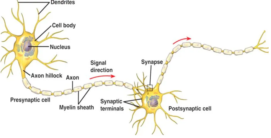
con-siderably in shape, size, chemical composition, and function. Despite these differences, neurons consist of the three main parts viz. a) soma or cell body, b) an axon, and c) dendrites. Figure 1 shows the structure of a neuron. The soma or cell body is the main body of the nerve cell that contains the nucleus of the cell as well as other structures common to living cells of all types. The genetic material of the neuron is stored in-side the nucleus and it becomes actively engaged during cell reproduction and protein synthesis. The soma also contains most of the cell fluid i.e. cytoplasm of the neuron. Dendrites are the branchlike specialized structures emanating
from the soma. They are the receiving ends of a neuron. Their function is to receive the incom-ing neural impulses from adjacent neurons or directly from the sense organs. On dendrites, there are the specialized receptors, which be-come active when a signal arrives in electro-chemical or bioelectro-chemical form. The received sig-nals are passed on to soma and then to axon so that the information is relayed to another neu-ron or to muscles. The axon conducts the infor-mation along its length, which can be several feet in the spinal cord and less than a millimeter in the brain. At the terminal point, the axon branches into small structures known as the ter-minal buttons. These buttons have the capability for transmitting information to another neuron, gland and muscles.
Neurons generally conduct information in one direction, that is, from the dendrites through soma and axon to the terminal buttons. The conduction of information from one place to another in the nervous system is done through projections of the nerves, which are bundles of axons. The longest axon in human is of diame-ter 10-20 µm and length ~1 m that extends from the brain to low in spinal cord or from spinal
cord to fingers, etc. Neuron cells are mainly of two types: sensory and motor. Sensory nerves, also called afferent nerves, carry information from sense organs to central nervous system. On the other hand, motor nerves, also called effer-ent nerves, carry information from ceffer-entral nerv-ous system to muscles or glands. A motor nerve conducts neural commands, which direct, con-trol, and regulates our movements and other responses. There are some mixed nerves also, but sensory and motor fibers in these nerves are separate. Dendrites carry information towards the cell body, whereas axons carry information away from it. Thus, in a sense, neurons are one-way channels of communication. Information usually moves from dendrites or the cell body towards the axon and then outward along the structure of neuron. Fatty material, called mye-lin sheath, produced by glial cells within the nervous system covers each axon. Glial cells surround, support, and protect neurons. Each axon ends with axon terminals that contain neu-rotransmitters. A region of gap where the axon of one neuron approaches closely other neuron or the cell membrane of other types of cells such as muscle cells is usually termed as synapse. The axon tips of a preceding neuron do make direct connections but form synaptic cleft.
There are two types of communications i.e. communication within neurons and between neurons that take place in all organisms.
C
OMMUNICATION WITHIN NEURONSInformation travels within the nervous sys-tem in the form of a nerve impulse. When stimulus energy exposes the receptors, electrical changes in the nerve potential occurs. Nerve potential is a sudden change in the electrical po-tential of the surface of a neuron. When the stimulus energy is relatively weak, the electrical changes are so small that the nerve impulse is not generated, and we do not feel that stimulus. If the stimulus energy is relatively strong, electri-cal impulses are generated and conducted to-wards the central nervous system.2 The strength
of the nerve impulse, however, does not depend
on the strength of the stimulus that caused the impulse. The nerve fibers work according to the all or none principle, which means that they ei-ther respond completely or do not respond at all. The strength of the nerve impulse remains con-stant along the nerve fiber.
Electrical potential of a neuronal membrane is due to chemical ions. Several types of ions e.g. Na+ and K+ exist in their different
concentra-tions. A relaxed neuronal cell membrane, so called resting membrane, shows higher perme-ability for small Na+ ions and poor permeability
for large K+ ions on one hand. While it behaves
like a sodium pump on the other hand that ac-tively pumps Na+ ions out from cytoplasm to
the ECF and transfers less K+ ions in exchange
from the ECF to the cytoplasm. These happen due to different diffusion rates depending on the concentration. There are other factors such as H+ pumps, uniporters and cotransportes
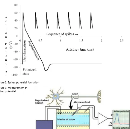
result-ing in a higher cationic concentration outside than inside the cell membrane and thus, inner side of cell membrane becomes relatively elec-tronegative to its outer side. As a result, cyto-plasm of each cell acquires slight large negative charge inside relative to its outside extra cellular fluid that provides negative potential known as the resting membrane potential RMP of ap-proximately -70mV across the cell membrane of a neuron at rest (Fig. 2). The state of resting membrane at RMP is the polarized state or readiness state of the cell membrane. The RMP undergoes a change under the effective stimulus termed as the action potential.3 The Figure 3
shows a systematic diagram for the measure-ment of action potential.4
brain receives these neural impulses via nerves and thus, an organism collects various informa-tion signals from its different specialized sensory receptors.
Another basic category of signals within neu-rons, called graded potentials, results from the external stimulus of the dendrite or cell body. Unlike the all-or-nothing nature of the action potential, the magnitude of a graded potential varies in proportion to the size of the stimulus
that produced it. Thus, a loud sound produces a graded potential of greater magnitude than a softer sound. Because graded potentials weaken quickly, they function primarily to convey in coming information over distances, usually
along the dendrite towards the neuron’s cell
body.
Though the nervous system expends a great deal of energy to maintain this state of readiness yet leaks a little under the minimum strength of
Figure 2. Spikes potential formation Figure 3. Measurement of
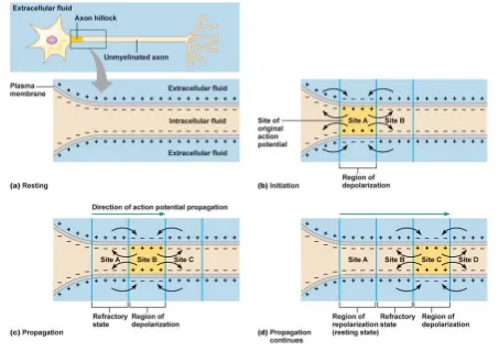
Figure 4. Formation of spikes and their propagation through neurons.
stimulus known as the threshold stimulus allow-ing some charged particles to slip in and other to slip out as these cell membranes are not a perfect barriers.5 When an effective external stimulus or
another neuron stimulates a neuron, positively charged particles enter the membrane through specialized ions channels, thereby shortly elimi-nating the negative charge just inside the neu-rons membrane that results the fluctuation in the electrical charges from negative to positive called depolarized state (Fig. 4b) and back again along the cell membrane through repolarization (Fig. 4d). The nerve fiber does not respond at all to the stimulus less than the threshold stimulus and responds a maximum action potential irre-spective of the strength of stimulus greater than the threshold stimulus. This is all-or-nothing principle of the action potential.
After a short period of about 1-2 ms, posi-tively charged particles acposi-tively pumped back
out side of the neuron’s membrane via the ions
channels. Because of this active process, the in-ner membrane of a neuron regains its negative charge to maintain potential of -70 mV relative to the outside and thus the cell is again ready to fire once more. Actually, the membrane poten-tial first rises sharply from about -55 mV to 50 mv under effective stimulus and almost immedi-ately declines sharply to 20 mV (Fig. 2). These changes produce a spikes-like record known as the spike potential. The spike first follows a slower decline in the potential (negative after potential) and then by a potential more negative than the resting potential (positive after poten-tial). The potential then gradually returns to the resting potential. These change in the electrical
disturbance in spikes’ along cell membrane,
serve as the basis for the communication in nervous system, travel along the nerve fibers in the form of neural impulses. The speed of neural impulses varies from 30 to 150 ms-1 (in human
0.6-120 ms-1) that depends not only on
myelina-tion but also on the fiber’s diameter and the ions
conduction. The impulses travel slower in thin-ner fibers than in thicker one.
Vertebrates like squids possessing unmyeli-nated fibers, possess very thick fibers for
con-ducting impulses rapidly to their distant long arms. In unmyelinated fibers, these ionic change repeat over the membrane all along the length of the fibers. Therefore, the action potential propa-gates all along the membrane over the entire length of the fiber. However, in myelinated fi-ber, the action potential jumps from one free space between the consecutive myelin sheaths so called as node of Ranvier to the next due to ionic conduction of nerve impulses (Fig. 5). This is the reason, nerve impulses conduct far more rapidly in myelinated fibers than in unmyeli-nated ones.6
C
APACITANCE OFCELL MEMBRANEThe conduction of neural impulses in living cells depends mainly on the conductivity of the membrane of the axon because the neurons op-erate mainly on the potential difference caused by ionic concentration difference between inte-rior and exteinte-rior sides of membrane. Let Vi and
Vo are the interior and exterior potentials of
membrane, then, in accordance with the Nernst equation, the ionic concentration ratio in equi-librium is
where z is the valence of ions, e = 1.6x10⁻¹⁹ C is
electronic charge, k = 1.38x10⁻²³ JK-1 is the
Boltz-mann constant, T is the absolute temperature, Ci
and Co are the interior and exterior
concentra-tions, respectively.
The thickness of cell membrane uncovered with myelin is x ~ 5-6 nm and covered with myelin it is ~ 2 µm. The strength of electric field E in the cell membrane under potential
differ-ence ∆ V= Vi– Vo along the interior of the axon
due to depolarization is i o/ ze V V kT i
o
C e C
13.7;
0.073; i
o
i
o
C
For ve ions
C C
For ve ions
C
The accumulation of electric charge over the surface S of the cell membrane is
where K ≈ 7 is the dielectric constant and Cm is
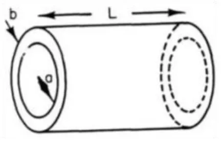
the neural capacitance of the cell membrane. The extracellular and intercellular fluids are the two conducting fluids surrounding the insulator membrane make the axon membrane as cylin-drical capacitor as shown in Figure 6.
The neural capacitance of cell membrane of thickness b is
Therefore, neural capacitance per unit sur-face area of the cell membrane is
The surface charge density of the cell mem-brane is now given as
For myelinated fiber, the thickness of mem-brane is much larger which reduces the neural
capacitance per unit area by a factor of nearly 300.
L
EAKAGE OF ELECTRICCHARGE ACROSSCELL MEMBRANE
The cell membrane is not a perfect insulator and hence the electric charge will leak across the membrane as it offers a resistance equal to
is the electrical resistivity of the cell membrane material.
Neural capacitance and resistance are in par-allel to each other as in Figure 7.
Therefore, the leakage of current through the neural resistance discharges the neural capaci-tance. Now, we have
Integration operation on both sides results to
where time constant 1
72
12 6
V mV
E MVm
x nm
0
m
QC V
KSE0 0 0
m
KSE KSE KS
Q C
V V Eb b
2 0 0.01
m
C K
Fm
S b
2 0 700
m Q
KE Cm
S
Figure 6.A cylindrical neural capacitance of thickness b, radius a and length L.
m m
b r
S
m
m m dC V
dQ V
i dt dt r
Figure 7. Each unit of parallel couple of membrane capacitance and resistance are in series with interior resistance.
m m
dV dt
V r C
/ 0
t
V
V e
3
0 10
m m m
r C K s
The resistance and the capacitance of the portion of axon membrane in terms of the axon radius a and length L using S = 2πaL is
Clearly, the resistance of the axon decreases with increases in length while its capacitance increases with increasing length.
R
ESISTANCE ALONG THE AXONThe interior fluid resists the current flowing through it. The resistivity ρi of fluid, the radius a
and the length L of the axon determine the resis-tance as below:
The axoplasma resistivity is ρi ≈ 0.5 Ωm.
The longitudinal internal resistance per unit length is
P
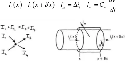
ULSE PROPAGATION THROUGH AXONMEMBRANE
When a section of membrane is excited by electrical current or external current or external stimulus, the membrane permeability changes, ion exchange takes place across the membrane causing a rapid change in potential according to Nernst equation. This changes the polarity be-cause positively charged ions enter the interior of the axon. The time constant determines the polarization time scale. Because of the potential difference along the outer surface of the axon, electrical currents are induced in the internal conductive fluids. Therefore, the depolarization moves along the axon and application of
Kirchhoff’s law for current conditions in the
axon describe the process of movement. Current across the membrane
Current along the inside of the axon
In accordance with the Kirchhoff’s current
law KCL, the total out going currents is equal to total incoming currents (Fig. 8) i.e.
Dividing both sides by elemental surface area dS = 2πadx results to
which describes the propagation of a pulse along a conductive material.
The change of potential with time is a func-tion of the leakage current through the surface of the membrane and the change of voltage along the axis. The neural capacitance Cm and
resis-tance rm, and internal resistance ri determine the
speed of the neural impulses. As larger the ca-pacitance, as longer the time constant it takes the cell membrane to discharge, therefore, a slower propagation speed of the neural im-pulses. Similarly, as larger the internal resis-tance, as smaller the axial current ii is. However,
the resistance is in inverse to the axon radius i.e. as larger the radius, as faster the propagation velocity of the neural impulses.
2 m m b r aL
0 2 m KaL C b
2 i iL
R
a
2 i i lR
r
L
a
m m m dV dQ i C dt dt i i l V dV iR r dx
i i m i m m
dV
i x
i x
x
i
i
i
C
dt
Figure 8. Kirchhoff’s current law is algebraic sum of all
current at a junction is zero.
2 2 1 2 m m l
C dV i d V
S dt S
ar dxThe cable equation can be reformulated as
with , , ,
and
Multiplying with the time constant yields to
Introducing the space constant
which is the node of Ranvier and using the time constantτ results to the final formulation for the motion of the signal along the axon with time as
For an unmyelinated axon of a = 2.5 µm and b = 6 nm, and a myelinated axon of a = 2.5 µm and b = 2 µm, the space constants are λum = 0.49
mm and λum =8.9 mm, respectively along with
time constant τ = 1 ms.
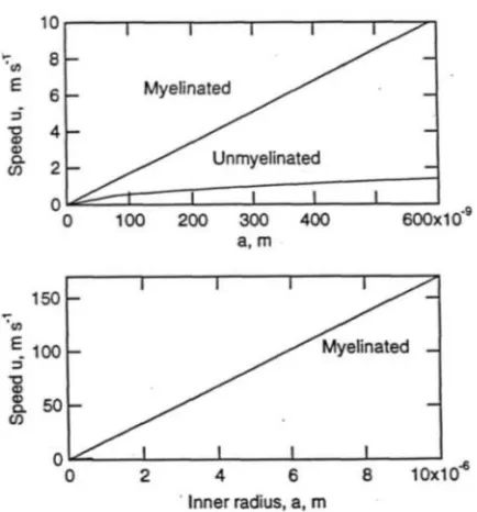
The speed of impulse v in an axon fiber can be determined as the ratio of space constant to the time constant i.e.
The signal speed in unmyelinated axon fiber with b = 6 nm is . Clearly, the neural impulse speed increases linearly with square root of the radius (Fig. 9). There are se-vere limitations as the radius of unmyelinated human fiber axons are typically smaller than 1µm.
For a myelinated axon fiber, there is a fixed relation between the radius a of the axon and the thickness b of the myelin layer i.e. b = ≈ 0.4a. This simplifies the expression for the space constant λ. Thus,
This yields for the speed of the impulse sig-nals in a myelinated axon as:
Thus, living cells communicate with finite speed to the brain via electrical systems of nerves.
C
ONCLUSIONComplex nets of nerves are like an electrical system in the living body through which living cells communicate the information to the brain via neural impulses. The transmission of electri-2
2
1
2
mm l m
i
dV
S
d V
dt
C
a r C
dx
0 m C K S b
2 i l r a
m m V i r 70
peak restV
V
V
mV
2 2 0
2 i
dV V ba d V
dt
K dx 0 m K 2 2 2 2 0 2 2 m i i
dV ba d V ba d V
V V
dt K dx dx
2 m i ba
2 2 2d V dV
V dt dx
0 12 i m ba v K
1 313 v amsFigure 9.Speed versus radius graph for myelinated and unmyelinated neural fibers. The impulse speed increases linearly with axon radius a.
7
1.6 10
0.2
0.2
2530
0.5
mi
a
a
a
cal pulses into neuronal sensors and motor nerve system create electric potential difference whose measurements are the clinical tool to understand the human body functions e.g. electrocardio-gram ECG for heart function, electroencephalo-gram EEG for brain function, electromyoelectroencephalo-gram EMG for muscles activity, etc.
R
EFERENCES1. Silverthorn, DU (2010). Human Physiology: An Integrated
Approach. Pearson, p. 253.
2. Deutsch S & Micheli-Tzanakou E (1987). Neuroelectric
Systems. New York University Press.
3. Barnett MW & Larkman PM (2007). The action potential.
Practical Neurol, 7, 192–197.
4. Brock LG, Coombs JS & Eccles JC (1952). The recording of potentials from motoneurones with an intracellular
electrode. J Physiology (London), 117, 431–460.
5. Leys SP, Mackie GO & Meech RW (1999). Impulse
con-duction in a sponge. J Expl Biol, 202, 1139–1150.
6. Poliak S & Peles E (2006). The local differentiation of
myelinated axons at nodes of Ranvier. Nat Rev Neurosci,