ABSTRACT
Molecular diagnostics is a promising scientific sector on the rise, enhanced to a great extent by technological developments. Massive mutation screening methods and direct automated DNA sequencing have enabled researchers and diagnostics laboratories to identify frequent and rare genetic defects, thus contributing to medical translational research. Despite all the progress, the complexity of gene systems, even of monogenic disorders, makes molecular testing a formidable task, espe-cially for relatively small molecular medicine laboratories. Nephrogenetics is one such field where genetic and consequent phenotypic heterogeneity are particularly complex, with tens of genes involved and interacting, often times in unpredictable ways, to cause disease. It turns out that useful approaches of the older generation will continue to be useful for many years to come, while supported and facilitated by more recent and advanced high throughput technologies.
INTRODUCTION
The results of market research by «Frost and Sullivan», a company specializing in market research of various sec-tors, including health sector, show that the genetic testing market is on the rise with a rate of 11% per year, while pharmacogenetics diagnostics is expected to achieve a rise by 26-36% until 2011. At the same time, Kenneth I. Berns, MD, PhD (Editor-in-Chief of Genetic Testing, and Director of the University of Florida’s Genetics Institute, College of Medicine, Gainesville, FL), has stated:
“As we learn more about the underlying causes of dis-ease and link this knowledge to the emerging realization that in the not too distant future good healthcare will include information about one’s genomic profile, the importance of genetic testing in clinical medicine will continue to grow”.
For this continuous and endless development several factors have had significant contribution, most decisive of which has been the improvement or upgrading of biotechnology/bioinformatics and the equipment or hard-ware tools that enabled easier detailed negotiation and analysis of the genetic material. To this end, the reading of the entire sequence of the human genome has been an
unprecedented success with still unimaginable impact.1, 2
However, even well before the 2000 feat, but nevertheless to a greater extend afterwards, the various genes, muta-tions in which are responsible for hundreds of monogenic (single gene) inherited diseases, have been identified and cloned and subsequently were characterized in detail one by one. This procedure has become a routine to a number of laboratories around the world and nearly every week
Recent Advances in Nephrogenetics and Molecular
Diagnostics: Are Current Approaches
Becoming Obsolete?
Constantinos Deltas, PharmR, PhD Professor of Genetics and Chair, Department of Biological Sciences Head, Laboratory of Molecular and Medical Genetics, University of Cyprus
Keywords: Molecular diagnostics, nephrogenetics, genetic
there are publications in various relevant journals that report on new genes or new functions, in association with heritable conditions. The discovery of new genes and specific mutations that are responsible for diseases with a strong genetic component, either monogenic, oligogenic or polygenic, is always the first crucial step for deciphering the molecular pathomechanism and aim for a targeted therapy. It is my personal opinion that the sacred purpose of our work as geneticists, is to make the link from the dramatic picture of the affected individual-the patient as a human macro-entity-to the patient as a molecular biological micro-entity. The next feat is the prevention or correction of the molecular malady.
For the field of our interest, nephrology and nephro-genetics, during the past less than two decades, medical and molecular investigations have shed light and led to the discovery and detailed characterization of very many genes, mutations in which result in X-linked, autosomal dominant or autosomal recessive renal diseases, thereby improving substantially the understanding of their clinical and molecular pathology, the diagnosis or prognosis and naturally the clinical intervention for a targeted design
of better medications.3
Molecular medicine, in its nature as a field of labora-tory non-clinical medicine, is often times applied by non-medical doctors, however nowadays it is imperative that clinicians understand and interpret the results of molecular examinations that are frequently performed in the course of investigating a patient. This is one form of translational medical research and elementary knowledge of the theory and the techniques hidden behind the molecular diagnostics approach certainly is helpful in better interpreting and applying the results. It is obvious therefore, that a synergy has been created and is being cultivated even further daily, between clinical and molecular medicine when the differential diagnosis calls for molecular investigations of the genetic mate-rial, aimed specifically at the following immediate and practical goals:
1. Confirmation of the clinical diagnosis or suspected diagnosis
2. Exclusion of the clinical suspicion 3. Presymptomatic genetic diagnosis
4. Investigation of the inheritance of a genetic predis-position for a certain disease
5. Pharmacogenetic application (many are in place and more are being developed)
6. Prenatal or preimplantation genetic diagnosis 7. Investigation for purely research purposes within the
framework of an approved clinical protocol
For 1-5 above, molecular investigation and production of genetics data should be destined to assist clinical doc-tors to resume in a definite diagnosis and to choose the best possible therapeutic intervention among several. At the same time these data could be useful for the interrup-tion of a non-optimum therapy that had been instituted already, for the reduction of the general morbidity and mortality or the risk for the specific patient. It could help to reduce the likely adverse reactions and avoid the suf-fering owing to uncertainty, to avoid the unsuccessful or inadequate drug trials and interventions, and so on and so forth. And all these should be aimed specifically and always to the benefit of the patient.
As regards the biological material used for molecular diagnostics, most frequently, by far, it requires DNA that can be obtained from various sources such as whole blood, saliva, tissue biopsy or cells from tissue culture that has been established in the framework of the investigation of the patient. In the overwhelming majority of cases the source of DNA is whole blood, from which it is isolated easily after selective lysis of the red blood cells and removal of the proteins, that is followed by the lysis of the peripheral blood mononu-clear cells (PBMC) that contain the DNA, and isolate
it in clean form by a popular salting out procedure,4 or
into complementary DNA (cDNA) with the use of a viral reverse transcriptase.
CLINICAL AND GENETIC
HETEROGENEITY
The genetic investigation of an individual for any reason, can be a relatively simple procedure of collecting peripheral blood, isolating DNA and examining a specific gene, known before hand. It is even simpler when the individual has a known family history, for example of Au-tosomal Dominant Polycystic Kidney Disease (ADPKD,
a nephropathy with severe symptoms usually after the 4th
or 5th decade) and he/she belongs to a family that had
been previously investigated fully and a mutation had been identified, for example in the PKD1 gene. Under these circumstances things are rather clear and easy and a definite diagnostic result can be produced within a few hours, a negative result relieving the existing anxi-ety and sparing the patient from frequent doctor visits, while in affirmative will direct the doctor to the correct decisions for close follow up and perhaps intervention, either through the administration of proper medication, if available, or otherwise.
Unfortunately, however, in many cases things are more complex and complicated and it is required to have a close collaboration and exchange of information between the nephrologist and the geneticist, who needs to be an expert in his field if he is to be of help to the clinician. As in every other scientific field, the good geneticist will be evaluated and “earn his money” while handling the difficult cases that require multiple approaches and perhaps even further advice of the existing literature. The performance of a molecular test and the reporting of a negative test is an action which shows to the doctor and the person under investigation and his family, that something has been done, and a result has been issued. This report however, may give an erroneous impression especially if it is not accompanied by adequate interpretation of the result or if the clinician is unable to comprehend the result and he does not seek further detailed explanation. For example, if a patient with a suspicion for Cystinuria without a fam-ily history (autosomal recessive disease), is investigated in great detail by DNA sequencing of the SLC3A1 gene and no mutation is detected, the diagnosis for Cystinu-ria is still not excluded. This is so because CystinuCystinu-ria is genetically heterogeneous, caused by mutations in either of two genes, SLC3A1 or SLC7A9, resulting in similar phenotypes, even though the cystine concentration in the urine of heterozygous individuals can produce suspicion
in favour of one or the other gene.5 To make things more
complex, digenic inheritance, although rare, cannot be
excluded (see also below). Also, usually the screening for mutations is focused within the coding exonic regions and the flanking sequences that contain the splicing signals, however occasionally there are mutations in the promoter sequences or elsewhere within the introns, perhaps lead-ing to activation of cryptic splice sites.
Similar scenarios apply for numerous other gene systems and heritable conditions, including genes that are impli-cated in several inherited renal diseases. A big challenge is the heterogeneous group of childhood syndromes of Focal Segmental Glomerulosclerosis (FSGS) that result in steroid resistant nephrotic syndromes. The number of genes involved is already large and it is more than certain that more will be identified. It is hoped that the specific clinical or histological characteristics or yet other biomarkesrs of each one, will direct the geneticist in his investigations, a situation nevertheless that emphasizes again the necessity for close collaboration between the genetics laboratory and the clinic (Table 1).
In cases where the clinical presentation is the result of di-genic inheritance there is indeed an interesting phenomenon that attracts added attention. This has been documented to
be the case in Bardet Beadle syndrome,6 and more recently
in patients with steroid-resistant nephrotic syndrome where heterozygous mutations in different genes account for the
phenotype. More specifically, Löwik et al,7 in a cohort of
19 non-familial childhood-onset steroid-resistant FSGS patients, reported that two patients showed mutations in the CD2AP gene, one combined with an NPHS2 mutation. Another patient carried three mutations, as the patient was compound heterozygous for NPHS2 (podocin) mutations and heterozygous for a NPHS1 (nephrin) mutation. Yet another patient carried a de novo WT-1 (Wilm’s tumor 1) mutation that was combined with a heterozygous NPHS1 mutation, while two other patients showed three hetero-zygous PLCE1 (phospholipase C epsilon 1) mutations. All above mutated genes are expressed in the podocytes that are crucial cells for the maintenance of the glomerular filtration barrier and especially the slit diaphragm between intergititat-ing podocyte processes. These findintergititat-ings therefore, emphasise that combined gene defects are capable of causing FSGS and consequently complicating things for the genetics laboratory.
Figure 1: Family CY 5304 (mutation G1334E in COL4A3 ). The mutation in this family from Cyprus segregates with Thin Basement Membrane Nephropathy and microscopic hematuria. It is a good example of great phenotypic heterogeneity , including reduced penetrance, as individual III16, at the age of 73 was healthy , with negative urine findings on repeated testing . P atients IV23 and IV25 at ages 56 and 49-y respectively had isolated microscopic hematuria, patient IV20 at age 41-y had proteinuria, patient III25 died at 83-y with chronic kidney disease, patient III1 reached end-stage kidney disease and died at 77-y , and patient III23 reached ESKD at 68-y . The mutation in this family was present also
in additional families, evidently as a result of a founder effect. Squares
and circles represent males and females respectively , filled symbols denote affected status. A diagonal line denotes deceased subjects. The arrow points to the proband.
All individuals are identified with generation and family numbers below
PKD2 gene cause a milder form of ADPKD compared to mutations in the PKD1 gene, as evidenced by later age of onset of end-stage kidney disease and a smaller overall
percentage of patients reaching ESKD.8, 9 This explains,
to some extent, the significant interfamilial clinical vari-ability observed among the population of patients with ADPKD but certainly cannot explain the variability among patients with type 1 or type 2 ADPKD and even worse, among patients within the same family who share the exact same mutation. Several works attempted to
address these important issues. Magistroni et al,10 showed
that the site of the PKD2 mutation does not play a role, while surprisingly patients with splice site mutations appeared to have milder renal symptoms compared to patients with other types of mutations. The group of Peter Harris at Mayo Clinic, MN, USA, showed that mutations in the 5’-half of the PKD1 gene confer more severe disease
compared to mutations in the 3’-half,11 while Hateboer
et al,12 had shown that different groups of mutations and
the location of the mutation within the PKD2 gene does influence clinical outcome.
In addition, in recent times, the implication of modifier genes that somehow affect the function or the outcome of the primary genes, has become a necessary prerequisite for interpreting the intrafamilial variable expressivity or phenotypic heterogeneity of many diseases. In these cases, it is obvious that the disease inheritance is dependent on a single gene with a defined mode of inheritance, yet one or more genes, perhaps in concert with environmental parameters, have an effect on the severity of symptoms or the age at onset of the disease. In a way, even classical monogenic disorders have an element of multifactorial or
polygenic inheritance. Fain et al13 demonstrated that up
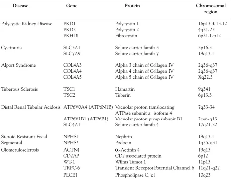
Table 1: Examples of inherited monogenic nephropathies that present genetic and allelic heterogeneity. That is, mutations in one of several genes cause identical or similar phenotype, while in every gene tens of different mutations have been identified that lead to a spectrum of phenotype severity.
Disease Gene Protein Chromosomal
region
Polycystic Kidney Disease PKD1 Polycystin 1 16p13.3-13.12
PKD2 Polycystin 2 4q21-23
PKHD1 Fibrocystin 6p21.1-p12
Cystinuria SLC3A1 Solute carrier family 3 2p16.3
SLC7A9 Solute carrier family 7 19q13.1
Alport Syndrome COL4A3 Alpha 3 chain of Collagen IV 2q36-q37
COL4A4 Alpha 4 chain of Collagen IV 2q36-q37
COL4A5 Alpha 5 chain of Collagen IV Xq22.3
Tuberous Sclerosis TSC1 Hamartin 9q341
TSC2 Tuberin 6p13.3
Distal Renal Tubular Acidosis ATP6V0A4 (ATP6N1B) Vacuolar proton translocating 7q33-34
ATPase subunit a isoform 4
ATP6V1B1 (ATP6B1) Vacuolar proton pump subunit B1 2cen-q13
SLC4A1 Solute carrier family 4 17q21-22
Steroid Resistant Focal NPHS1 Nephrin 19q13.1
Segmental NPHS2 Podocin 1q25-q31
Glomerulosclerosis ACTN4
α
-Actinin 4 19q13CD2AP CD2 associated protein 6p12
WT-1 Wilms Tumor 1 11p13
TRPC-6 Transient Receptor Potential Channel 6 11q21-q22
to 18-59% of the phenotypic expression of PKD1 muta-tions can be attributed to genetic modifiers. A similar
study by Paterson et al,14 showed that for patients before
reaching ESKD the heritability of phenotypic variation was 42% whereas for patients with ESKD it was estimated
to 78%. Also, Persu et al,15 first and Lamnisou et al,16
subsequently, suggested a modifier role for a ENOS (en-dothelial nitric oxide synthase) polymorphism on the age at onset or rate of progression of ADPKD. However other publications had mixed results. Finally, a meta-analysis for the role of the ACE I/D (angiotensin converting enzyme, insertion/deletion) polymorphism failed to confirm any
association with ADPKD phenotype.17 The search for
identifying true modifier genes has added difficulties, similar to the ones encountered when searching for genes implicated in polygenic conditions. In such studies there is a need for large patient cohorts and accurate detailed diagnosis of patients who may develop some of the more severe symptoms at older ages.
Another striking example of the wide spectrum of symptoms caused by mutations in the same gene is the one involving mutations in the COL4A3/COL4A4 genes on chromosome 2q36-37, that encode the alpha 3 and alpha 4 chains of glomerular basement membrane collagen IV molecules. Homozygous mutations in either of these genes cause classical full blown autosomal reces-sive Alport syndrome whereas heterozygous mutations were shown and considered to be responsible for Benign Familial Microscopic Hematuria, most of the times as an isolated symptom, as a consequence of Thin Basement
Membrane Nephropathy (TBMN).18-21 Apart from
oc-casional episodes of macroscopic hematuria and low grade proteinuria, heterozygous patients have been considered to follow a benign course with excellent prognosis. Earlier reports had alluded to patients who had developed more severe glomerulopathy in the presence of TBMN, but no molecular testing had been performed to document the
cause of TBMN.22 A recent work by Voskarides et al,23,
24 (and unpublished results) in my laboratory, described a
large number of Greek-Cypiot families with TBMN where in 59 patients over the age of 51 years, approximately two in three had developed proteinuria and chronic kidney disease, while about one in three had progressed to ESKD, as evidenced by the need for hemodialysis or kidney trans-plantation. Based on selected renal biopsies from patients who had developed proteinuria and renal failure in the studied families, the cause of the severe kidney function impairment was focal and segmental glomerulosclerosis, thereby satisfying a dual diagnosis. This obscure finding cannot be explained by the kind of the mutation as most patients share the same exact heterozygous mutation in the COL4A3 gene, G1334E (substitution of glycine by
glutamate), as a result of a documented strong founder effect (Figure 1). It is more than apparent that a modi-fier gene (or genes) coinherited with the above mutation predisposes a subset of these patients to a more severe clinical outcome. It is reasonable to assume that the putative modifier might be a gene playing a role in the glomerular filtration barrier environment, affecting the podocyte supporting function. Without knowledge about the identity and the exact role of a putative modifier gene that somehow predisposes to more severe phenotype, it is impossible to make predictions of the phenotype of a monogenic disorder, which is primarily caused by a causa-tive mutation in a responsible gene.
UNCERTAINTY OF THE
PATHOLOGICAL ROLE OF CERTAIN
MUTATIONS OR VARIANTS
The dramatic improvement of technology and the reduction of cost of molecular testing have permitted the rather easy and effective analysis of multiple genes, a practice that has led to the description and identification of many DNA sequence changes. Not withstanding this very fact, it is frequently difficult to associate with safety and a fair degree of certainty the gene variation with a specific phenotype. It is often times said or expected by non-experts that so far as the gene for an inherited disease is known then the identification of disease causing muta-tions is readily available, or even better, already known. It is not unusual to run into a confusion between a gene itself and the pathogenic mutations. In the overwhelming majority of cases, a specific monogenic inherited condition is caused by one of many different mutations within the same gene, with nearly every patient or family to carry their own mutation. This extensive allelic heterogeneity is the result of a host of molecular defects, frequently single nucleotide substitutions that result in missense changes or aminoacid residue substitutions, nonsense changes by introducing premature termination codons, splice site defects often resulting in whole exon deletions or inser-tions, or single or few nucleotide deletions or inserinser-tions, often causing severe translation frameshifts. Of course depending on the particular gene, there might be larger defects such as multiexon deletions or duplications, etc. It is interesting and quite challenging that some times single nucleotide substitutions that apparently are neutral without affecting the coded aminoacid, turn out to be very severe as they cause activation of cryptic splice sites. A classical example is a mutation in the LMNA gene (Lamin A) where one such recurrent de novo mutation in exon 11 results in aberrant alternative splicing and causes a rare form of premature aging, known as Hutchinson-Gilford
of such DNA variants as regards their putative role in
alternative splicing has been developed by Tosi et al.26,
27 This functional test involves in vitro cloning of the
se-quence area with the variation, followed by expression in tissue culture and RT-PCR (reverse transcription-PCR) as an ex vivo assay. They applied this approach for evaluating the effect of intronic and synonymous exonic variations on RNA splicing, in genes such as BRCA1, BRCA2 (mutated in inherited breast cancer) and MLH1, MSH2 (mutated
in hereditary non-polyposis colorectal cancer).26, 27
Based on the reported allelic heterogeneity, the finding of a specific mutation in a patient does not necessarily mean that our patient carries also this one mutation. Especially in cases of autosomal dominantly inherited diseases, the rule is that nearly every patient has his own mutation and consequently all his or her affected relatives. While this is the rule there are also exceptions either because of the appearance of the same mutation as a separate event in unrelated patients due to recur-rence by chance, or because of the Founder Effect where apparently unrelated patients share a common ancestry in the distant past. This phenomenon is more prevalent in communities that were relatively isolated for many generations. Prime examples of such diseases where a few founder mutations account for a large percentage of
the patients are β-thalassaemia28, Cystic Fibrosis,27
Finn-ish Type Nephrotic Syndrome29, Cystinuria5 and more
recently Microscopic Hematuria and Thin Basement
Membrane Nephropathy in Cyprus,23,31,32 among many.
The interpretation of the molecular finding itself repre-sents another level of complexity in the every day routine delivery of molecular services. When the molecular test-ing results in the identification of a nucleotide change that causes the substitution of an aminoacid codon by a termination of translation codon, it is easy and straightfor-ward to accept it as the responsible pathogenic mutation. However, when we detect a DNA missense change that alters the nature of the coded aminoacid, for example from arginine to proline, even though biochemically this may be viewed as a serious substitution, it is neverthe-less not enough to hold it responsible for the phenotype and proceed to further actions either for the patient or his relatives, without further experimentation. In cases where there are additional affected and healthy family members and the family is rather large extending to 3-4 generations, it may be possible to examine more subjects and document with confidence that the putative muta-tion segregates with disease, carried by all patients and by none of the healthy relatives. Even in this case however, one cannot exclude that he has been misled by linkage disequilibrium, where the real pathogenic mutation was
only closely linked to the identified variant. A well known variant in the PKD2 gene, mutations in which cause the type 2 autosomal dominant polycystic kidney disease, is indeed at position 28 of the polycystin 2 protein which is occupied by either arginine (60%) or by proline (40%),
R28P.33 Biochemically this is indeed a serious change,
therefore great caution should always be exercised when applying molecular diagnostics. In addition, when novel mutations are identified during patient investigation, and in the absence of a functional test, its absence from a number of healthy individuals of the tested population should be demonstrated (at least 50 subjects).
In a recent study of a large number of patients with autosomal dominant polycystic kidney disease in the laboratory of Peter Harris at Mayo Clinic, the research-ers concluded that without further negotiation of the data with the use of specific algorithms that theoretically evaluate the gene variants based on the change and the neighbourhood, only one third of the DNA changes can
be considered with confidence as causative mutations.34
In this case as well as in others with similar findings, changes that cannot be tested by functional assays are
classified as “Unclassified Variants”.34, 35 Fortunately, in
small countries such as Cyprus, where the population we serve, usually patients are not isolated cases but we have access to additional family members for extending the molecular examinations and investigating in more depth every case in order to document or exclude inheritance from one generation to the next. Exceptions, of course,
are sporadic de novo cases we occasionally are faced with.36
It is useful to mention that in cases where we estimate a degree of predisposition for a certain disease, there is a need for familiarization with the condition in order to avoid being trapped into erroneous conclusions. From
work initially performed in my laboratory,37 as well as in
another similar study by Goforth et al,38 it was shown
be associated and increased risk for micro-albuninuria in the Brazilian population, although a more recent work with larger numbers did not corroborate those data in a
USA adult population.39, 40 Additionally, variant R229Q
was examined further for its association with measures of kidney dysfunction in diabetic individuals or for altering the risk for proteinuria in the general population with
negative results.41
As regards the role of this same variant Tonna et al,42
reported on 56 TBMN patients (on the basis of persistent hematuria), some of whom also had proteinuria of ≥300 mg/l, or ≥ 500 mg/l. Among these, 3 of 5 (60%) who
car-ried the R229Q variant had proteinuria of ≥500 mg/l,
whereas only 7 of 51 (14%) who did not carry R229Q had a similar level of proteinuria. This showed statistical significance, although the numbers are somewhat small, clearly needing confirmation by independent larger studies. This implies that TBMN patients who are also heterozygous carriers of the R229Q variant in podocin are predisposed to a somewhat more severe phenotype. However, in the same cohort, none of nine patients with TBMN and coincidental glomerular or tubulointerstitial disease carried R229Q, thereby not supporting the notion
of a confounding negative role. Also, from work in my laboratory, none of 43 patients over 45 years of age with TBMN and a proven COL4A3/COL4A4 mutation and documented proteinuria or chronic kidney disease carried this podocin variant. It becomes obvious therefore, that the association of a DNA variant with the pathogenic cause of an inherited disease or even the association with an increased predisposition, requires careful ex-perimentation and interpretation of the results, which certainly require correct statistical power. Confirmation by independent studies adds confidence to the results. Finally, in cases of suspicion for a heritable condition, it is of utmost importance and usefulness to construct a detailed family pedigree, after having asked the right questions. Perhaps the most powerful, less expensive, painless and non-invasive and most informative genetic test-investigation is the preparation of a detailed family history along with a family pedigree.
Example of Molecular Investigation (A)
A male patient from Greece with a clinical diagnosis of ADPKD was referred to my laboratory for genetic counselling and a probable molecular investigation due
Figure 2: The proband has sustained a sporadic de novo mutation, W3841S, in the PKD1 gene, probably during his early intrauterine life, shortly after conception. Our inability to detect this mutation in his sperm DNA reduces effectively the risk to bear children with this mutation and polycystic kidney disease.
to his interest for preimplantation genetic diagnosis. In-terestingly there was no family history. Both parents of the affected man had been examined and found to be healthy in their 60’s, thereby allowing the conclusion of a sporadic de novo mutation (Figure 2). Mutations in either of two genes, PKD1 by 85% or PKD2 by 15%, are responsible for ADPKD, and especially the PKD1 is very large with 45 exons and unique features that make it difficult to analyse by direct DNA sequencing in a routine molecular
genet-ics laboratory.43 Genetic linkage analysis as an indirect
method for diagnosis is impossible due to the presence
of a single sample.44 Upon the agreement of the couple
we sent a sample of DNA isolated from peripheral blood leukocytes to Athena Diagnostics in the USA, a private molecular testing laboratory, which performs blindly mas-sive DNA sequencing of both genes. The result was that 21 variations were found in the PKD1 gene, compared to the control sequence. Twenty of them are known
reported polymorphisms whereas the 21st resulted in the
novel substitution of aminoacid serine for tryptophane at position 3841 of the polycystin 1 protein. As mentioned before, even though this substitution is biochemically severe, as a polar small serine residue substituted a bulky non-polar aromatic residue, it is impossible based on that alone to implicate it as the causative defect. The role of the commercial company ended by announcing to us this finding. We tested the two parents of the affected male and none carried this novel change. Consequently, it was a de novo mutation for which the patient might have been somatically and/or germinally mosaic, carrying it only in a subset of his somatic and/or germinal cells. We tested DNA from his sperm and it turned out that our method was not sensitive enough to detect the mutation. We concluded, therefore, that either the mutation was completely absent from his sperm or it was represented in a very small fraction. This can be explained if the de novo mutation occurred in our patient shortly after his conception, during early intrauterine life and contributed substantially to the somatic lines but not as significantly, if
at all, to the germinal line.36, 45-47 The message I am trying
to convey is that detailed and in-depth molecular analysis showed that this patient with ADPKD ran a much lower than 50% risk to bear a child with the same condition, as is expected for an autosomal dominant disease. The risk actually dependent entirely on the percentage of sperm cells that carried the mutant allele.
Example of Molecular Investigation (B)
The proband in Fig. 3 after a renal biopsy, was diag-nosed with Alport Syndrome, a serious glomerulopathy, causing proteinuria and impairment of renal function. He developed ESKD at 46 and was transplanted at 52.
His brother had proteinuria, without hematuria, and FSGS accompanied by reduced glomerular filtration rate, at the age of 54. His two daughters have microscopic hematuria in their 20s, while one daughter also has low grade proteinuria. The information on his parents was not particularly useful.
There is a clinical suspicion for the X-linked Alport Syndrome, however based on the pedigree autosomal recessive Alport cannot be excluded. According to the literature patients with Alport Syndrome develop ESKD at much younger age and certainly before the age of 30, in most cases. It crossed our mind that it could be the result of a heterozygous mutation in one of the COL4A3/ COL4A4 genes that encode for the alpha 3 and alpha 4 chains of type IV collagen, found exclusively in base-ment membranes, including the glomerular basebase-ment membrane. This genetic make up is usually characterised by microscopic hematuria and TBMN with excellent prognosis (Benign Microscopic Hematuria), but occa-sionally it progresses to renal failure of variable degree
as age advances.23
Genetic linkage analysis excluded linkage to the CO-L4A3/COL4A4 genes on chromosome 2q36-37 (Figure 3A) but not to chromosome X, as both affected brothers inherited the same X chromosome from their mother (Fig-ure 3B, common blue haplotype). The rather small size of this family tree and the impressive difference in the disease course of the two brothers allows for disputes regarding the true diagnosis. Since linkage to the X-chromosome has not been excluded the DNA sequencing of the entire COL4A5 gene has been in progress in my laboratory, in the hope that a putative mutation will shed light on the disease history in this family and will subsequently permit proper genetic counselling.
MUTATIONS DETECTION
Figure
3:
The
proband
was
diagnosed
with
Alport
syndrome
after
a
renal
biopsy
. He
developed
end-stage
kidney
disease
at
46-y
and
was
transplanted
at
52-y
. His
course
of
disease
is
unusual
as
normally
patients
with
X
-link
ed
or
autosomal
recessive
Alport
syndrome
reach
end-stage
kidney
disease
much
earlier
, until
the
age
of
30-y
. The
family
structure
cannot
ex
clude
either
of
the
X
-link
ed
or
autosomal
inheritance.
Genetic
link
age
analysis
with
mark
ers
flanking
the
relevant
genes
ex
cluded
link
age
to
COL4A3/COL4A4
at
2q36
(A) but not to the X
-link
ed
COL4A5
gene (B). Sequencing of
COL4A5
is in progress.
All symbols as in F
igure 1.
A
the at-risk individual that he/she has not inherited the responsible mutation that segregates with the disease in the family, thereby relieving much of the anxiety and sparing the individual of unnecessary frequent visits to the doctor. In cases of autosomal recessive conditions the molecular diagnosis is useful for identifying healthy carriers for informing them and guiding for proper family programming in agreement with the spouse. Especially in relation to inherited renal disorders, one additional ap-plication of molecular testing that bears great usefulness, is for identifying healthy potential donors of a kidney transplant. Unfortunately clinical examination alone may not always be adequate to diagnose with absolute confidence that a relative who is also at risk for the dis-ease is definitely healthy. There are paradigms of disdis-eases, such as ADPKD and Medullary Cystic Kidney Disease, with age-dependent penetrance, thereby rendering the clinical approach unable to reach a definite conclusion at younger age. Molecular testing can verify that a potential donor is indeed negative for the mutation segregating in the family, either by indirect DNA linkage analysis or by directly detecting the causative mutation, when known.
Molecular testing technology has improved tremen-dously and this allows even relatively small laboratories to engage in this adventure for the benefit of their patients. Even for genetically heterogeneous diseases where one mutation in one of several genes is responsible for the inheritance, which is not uncommon, an attempt should be made in a systematic way for identifying the causative mutation. It is very important and emphasised by many eponymous experts that clinical nephrologists should have easy access to modern laboratories engaged profes-sionally into molecular diagnostics for delineating cases of disputed diagnosis or for early presymptomatic testing, where possible. The sequencing of the entire genome on a personal basis is not far away in the future. The 1000-dollar or even less, personal genome sequence may not be just around the corner, but undoubtedly it is coming soon. The medical community will need to prepare for this develop-ment, as the need for such a test and most importantly the interpretation of the results will be a challenge. For purely scientific as well as ethical, legal and social reasons, it is also of paramount importance to cultivate a close and frank communication and collaboration between clinicians and genetics laboratory experts for the best benefit of the patients and their relatives.
PHARMACOGENETICS APPROACH
AND USEFULNESS
There are cases where, for instance, the patient is diagnosed with nephrotic syndrome which is resistant to
steroids treatment. It is known that homozygous muta-tions in NPHS2 account for 10-30% of childhood onset steroid resistant Focal Segmental Glomerulosclerosis while heterozygous mutations in ACTN4, CD2AP and TRPC6 are causes for inheritance of rare forms of this
condition41 (and refs therein). Assistance in differential
diagnosis through the use of molecular genetics will direct the clinician to the correct immediate action or avoid unnecessary delays with steroid treatment. Equally important pharmacogenetic approach refers to the use of Tacrolimus, an immunosuppressant administered after kidney transplantation. Tacrolimus is metabolized partly by an enzyme encoded by CYP3A5 of cytochrome P450 large family of genes, which exists in several isoforms due to polymorphic variations or mutations that affect the enzyme function. Rapid metabolizers remove the active immunosuppressant quite faster thereby running the risk for transplant rejection, especially during the early days af-ter transplantation. On the other hand slow metabolizers run the risk of higher toxicity, especially nephro-toxicity, neuro-toxicity, hypertension and disturbances of metabo-lism, owing to the rise in the concentration of Tacrolimus, a drug with relatively narrow therapeutic window. It has been reported that up to 52% of kidney transplanted patients who use Tacrolimus as part of their medication for immunosuppression, develop some kidney toxicity. It would be useful therefore, in a kidney transplanted patient that Tacrolimus is to be administered, to have a molecular test to determine the recipient’s genotype as regards CYP3A5 and decide accordingly for the initial dose, taking also into consideration other parameters that affect the pharmacokinetics of Tacrolimus. Research has shown that up to 45% of the blood level fluctuation can be attributed to the genetic variants of CYP3A5. Such pharmacogenetic approach is a step forward in
adminis-tering personalized medicine.48
CONCLUDING REMARKS
the patients and their relatives. For more serious medical conditions, usually but not exclusively, with congenital or neonatal to childhood onset, the identification of the molecular defect will make far easier the option for a prenatal diagnosis in future pregnancies, after close communication between the nephrologist, the gynaecolo-gist and the geneticist. It is of utmost importance if not imperative, to inform or have a consultation with the geneticist even before the concerned couple proceeds to a pregnancy because in many occasions there might be a need for preparations or preliminary tests, especially in cases where the mutation had not been identified during previous investigations, rather than involving him when the pregnancy is already in progress.
The ethical, social and perhaps legal implications of genetic testing should not be ignored, instead they should be given serious consideration while extreme respect should be paid to the desire and opinions of the patient or the couple under consultation. In cases that a genetic investigation is not underscored by adequate knowledge but is rather part of a research protocol, it is imperative and a stipulation by law that all participants are well informed before hand and their written informed consent is sought.
Finally, a word should be cast on very recent biotech-nology developments that may soon enable quick and cost effective negotiation of the whole human genome, either through genome wide SNP analysis and whole transcriptome microchip array analysis, as well as through deep sequencing that might overcome the previous two. Such developments will assist and expedite progress in the field of multifactorial conditions such as chronic kid-ney disease, diabetic nephropathy, hypertension, obesity, coronary artery disease and various cancers, to name a few. Until then, however, proven current approaches will not be rendered obsolete but will be around for many years to come, especially in smaller laboratories that serve regional well defined populations.
It is worth mentioning that along with the significant and undeniable contribution of molecular genetics as a tool for diagnosis, prognosis and in-depth investigation of innumerable inherited diseases, the advent of new high tech tools in proteomics research is promising to identify peptide biomarkers as early signs of disease onset. Such tools are sophisticated modern 2D-gel electrophoresis systems or liquid chromatography in combination with high throughput mass spectroscopy, an advance that will certainly complement and enhance our armamentarium in medical laboratory diagnostics. Relevant publications have already associated such protein early biomarkers
with the high risk for renal transplant rejection.49
ACKNOWLEDGEMENTS
The research work performed in the author’s laboratory was funded partly by a grant from the Cyprus Research Pro-motion Foundation, ENI∑X/0505/02, the Cyprus Ministry of Health, the Cyprus Kidney Association, the University of Cyprus Research Activities 3/311.
REFERENCES
1 International Human Genome Sequencing Consortium (2001) Initial sequencing and analysis of the human genome. Nature; 409: 860-921.
2 Venter JC, et al. (2001) The sequence of the human genome. Science; 291: 1304-1351.
3 NephSAP 6(1) (2007). Genetic Diseases of the Kidney. Editor in Chief: RJ Glassock. A publication of the American Society of Nephrology.
4 Miller SA, Dykes DD, Polesky HF (1988) A simple salting out procedure for extracting DNA from human nucleated cells. Nucleic Acids Res; 16: 1215.
5 OMIM #220100, http://www.ncbi.nlm.nih.gov/entrez/dispomim. cgi? id=220100 (Access 16 Nov. 2008)
6 Gropman AL, Adams DR. Atypical patterns of inheritance. Semin Pediatr Neurol (2007); 14: 34-45.
7 Löwik M, Levtchenko E, Westra D, Groenen P, Steenbergen E, Weening J, Lilien M, Monnens L, van den Heuvel L (2008) Bigenic heterozygosity and the development of steroid-resistant focal segmental glomerulosclerosis. Nephrol Dial Transplant. Apr 28. [Epub ahead of print].
8 Hateboer N, v Dijk MA, Bogdanova N et al. Comparison of phenotypes of polycystic kidney disease types 1 and 2 (1999) European PKD1-PKD2 Study Group. Lancet; 1999: 353: 103-107.
9 Demetriou K, Tziakouri C, Anninou K, Eleftheriou A, Koptides M, Nicolaou A, Constantinou Deltas C, Pierides A (2000) Autosomal dominant polycystic kidney disease-type 2. Ultrasound, genetic and clinical correlations. Nephrol Dial Transplant; 15: 205-211. 10 Magistroni R, He N, Wang K, Andrew R, Johnson A, Gabow P,
Dicks E, Parfrey P, Torra R, San-Millan JL, Coto E, Van Dijk M, Breuning M, Peters D, Bogdanova N, Ligabue G, Albertazzi A, Hateboer N, Demetriou K, Pierides A, Deltas C, St George-Hyslop P, Ravine D, Pei Y (2003) Genotype-renal function correlation in type 2 autosomal dominant polycystic kidney disease. J Am Soc Nephrol; 14: 1164-74.
11 Rossetti S, Burton S, Strmecki L, Pond GR, San Millan JL, Zerres K, Barratt TM, Ozen S, Torres VE, Bergstralh EJ, Winearls CG, Harris PC (2002) The position of the polycystic kidney disease 1 (PKD1) gene mutation correlates with the severity of renal disease. J Am Soc Nephrol; 13: 1230-1237.
12 Hateboer N, Veldhuisen B, Peters D, Breuning MH, San-Millán JL, Bogdanova N, Coto E, van Dijk MA, Afzal AR, Jeffery S, Saggar-Malik AK, Torra R, Dimitrakov D, Martinez I, de Castro SS, Krawczak M, Ravine D (2000) Location of mutations within the PKD2 gene influences clinical outcome. Kidney Int; 57:1444-1451.
13 Fain PR, McFann KK, Taylor MR, Tison M, Johnson AM, Reed B, Schrier RW (2005) Modifier genes play a significant role in the phenotypic expression of PKD1. Kidney Int; 67: 1256-1267. 14 Paterson AD, Magistroni R, He N, Wang K, Johnson A, Fain PR,
loss of renal function Is an age-dependent heritable trait in type 1 autosomal dominant polycystic kidney disease. J Am Soc Nephrol; 16: 755-762.
15 Persu A, Stoenoiu M, Messiaen T, Davila S, Robino C, El-Khattabi O, Mourad M, Horie S, Feron O, Balligand L, Wattiez R, Pirson Y, Chauveau D, Lens XM, Devuyst 0 (2002) Modifier effect of ENOS in autosomal dominant polycystic kidney disease. Hum Mol Genet; 11: 229-241.
16 Lamnissou K, Zirogiannis P, Trygonis S, DemetriouK, PieridesA, KoptidesM, Constantinou Deltas C (2004) Evidence for association of endothelial cell nitric oxide synthase gene polymorphism with earlier progression to end-stage renal disease in a cohort of Hellens from Greece and Cyprus. Genet Test; 8: 319-324.
17 Pereira TV, Nunes ACF, Rudnicki M, Magistroni R, Alertazzi A, Pereira AC, Krieger JE (2006) Influence of ACE I/D gene polymorphism in the progression of renal failure in autosomal dominant polycystic kidney disease: a meta-analysis. Nephrol Dial Transplant; 21: 3155-3163.
18 Mochizuki T, Lemmink HH, Mariyama M, Antignac C, Gubler MC, Pirson Y, Verellen-Dumoulin C, Chan B, Schroder CH, Smeets HJ,
et al (1994) Identification of mutations in the alpha 3(IV) and alpha
4(IV) collagen genes in autosomal recessive Alport syndrome. Nat Genet; 8: 77–81.
19 Lemmink HH, Nillesen WN, Mochizuki T, Schroder CH, Brunner HG, van Oost BA, Monnens LA, Smeets HJ (1996) Benign familial hematuria due to mutation of the type IV collagen _4 gene. J Clin Invest; 98: 1114–1118.
20 Badenas C, Praga M, Tazon B, Heidet L, Arrondel C, Armengol A, Andres A, Morales E, Camacho JA, Lens X, Davila S, Mila M, Antignac C, Darnell A, Torra R (2002) Mutations in the COL4A4 and COL4A3 genes cause familial benign hematuria. J Am Soc Nephrol; 13: 1248–1254.
21 Rana K, Wang YY, Buzza M, Tonna S, Zhang KW, Lin T, Sin L, Padavarat S, Savige J (2005) The genetics of thin basement membrane nephropathy. Semin Nephrol; 25: 163–170.
22 Deltas C (2008) Thin Basement Membrane Nephropathy: Is there genetic predisposition to more severe disease? Pediatr Nephrol. In Press.
23 Voskarides K,Damianou L,Neocleous V, Zouvani I, Christodoulidou S, Hadjiconstantinou V, Ioannou K, Athanasiou Y, Patsias C, Alexopoulos E, Pierides A, Kyriakou K, Deltas C (2007) COL4A3/
COL4A4 mutations producing focal segmental glomerulosclerosis
and renal failure in thin basement membrane nephropathy. J Am Soc Nephrol; 18: 3004-3016.
24 Voskarides K, Pierides A, Deltas C (2008) COL4A3/COL4A4 mutations link familial hematuria and focal segmental glomerulosclerosis. glomerular epithelium destruction via basement membrane thinning? Connect Tissue Res; 49: 283-288. 25 Eriksson M, Brown WT, Gordon LB, Glynn MW, Singer J, Scott L,
Erdos MR, Robbins CM, Moses TY, Berglund P, Dutra A, Pak E, Durkin S, Csoka AB, Boehnke M, Glover TW, Collins FS (2003) Recurrent de novo point mutations in lamin A cause Hutchinson-Gilford progeria syndrome. Nature; 423: 293-298.
26 Bonnet C, Krieger S, Vezain M, Rousselin A, Tournier I, Martins A, Berthet P, Chevrier A, Dugast C, Layet V, Rossi A, Lidereau R, Frébourg T, Hardouin A, Tosi M (2008) Screening BRCA1 and BRCA2 unclassified variants for splicing mutations using reverse transcription PCR on patient RNA and an ex vivo assay based on a splicing reporter minigene. J Med Genet; 45: 438-446. 27 Tournier I, Vezain M, Martins A, Charbonnier F, Baert-Desurmont
S, Olschwang S, Wang Q, Buisine MP, Soret J, Tazi J, Frébourg T, Tosi M (2008) A large fraction of unclassified variants of the mismatch repair genes MLH1 and MSH2 is associated with splicing defects. Human Mutat. In Press.
28 OMIM #141900, http://www.ncbi.nlm.nih.gov/entrez/dispomim. cgi?id=141900 (Access 16 Nov. 2008)
29 OMIM #602421, http://www.ncbi.nlm.nih.gov/entrez/dispomim. cgi?id=602421 (Access 16 Nov. 2008)
30 OMIM #256300, http://www.ncbi.nlm.nih.gov/entrez/dispomim. cgi?id=256300 (Access 16 Nov. 2008)
31 OMIM #141200, http://www.ncbi.nlm.nih.gov/entrez/dispomim. cgi?id=141200 (Access 16 Nov. 2008)
32 OMIM #203780, http://www.ncbi.nlm.nih.gov/entrez/dispomim. cgi?id=203780 (Access 16 Nov. 2008)
33 Constantinou Deltas C (2001) Mutations of the human Polycystic Kidney Disease 2 (PKD2) gene. Hum Mut; 18: 13-24.
34 Rossetti S, Consugar MB, Chapman AB, Torres VE, Guay-Woodford LM, Grantham JJ, Bennett WM, Meyers CM, Walker DL, Bae K, Zhang QJ, Thompson PA, Miller JP, Harris PC (2007) CRISP Consortium. Comprehensive molecular diagnostics in autosomal dominant polycystic kidney disease. J Am Soc Nephrol; 18: 2143-2160.
35 Garcia-Gonzalez MA, Jones JG, Allen SK, Palatucci CM, Batish SD, Seltzer WK, Lan Z, Allen E, Qian F, Lens XM, Pei Y, Germino GG, Watnick TJ (2007) Evaluating the clinical utility of a molecular genetic test for polycystic kidney disease. Mol Genet Metab; 92: 160-167.
36 Koptides M, Mean R, Demetriou K, Constantinides R, Pierides A, Harris PC, Constantinou Deltas C (2000) Screening of the PKD1 Duplicated Region Reveals Multiple Single Nucleotide Polymorphisms and a de novo Mutation in Hellenic Polycystic Kidney Disease Families. Hum Mut; 16: 176 (On Line). 37 KoupepidouP, Deltas C, Christofides TC, Athanasiou Y, Zouvani
I, Pierides A (2005) The MTHFR 677TT and 677CT/1298AC genotypes in Cypriot patients may be predisposing to hypertensive nephrosclerosis and chronic renal failure. International Angiology; 24: 287-294.
38 Goforth RL, Rennke H, Sethi S (2006) Renal vascular sclerosis is associated with inherited thrombophilias. Kidney Int; 70: 743-750. 39 Pereira AC, Pereira AB, Mota GF, Cunha RS, Herkenhoff FL, Pollak
MR, Mill JG, Krieger JE (2004) NPHS2 R229Q functional variant is associated with microalbuminuria in the general population. Kidney Int; 65: 1026–1030
40 Köttgen A, Hsu CC, Coresh J, Shuldiner AR, Berthier-Schaad Y, Gambhir TR, Smith MW, Boerwinkle E, Kao WH (2008) The association of podocin R229Q polymorphism with increased albuminuria or reduced estimated GFR in a large population-based sample of US adults. Am J Kidney Dis. doi:10.1053/j. ajkd.2008.02.306
41 Tonna SJ, Needham A, Polu K, Uscinski A, Appel GB, Falk RJ, Katz A, Al-Waheeb S, Kaplan BS, Jerums G, Savige J, Harmon J, Zhang K, Curhan GC, Pollak MR (2008) NPHS2 variation in focal and segmental glomerulosclerosis. BMC Nephrol; 9: 13
http://www.biomedcentral.com/1471-2369/9/13
42 Tonna S, Wang YY, Wilson D, Rigby L, Tabone T, Cotton R, Savige J (2008) The R229Q mutation in NPHS2 may predispose to proteinuria in thin-basement-membrane nephropathy. Pediatr Nephrol. doi:10.1007/s00467-008-0934-7
43 Koptides M, Deltas CC (2000) Autosomal dominant polycystic kidney disease: molecular genetics and molecular pathogenesis. Hum Genet; 107: 115-126.
44 Deltas CC, Christodoulou K, Tjakouri C, Pierides A (1996) Presymptomatic molecular diagnosis of autosomal dominant polycystic kidney disease using PKD1- and PKD2-linked markers in Cypriot families. Clin Genet; 50: 10-18.
45 Constantinou CD, Nielsen KB, Prockop DJ (1989) A lethal variant of osteogenesis imperfecta has a single base mutation that substitutes cysteine for glycine 904 of the alpha 1(I) chain of type I procollagen. The asymptomatic mother has an unidentified mutation producing an overmodified and unstable type I procollagen. J Clin Invest; 83: 574-584.
Phenotypic heterogeneity in osteogenesis imperfecta: the mildly affected mother of a proband with a lethal variant has the same mutation substituting cysteine for alpha 1-glycine 904 in a type I procollagen gene (COL1A1). Am J Hum Genet; 47: 670-679. 47 Cohn DH, Starman BJ, Blumberg B, Byers PH (1990) Recurrence
of lethal osteogenesis imperfecta due to parental mosaicism for a dominant mutation in a human type I collagen gene (COL1A1). Am J Hum Genet; 46: 591-601.
48 Op den Buijsch RA, Christiaans MH, Stolk LM, de Vries JE,
Cheung CY, Undre NA, van Hooff JP, van Dieijen-Visser MP, Bekers O (2007) Tacrolimus pharmacokinetics and pharmacogenetics: influence of adenosine triphosphate-binding cassette B1 (ABCB1) and cytochrome (CYP) 3A polymorphisms. Fundam Clin Pharmacol; 21: 427-435.