Variational Level Set Formulation and
Filtering Techniques on CT Images
Shweta Gupta
Assistant Professor, Dept. of Electronics and Communication
Dronacharya College of Engineering, Khentawas, Farrukhnagar, Gurgaon-122506, Haryana (India) M.D.U, University, Rohtak-124001, Haryana (India)
[email protected] http://www.dronacharya.info
Sumit Kumar
Assistant Professor, Dept. of Electronics and Communication
Dronacharya College of Engineering, Khentawas, Farukhnagar, Gurgaon-122506, Haryana (India) M.D.U, University, Rohtak-124001, Haryana (India)
[email protected] http://www.dronacharya.info
Abstract - This paper aims at studying the level set segmentation technique using Variational Level Set Formulation techniques without reintialisation with various filtering methods applied on biomedical images and analyzing the results obtained after applying various filters to the segmented images. The various steps taken in the development of the program and then the testing of the simulation program with various biomedical are described and the test samples are obtained from set of CT images using MATLAB simulation programs. With the comparison of various filtering techniques on the images sets, it is found that maximum filter provides the best results on the samples of the segmentation of CT images.
Keywords: Level Set Segmentation, Reinitialisation, CT, Filtering.
1. Introduction - Variational Level Set Formulation of curve evolution without re-initialization
Re-initialization has been extensively used as a numerical remedy in traditional level set methods. The standard re-initialization method is to solve the following reintialisation equation:
∅= sign (∅0) (1-| ∇∅|) (1)
Where ∅0 is the function to be re-initialized, and sign ∅ is the sign function. But problem is there if ∅0 is not smooth
or ∅0 is much steeper on one side of the interface than the other, the zero level set of the resulting function ∅0 can be
moved incorrectly from that of the original function. For removing this limitation we use new approach of Variational Level Set Formulation of Curve Evolution without Re-initialization [1], [2]. The evolving level set function can deviate greatly from its value as signed distance in a small number of iteration steps, especially when the time step is not chosen small enough. So far, re-initialization has been extensively used as a numeric remedy for maintaining stable curve evolution and ensuring desirable results but re-initialization process is quite complicated, expensive and has subtle side effects. In Variational level set formulation, the level-set are dynamic curves that move toward the object boundaries. Therefore we define an external energy that can move towards the edges. If I be the image, then edge indicator function (g) is defined by:
(2)
Where, Gσ - Gaussian kernel with standard deviation σ, we define an external energy for a function (x, y) as
below:
Eg, λ, α (∅) = λ Lg (∅) + α Ag (∅) (3) Where, λ > 0 and are constants, and the terms Lg ( ) and Ag ( ) are defined by
Lg (∅) = ∫Ω gε (∅) |∇∅| dx dy (4)
Ag (∅) = ∫gH (-∅) dx dy (5)
Respectively, where is the unvariate Dirac Function, and H is the Heaviside Function. Now, the following total energy functional.
The external energy Eg, λ, drives the zero level set towards the object boundaries, while the internal energy μP (∅) penalizes the deviation of from a signed distance function during its evolution which is give in equation given below:
P (∅) = ∫Ω (|∇∅|-1) 2 dx dy (7)
The variational formula derives from the penalize energy equation:
E (∅) = μP (∅) + Em (8)
Where, μ > 0 is a parameter controlling the effect of penalizing the deviation of from a signed distance function, and Em (∅) is a certain energy that would drive the motion of the zero level curve of ∅. The energy functional Ag (∅) introduced to speed up curve evolution. The coefficient ∝ of Ag can be positive or negative, depending on the relative position of the initial level-set to the object of interest. If the initial level-sets are placed inside the object, the coefficient ∝ should take negative value to speed up the expansion of the level-sets. By calculus of variations, the Gateaux derivative of the functional E in can be written as-
E
∅ = -µ [∆∅-div ( ∇∅
|∇∅| )] - λδ (∅) div (g) - ∝gε (∅) (9) Where, ∆ is the Laplacian operator, Therefore, the function ∅ that minimizes this functional satisfies the Euler-Lagrange equation E∅ = 0. The gradient flows of the energy function λLg (∅) and ∝Ag (∅), are responsible of driving the zero level curve towards the object boundaries. So this new approach of level-sets is tested on medical images like X-Ray and CT. It shows good result on medical images even on more noisy images. But one problem is there that is we have to make the level-set optimized to the particular image, and if images changes than topology has to change by user itself.
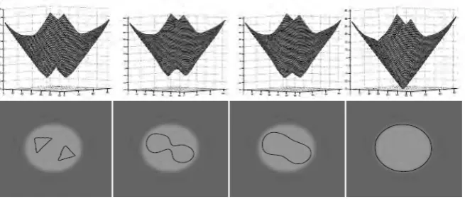
Figure 1.2: Evolution of zero level curve of the corresponding level set function
Figure1.3: (a) CT image (b) Segmentation of CT image
Implementation of Algorithm and Simulation
The algorithm was originally developed by Chumming Li [1] for his MATLAB code for level-set without re-initialisation. However the algorithm is complicated, expensive to implement and images result as obtained are also not smooth. The modified steps include specialized filtering methods used at various levels of image processing. Step 1: Image acquiring and reading
Step 2: Processing the image through desired filter Step 3: Processing the image through Gaussian Filter Step 4: Select the region of interest from the input image Step 5: Finding the gradient of the image
Step 6: Set the parameter of level-set Step 7: Set the intensity of the image
Following changes have been incorporated in the algorithm:
1. The different filters were used to filter the image. The filters were used before the Gaussian filter. This technique is used to modifying or enhancing an image. It helps to emphasize certain features or remove other features of the image .It smoothness, sharpening and enhancement the edge of the image. These filtering techniques include Linear filtering and Non Linear Filtering [3] [4].
2. To increase the intensity of the image the parameters controlling the intensity has been adjusted.
3. Now call the selection base program. This program chooses the appropriate values of level-set parameters form its database according to the if-else rules .The output of this control strategy are the input for the segmentation program. The parameters alfa, lamda, sigma, epsilon are the optimized parameter for the level set program.
4. The height, length and the area of the segmented part can be calculated. Segmented-area equals to the area of the closed curve when it is in anti-clockwise and equals to the negative area when it is in clockwise. Negative area means equal to area in magnitude but negative in sign. It used to judge the direction of a closed curve. C provides the coordinates of the nodes of the curve Area = varea (C); Area returns the area of the curve (>0) when it is in anti-clockwise and negative area of the curve (<0) when it is in anti-clockwise.
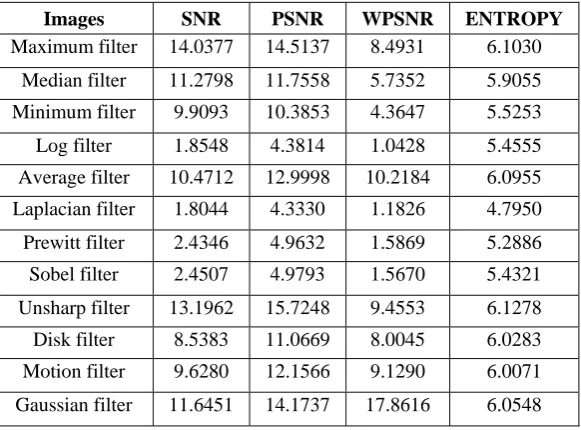
5. Next step is to calculate SNR, PSNR, WPSNR and Entropy parameters of the filtered image and the original image.
Test results for CT image shown for ‘Level Segmentation’ Using different filters for obtaining various parameters -
Dimension of segmented area when no filter is used: Width= 43.6 pixels; Height= 26.9 pixels;
Area of closed curve = 1.8067e + 003.
Type of Filter
Images Width Height Segmented part Area
Result
Maximum filter
42.3 25.7 1.7969e + 003 The area of segmentation has increased and nearly entire desired area is segmented.
Median filter 38.9 23.3 1.6161e + 003 The area of segmentation has increased but fails to segment the entire desired area.
Minimum filter
40.1 21.8 1.5924e + 003 This filter fails to segment the entire desired area.
Log filter 5.2 19.8 1.2468e + 003 This filters segment very less area therefore the filter is not suitable for such type of segmentation
Average filter
38.6 22.7 1.4812e + 003 The area of segmentation has increased but filter fails to segment the desired area.
Laplacian filter
38.9 28.2 1.9782e + 003 The filters crosses the boundaries of segmented area therefore it is not suitable for such type of segmentation
Type of Filter
Images Width Height Segmented part Area
Result
Sobel filter 35.2 19.1 1.1365e + 003 The area of segmentation is very small therefore the filter is not suitable for such type of segmentation.
Un-sharp filter
40.7 23.6 1.6483e + 003 The area of segmentation is very small therefore this filter is not suitable for such type of segmentation.
Disk filter 36.9 21.7 1.3748e + 003 The area of segmentation has increased but filter fails to segment the desired area.
Motion filter 34.5 19.1 3.8522e + 003 By the use of this filter the area of segmentation has decreased.
Gaussian
filter
37.3 23.3 1.4265e + 003 This filter fails to segment the entire desired part.
Table 1.1: Dimensions of segmented area of CT image 1 with various filters
Images SNR PSNR WPSNR ENTROPY
Maximum filter 14.0377 14.5137 8.4931 6.1030
Median filter 11.2798 11.7558 5.7352 5.9055
Minimum filter 9.9093 10.3853 4.3647 5.5253
Log filter 1.8548 4.3814 1.0428 5.4555
Average filter 10.4712 12.9998 10.2184 6.0955
Laplacian filter 1.8044 4.3330 1.1826 4.7950
Prewitt filter 2.4346 4.9632 1.5869 5.2886
Sobel filter 2.4507 4.9793 1.5670 5.4321
Unsharp filter 13.1962 15.7248 9.4553 6.1278
Disk filter 8.5383 11.0669 8.0045 6.0283 Motion filter 9.6280 12.1566 9.1290 6.0071
Gaussian filter 11.6451 14.1737 17.8616 6.0548
Table 1.2: Parameters of CT image 1 of various filters
Conclusions
The process of segmentation of biomedical images requires a very high degree of accuracy. A similar effort has been made in this work to analyze the various filtering techniques and to find out the best among the twelve filters. The setup has been tested for a given set of biomedical images such as CT images and can also be used for X- rays and MRI images. In the process of final evaluation, we found that the results using the variational level set segmentation techniques on CT images are better. Out of the twelve filters used, maximum filter did the best job as far as segmentation of CT images is concerned. It may be concluded that the algorithm applied has been by and far successful.
References
[1] Chunming Li , Chenyang Xu and Changfeng Gui, (2005): Level set evolution without re-initialization: a new variational formulation, IEEE Computer Conference on Computer Vision and Pattern Recognition, Vol:1, pp: 430-436.
[2] Shaojun Liu and Jia Li, (2006): Automatic Medical Image Segmentation Using Gradient and Intensity Combined Level Method, IEEE Annual International Conference on Medical Imaging, pp: 78-82.
[3] Xujia Qin, Jionghui Jiang, Weihong Wang and Fan Zhang, (2007): Canny Operator Based Level Set Segmentation Algorithm for Medical Images, International Conference on Bioinformatics and Biomedical Engineering, pp: 892-895.