R E S E A R C H
Open Access
High
in vitro
survival rate of sheep
in vitro
produced blastocysts vitrified with a new
method and device
Sergio Ledda
1*, Jen M. Kelly
2, Stefano Nieddu
1, Daniela Bebbere
1, Federica Ariu
1, Luisa Bogliolo
1,
Dity Natan
3and Amir Arav
3Abstract
Background:To advance the use of embryo vitrification in veterinary practice, we developed a system in which embryo vitrification, warming and dilution can be performed within a straw. Ovinein vitroproduced embryos (IVEP) were vitrified at either early (EBs:n= 74) or fully expanded blastocyst stage (FEBs:n= 195), using a new device named“E.Vit”, composed by a 0.25-mL straw with a 50-μm pore polycarbonate grid at one end. Embryos at each stage (EBs and FEBs) were vitrified by either Two-step (TS) or Multi-step (MS; 6 different concentrations of vitrification solutions) protocol. Non-vitrified embryos (n= 102) were maintained inin vitroculture as a control. Warming consisted of placing the straws directly into 1.5 mL tubes containing a TCM-199 solution with three decreasing concentrations of sucrose. Blastocyst re-expansion, embryo survival and hatching rate were evaluated at 2, 24 and 48 h post warming. The number of apoptotic cells was determined by TUNEL assay.
Results:Blastocyst re-expansion (2 h) after warming was higher (P< 0.05) in FEBs group, vitrified with the MS and TS methods (77.90% and 71.25%, respectively) compared with the EBs group (MS: 59.38% and TS: 48.50%,
respectively). Survival rates of vitrified FEBs after 24 h IVC were higher (P< 0.001) in both methods (MS and TS) than vitrified EBs (MS: 56.25%; TS: 42.42%) and was higher (P< 0.05) in the MS method (94.19%) compared with those in TS (83.75%). After 48 h of culture the hatching rate for FEBs vitrified in MS system (91.86%) was similar to control (91.89%), but higher than FEB TS (77.5%) and EBs vitrified in MS (37.5%) and TS (33.33%). Number of apoptotic cells were higher in EBs, irrespective of the system used, compared to FEBs. The number of apoptotic cells in FEBs vitrified with MS was comparable to the control.
Conclusions:A high survival rate of IVP embryos can be achieved by the new“E.Vit”device with hatching rates
in vitrocomparable with control fresh embryos. This method has the potential for use in direct embryo transfer in field conditions.
Keywords:Blastocysts, High survival, In-straw,In vitroembryo produced, Ovine
Introduction
Over the past decades, the worldwide increase in small ru-minant breeding has been supported by the development and improvement of assisted reproductive technologies
(ART) [1, 2]. However, while some ART including estrus
induction, estrus synchronization and artificial insemin-ation (AI) have reached widespread applicinsemin-ation, the adop-tion of ART, such as superovulaadop-tion and embryo transfer
(MOET),in vitro embryo production (IVEP) and embryo
cryopreservation have, to date, been limited.
New prospects offered by IVEP, repeated ovum pick-up from live adult and juvenile female donors, suggest that IVEP technology can be used as an alternative system to MOET programs, thus moving the use of this technology from exclusive research in the laboratory to the field [3]. Recent improvements of embryo produc-tion and cryopreservaproduc-tion technologies have the poten-tial to allow wider propagation of valuable genetics in small ruminant populations and establishment of flocks
© The Author(s). 2019Open AccessThis article is distributed under the terms of the Creative Commons Attribution 4.0 International License (http://creativecommons.org/licenses/by/4.0/), which permits unrestricted use, distribution, and reproduction in any medium, provided you give appropriate credit to the original author(s) and the source, provide a link to the Creative Commons license, and indicate if changes were made. The Creative Commons Public Domain Dedication waiver (http://creativecommons.org/publicdomain/zero/1.0/) applies to the data made available in this article, unless otherwise stated.
* Correspondence:[email protected]
1Department of Veterinary Medicine, University of Sassari, Sassari, Italy
without risk of disease transmission. In addition, these technologies could make a substantial contribution to the preservation of endangered species or breeds.
Embryo cryopreservation has become an integral part of the commercial embryo transfer industry, but its
ap-plication in small ruminants is still limited [4, 5]. From
the practical viewpoint, embryo cryopreservation, has many advantages, it facilitates i) distribution of superior genetics from dams of high genetic value, which acceler-ates the rate of genetic improvement and ii) inter-national transportation of valuable genetic stock, which is a financially feasible and safe alternative to live animal transport. To date, documented use and success rates for different cryopreservation techniques and devices in small ruminants are relatively scarce compared with cat-tle [6–8]. However, it has been observed that sheep
em-bryos are able to survive both “equilibrium cooling” or
controlled slow freezing and vitrification procedures [9]. Controlled slow freezing protocols require a biological freezer and need more time to be completed, while the ultra-rapid techniques such as vitrification require no spe-cial equipment and are time and cost effective, therefore, making this technology more adapted to routine field use [4]. Moreover, the viability rate for cryopreservingin vitro and in vivo derived morula and blastocyst stage ovine embryos by vitrification is significantly higher com-pared with embryos cryopreserved by slow freezing techniques [10–12].
Different factors can affect the efficiency of the cryo-preservation system used for embryo freezing. The origin of the embryo,in vivovs.in vitro, contributes to these
dif-ferences with in vivoproduced embryos having increased
viability and survival rates post thawing compared with
their in vitro counterparts [13, 14]. In addition, several
methods and devices have been proposed in order to improve and simplify embryo cryopreservation methodolo-gies of the different farm species [15–17]. In ovine, the 0.25 mL straw [18] and the open pulled straw (OPS) have
been successfully used for vitrifying in vivo [4, 9] or
in vitro[5] produced morula and blastocysts.
The successful application of IVEP embryo cryopreserva-tion and transfer in small ruminants is largely dependent on the efficiency of the techniques used.
Vitrification has become a viable and promising alter-native to traditional slow freezing approaches since is simple, rapid and fewer equilibration and cryopreserva-tion steps are needed. For the use of vitrificacryopreserva-tion tech-nology in veterinary practice, embryos would have to be warmed and diluted in the vitrification straw so that they could be directly transferred to the uterus of recipients animals. Several methods have been designed to facilitate the direct transfer of vitrified embryos in field conditions with a different level of complexity of the technical pro-cedures [19,20].
In this vein, we are proposing a new system,“E.Vit”in
which embryo vitrification, warming and dilution can be performed within a straw. An in-straw embryo cryo-preservation method reduces the need for equipment, technical skills and embryo handling and can facilitate direct embryo transfer to the uterus.
To evaluate the feasibility and efficiency of the new “E.Vit” system, we compared the post-warming survival rate of IVEP embryos (early or expanded blastocyst) vit-rified by Two- or Multi-step systems with fresh IVEP embryos (control).
Materials and methods Chemical
All chemicals were purchased from Sigma-Aldrich Chemical Co. unless otherwise specified.
Oocytes recovery
Ovaries from adult Sardinia sheep (Ovis Aries, 3-6 years old) were recovered from local abattoirs and transported within 3 h to the laboratory in Phosphate Buffered Saline (PBS) with penicillin (100 mg/mL) and streptomycin (100 mg/mL) at 27–30 °C.
Upon arrival at the laboratory, ovaries were cleaned of any tissue residues, washed twice in PBS and placed in Holding Medium (HM) at pH 7.22 ± 0.1 at 37 °C consisting of TCM-199 with Hepes (N-2-Hidroxyethylpiperazine-N-2-ethansulfonic acid) 25 mmol/L, 50 IU/mL of antibiotics (streptomycin and penicillin), sodium bicarbonate 0.005 mol/L and 0.1% (w/v) of polyvinyl alcohol (PVA). The ovar-ies were divided sagittally with the aid of a sterile micro-blade and cumulus oocyte complexes (COCs) were released from the follicles by slicing technique.
In vitromaturation (IVM),in vitrofertilization (IVF) and in vitroembryo culture (IVC)
Forin vitromaturation (IVM), COCs with several intact cumulus cells layers and homogenous cytoplasm were
selected. COCs were matured in 650μL ofin vitro
mat-uration medium consisting of TCM-199 containing 10% heat-treated estrous sheep serum (ESS), 0.36 mmol/L pyruvate, 100 mmol/L cysteamine, FSH 1 IU/mL and LH 1 IU/mL (Pluset; Bio98, Milan, Italy) under mineral oil, in 4-well dishes (Nunc Cell Culture, Thermo Fisher Sci-entific, Waltham, Massachusetts, USA) in a humidified
atmosphere of 5% CO2, at 38.5 °C.
After 24 h from IVM, COCs were partially stripped of cumulus cells as described by Bogliolo and co-authors [21] and co-incubated with frozen-thawed spermatozoa,
selected by the swim-up technique, (1 × 10⁶
humidified atmosphere of 5% CO2, 5% O2, 90% N2 in
four well Petri dishes.
The semen was preliminarily evaluated with the aid of the stereomicroscope (mass motility) and of the CASA system (Ivos, Hamilton Thorne, Biosciences).
At the end of IVF (approximately 24 h), presumptive
zy-gotes (n= 15-20) were transferred to 650μL IVC medium
(SOF supplemented with BSA (4 mg/mL), essential amino acids (EAA) and non-essential amino acids (NEAA) at oviductal concentration [23] in 4-well culture dishes and
incubated at 38.5 °C under 5% O2, 5% CO2, 90% N2 and
maximum humidity. At 30 h post fertilization cleavage rate was recorded and on day 6 and 7 (day 0 = day of IVF) embryos were evaluated for development.
Embryo vitrification
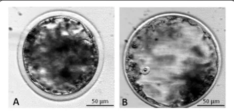
For the experiments of vitrification, blastocysts were clas-sified according to degree of expansion and hatching sta-tus as: early blastocyst (EB: blastocyst with a blastocoel less or equal to the half of the embryo volume), fully ex-panded blastocyst (FEB: a large blastocyst with a blasto-coel greater than half of the embryo volume) (Fig.1).
The“E.Vit”device (FertileSafe, Ness Ziona, Israel) used for vitrification consisted of a 0.25-mL straw with a
poly-carbonate grid (capsule) with pore diameters of 50μm
inserted at free end of the straw (Fig.2). At day 6 or 7 of IVC either EB or FEB were placed in embryo handling medium (EHM see below) before being loaded by aspir-ation into the straw.
Each 0.25 mL empty straw was loaded with up to 2
embryos in a volume range of 20–30μL, taking care to
leave an air bubble at the end of straw. Next, the capsule
was inserted by pressure into the straw end (Fig.2). The
insertion of the capsule allowed the removal of excess HM medium; the solution that contains the embryo is
reduced to 5μL. The straw loaded with the embryos and
with the inserted capsule is transferred between the
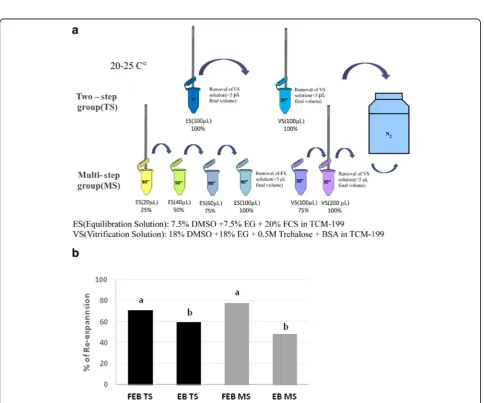
fol-lowing solutions (Fig. 3); the volume of the solution in
the straw is increased by capillary force and according to
the increasing volume of the different vials (Fig. 3). The
straws are evacuated by gentle blotting on sterile gauze in two times: the first after loading the 100% ES and the second after loading with 100% VS solution.
Three replicates of each embryonic stage and
vitrifica-tion system were performed. The experiment was
per-formed at room temperature (20-25 °C) and all media were at 25 °C using either:
a) Two-step system (TS), with the exposure of the embryos at only one equilibration and one vitrification solution or;
b) Multi-step system (MS), based on the exposure to 4 increasing concentrations of cryoprotectants for the equilibration procedure and two different concentrations for the vitrification solution.
Embryo handling medium (EHM) consisted of
TCM-199 + HEPES supplemented with 0.6% bovine serum albumine (BSA) and 0.5 mol/L trehalose.
Equilibrium solution (ES) consisted of EHM
supple-mented with 7.5% of ethylene glycol (EG) and 7.5% of dimethyl sulphoxide (DMSO) .
Vitrification solution (VS) consisted of EHM supple-mented with 18% of EG and 18% DMSO.
Two-step system (TS) (Fig.3a)
The TS involves the use of a single equilibrium solution (ES: 100%) and a single vitrification solution (VS: 100%). The embryos were loaded into the E.Vit device (as de-scribed above) and then the E.Vit device lowered into ES (1 mL of ES in 1.5 mL micro-centrifuge tube) for 5 min. After which the ES was gently removed from the E.Vit device be-fore being lowered into VS (1 mL of VS in 1.5 mL micro-centrifuge tube) for 30 s and then plunged into liquid nitro-gen. Between the passage from the VS and the immersion in liquid nitrogen, excess VS was quickly removed by gentle blotting on sterile gauze, from the E.Vit device.
Multi-step system (MS) (Fig.3b)
After loading embryos into the E.Vit device (described above), excess handling medium was removed from the E.Vit device before being exposed, in a step-wise man-ner, to increasing concentrations of ES cryoprotectants (1 mL of each of the following ES concentrations diluted with EHM) set up in 1.5 mL micro-centrifuge tubes; ES 1: 25%, ES2: 50%, ES3: 75%, ES4: 100%). The E.Vit device with the embryos was exposed to each ES concentration (with increasing volume) for 90 s for a total of 6 min. Be-tween the equilibrium phase and the exposure to the VS1 medium, excess ES4 was removed from the E.Vit device by gentle blotting on sterile gauze. The E.Vit de-vice containing the embryos was subsequently plunged into 1 mL of 75% of final VS concentration (VS1) for 30 s (1.5 mL micro-centrifuge tube), followed immediately Fig. 1Representative images ofin vitroproduced embryos at
Fig. 2Sample insertion into E.Vit device
by plunging into 100% VS concentration (VS2) for 30 s (1 min total exposure for the two VS concentrations). Excess V2 medium was removed from the E.Vit device by gently blotting on sterile gauze before being immersed into liquid nitrogen.
Warming of the embryos
Embryos were left in LN2storage for a minimum 7 days.
Warming for both the TS and MS systems was carried out as follows. The E.Vit device, with the embryos inside, im-mediately after removing from the liquid nitrogen, was plunged stepwise into decreasing concentrations of su-crose (1 mol/L, 0.5 mol/L and 0.25 mol/L in TCM-199 + 20% FCS at 38.5 °C). The E.Vit device was left immersed in each solution for 5 min.
Embryo recovery and post warming in vitro culture
After the warming procedure, the capsule was removed by cutting the end of straw and embryos released in HM. The number of embryos recovered after the remov-ing the capsule from each straw was recorded. The em-bryos were washed 2-3 times with HM and incubated in
IVC at 38.5 °C at 5% CO2at maximum humidity for the
subsequent evaluations after 2, 24 and 48 h of culture. For the evaluation we have used the following mor-phometric criteria [24]:
– Time of the start of re-expansion (the first appear-ance of the blastocoele cavity or increase in size). – Time of completion of re-expansion (the blastocyst
occupies the whole perivitelline space).
– Time of hatching (the trophectoderm blebbs out of the zona pellucida).
In particular, the following embryo development pa-rameters were recorded:
– after 2 h: number of embryos that showed a start of re-expansion of the blastocoel cavity;
– after 24 h: number of embryos with completion of expansion of blastocoel cavity;
– after 48 h: number of hatched blastocyst.
Analysis of apoptosis through the use of the TUNEL technique and confocal microscope
Apoptotic cell death in the embryos was evaluated by TUNEL using an In situ Cell Death Detection kit reac-tion mixture (Fluorescein; Roche Diagnostics Corp., In-dianapolis, IN, USA). This method is used to detect cells that contain single and double strand breaks (nick) more or less extensive along the nuclear DNA molecule.
The TUNEL kit consists of an enzyme, the TdT (Ter-minal deoxynucleotidyltransferase), which catalyzes the polymerase reaction of nucleotides (always added to the
mixture) labeled with fluorescein [25], at the free end 3´ OH of fragmented DNA molecules both at the single
and double stranded levels. The labeled nucleotides“fill”
the single or double strand breaks on the DNA and emit a typical fluorescence green light; the more intense the more the DNA breaks are extended. To determine apop-tosis of embryos a subset of embryos from Control fresh
IVP embryos (EBn= 13; FEBn= 15), and from vitrified/
warmed TS (EBn= 14; FEBn= 12) and MS (EBn= 12;
FEBn= 20) after 24 h of IVC were fixed in 4%
parafor-maldehyde in PBS at 37 °C for 1 h. Following fixation, they were washed 3 times per 15 min in PBS + 0.1% PVA, to remove the residual fixative. The embryos were then permeabilized with 0.1% Triton X 100 in 0.1% so-dium citrate for 5 min at 4 °C. The permeabilization aims to make permeable the zona pellucida at the entrance of the reagents that will be added later. The samples were incubated in TUNEL (Enzyme Solution + Label Solu-tion) for 1 h at 38.5 °C in the dark. As a negative control,
embryos (EB n = 2; FEB n = 3) from the control group
were incubated in the presence of Label Solution and subsequently processed, as described below for the other groups of vitrified embryos.
Following incubation, the embryos were washed 3 times for 15 min in PBS+ 0.1% PVA, stained with a
Glycerol-Hoechst 33342 solution (10μg/mL) for the
evaluation of the nuclear chromatin of the blastomeres. A drop of dye solution was placed on a glass slide and the embryos were transferred (3-5 embryos) to the drop. A coverslip was placed on the drop to slightly compress the embryos in order to facilitate the visualization of the nuclei. The slide was kept for 1 h in the dark at 4 °C and then read with the confocal microscope. The images were acquired with a laser scanning confocal microscope (Leica TCS SP5), equipped with 543 nm HeNe, 488 nm Argon and 405 nm 405-diode laser using an immersion objective (in oil) 40 × (NA = 1.25). The parameters re-lated to fluorescence intensity (laser energy, gain, offset and pinhole size) were maintained with constant values during all the acquisitions of images.
TUNEL’s green fluorescence was determined using ex-citation wavelengths of 488 and 543 nm and emission spectra of 515-565 nm (green).
The number of TUNEL positive nuclei in embryos was determined and the apoptotic index (No. of apoptotic cells/ total No. of cells × 100) was calculated [26].
Statistical analysis
Data were analyzed by StataIC 11.2 software (Stata Corp LP, USA). Recovery rate, blastocoel re-expansions after 24 h post warming and hatching (survival) after 48 h post warming were compared between embryonic stage EB and FEB and between the two methods of vitrification
post hocBonferroni test. Values ofP< 0.05 were consid-ered statistically different.
Results
Embryo recovery
The embryo recovery rates from the “E.Vit” system are
reported in Table 1 comparing the different embryonic
stages (EB vs. FEB) and the different methods of cryo-protectant exposure (TS vs. MS). No statistically signifi-cant differences were observed between the embryonic stages and methods of cryoprotectant exposure.
Blastocoel re-expansion after 2 h post warmingin vitro culture
After 2 h post warming, overall rates of embryos that showed a regular blastocoel re-expansion were not af-fected by TS or MS vitrification methods. However, a
significant difference (P< 0.05) was observed between
EB [TS: N= 16/33 (48.5%) and MS: N= 19/32 (59.38%)]
and FEB [TS: N= 57/80 (71.25%) and MS: N= 67/86
(77.91%)] within both methods (Fig.4).
Survival of embryos after 24 h of post warmingin vitro culture
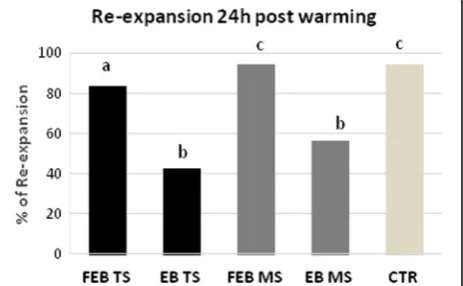
Survival rates were significantly (P< 0.001) higher after vitrification of fully expanded blastocysts (FEB) compared
with the early blastocyst (EB) in the TS method [FEB:N=
67/80 (83.75%) vs EB: N= 14/33 (42.42%), respectively]
and in the MS method [FEB: N= 81/86 (94.19%) vs EB:
N= 18/32 (56.25%), respectively). Rate of survival was also significantly different between the FEB vitrified in TS
method compared with the MS method (P= 0.031). The
non-cryopreserved control (CTR: N= 70/74; 94.59%)
dif-fered from all vitrified-warmed groups except for FEB (N= 81/86; 94.19%) vitrified with the MS method (Fig.5).
Hatching rate after 48 h post warmingin vitroculture
After 48 h of culture the hatching rates were higher in the vitrified fully expanded embryos (FEB), irrespective of the system used, compared with early blastocysts [FEB TS:
N= 62/80 (77.5%) vs EB TS:N= 11/33 (33.30%) and FEB
MS: N= 79/86 (91.86%) vs EB MS N= 12/32 (37.50%);
P< 0.001 in both cases]. The hatching rate of vitrified FEB
with the MS method was higher (P= 0.01) compared with
FEB for the TS method and similar to controls (N= 68/74;
91.89%; Fig.6).
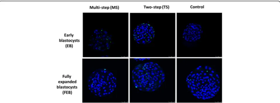
Apoptotic cell detection in vitrified and control blastocysts
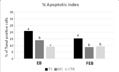
The results of DNA fragmentation evaluated by TUNEL technique using the two vitrification systems are shown in Fig.7.
The apoptosis index in vitrified EBs was higher in TS (26.11%) compared with the MS (16.48%) and control group (9.95%;P< 0.001; Fig.7).
FEBs cryopreserved with the MS method showed
apoptosis index (8.95%) significantly lower (P< 0.001)
than those vitrified with TS method (26.67%) and similar
to the Control group (10.28%; Fig.7).
Table 1Recovery rates of early blastocyst (EB) and fully expanded blastocyst (FEB) vitrified using E.Vit device with Multi-step or Two-Multi-step methods
Embryonic stage
No. of embryos
No. of recovered embryos
Recovery rate, % Two-step (TS)
method
EB 37 33/37 89.1%
FEB 90 80/90 88.89%
Multi-step (MS) method
EB 37 32/37 86,49%
FEB 95 86/95 90.5%
Fig. 4Rate of post warming blastocoel re-expansion of EB and FEB blastocysts vitrified in“E.Vit”device with Two-step (TS) and Multi-step (MS) method. Different letters above columns indicate significant differences (P< 0.05; Chi-square test). Survival rates of vitrified early (EB), expanded blastocysts (FEB) by Two-step (TS) and Multi-step (MS) methods and control (CTR) after culturein vitrofor 24 h. Different letters above columns indicate significant differences (P< 0.05; Chi-square test)
Confocal microscopy analysis allowed for identification of the positive fluorescent signal in the EB, FEB and
CTR groups (Fig.8).
Discussion
Although cryopreservation of preimplantation embryos has made great progress in recent years, the achieve-ments obtained for ovine embryo freezing, especially for in vitro produced embryos are still relatively low. The development of simplified and reproducible vitrification cryopreservation systems that allow gametes, embryos, and reproductive tissues to be successfully cryopreserved are important considerations if this technology is to be widely adopted. Moreover, to achieve a large application of vitrification technology in the field, the need to ma-nipulate embryos should be reduced to a minimum and be able to be performed by operators without a need for specialized skills and equipment.
Here, we report a new vitrification protocol and device
(E.Vit) that allowedin vitroproduced ovine embryos to be
efficiently vitrified and thawed in-straw. Our results showed that a step-wise exposure to cryoprotectants during vitrifi-cation positively affected embryo survival rates and that embryonic stage at vitrification correlated to embryo sur-vival after vitrification/warming. In addition, our results demonstrated that after vitrification of fully expanded blas-tocysts (FEB) in multi-step (MS) cryoprotectant exposure, no differences in the survival and hatching rates were ob-served between vitrified and fresh IVEP embryos after cul-ture in vitro. Several factors can affect the efficiency of
cryopreservation of ovine in vitro produced embryos;
among them, embryo survival after cryopreservation has been reported as mainly related to the source of embryos and to the methods used for the embryo freezing [18,27].
The reduction of survival after cryopreservation of IVP embryos is also due to osmotic and toxic effects exerted by the cryoprotectant exposure. This is more evident in the vitrification procedures that require higher cryopro-tectant concentrations. A correct balance between the for-mulation of cryoprotectant mixture, their concentration, time of exposure and temperature seems also crucial to reduce cryodamage [28]. The right interconnection of these factors could have more importance when chilling sensitive embryos, as the IVP ones, are subjected to cryo-preservation. To reduce toxicity and improve osmotic re-sponse to cryoprotectants, usually embryos are pretreated / equilibrated in a solution containing a lower concentra-tion of permeating cryoprotectants before being sus-pended in a vitrification solution. The pretreatment is effective to promote permeation of the cryoprotectant and at the same time reduce toxicity. The concentration of the permeating cryoprotectants used in the pretreatment ranges from 2% to 20%. Our results are in this lane and we have observed that a gradual exposure to increasing concentrations of cryoprotectants before suspending in the final vitrification solution increased the survival rates of vitrified expanded blastocysts, after 24 h of post warm-ing culture. Exposure of the same stage embryos to only one equilibration solution (ES 100%) yielded a significantly lower number of blastocysts able to complete blastocoel re-expansion. The reduction of potential adverse toxic effects depends not only on cryoprotectant solution con-centrations and the type of cell, but is also related to temperature. In our experiments, besides using a combin-ation of ethylene glycol (EG) and dimethyl sulfoxide (DMSO), which have high penetration rate and low tox-icity, we performed all procedures at room temperature to further reduce the toxicity of cryoprotectants. A similar approach has been reported in previous studies for the vit-rification of ovine [5] and bovine embryos [29].
Further key elements for embryos survival by reducing chilling injury are high cooling and warming rates, which are achieved by using a small-volume and a small-size car-rier. Various methods were developed to achieve these Fig. 6Hatching rates of vitrified early (EB) and expanded blastocysts
(FEB) by Two-step (TS) and Multi-step (MS) methods and control (CTR) afterin vitroculture for 48 h. Different letters among columns indicate significant differences (P≤0.01; Chi-square test)
features, including straws [4], electron microscope grids [30], fine capillaries [31,32], cryotops [33], cryoloops [34] or the tips of micropipettes [35]. The E.Vit system offers similar advantages in term of cooling rate. It has been pre-viously reported [28] that cooling and warming rates are very fast and similar to other largely used open systems [33–35]. The efficacy of embryo vitrification can be esti-mated by recording embryo morphokinetics after post warming culture. Previous studies have indicated that, following embryo vitrification/warming, blastocoel re-expansion and hatching is predictive of subsequent devel-opment, including ability to implant, establish a pregnancy and result in a live offspring [24, 36]. In addition, timing of blastocoelic cavity re-expansion after vitrification/ warming andin vitroculture is considered a reliable index of in vitro produced embryo quality and developmental potential [37]. Our results showed that embryonic stage can affect the start of re-expansion (2 h post warming), completion of re-expansion (24 h post warming) and hatching rate (48 h post warming). Fully expanded blasto-cysts consistently yielded better rates compared with early blastocysts, irrespective of the system used, and after 24 h of culture, we observed a higher survival rate of fully ex-panded blastocyst compared with those vitrified with the two-step (TS) protocol. The fully expanded blastocysts ex-posed to the MS cryoprotectant protocol and vitrified using E.Vit, gave a hatching rate comparable with fresh control embryos. It has been previously reported that early stages embryos are more sensitive to cryopreservation procedures and that their survival is significantly lower than embryos vitrified at later stages. The proportion of embryos that can survive after cryopreservation is in fact significantly lower in 2-8 cell stage embryos compared
with morula and blastocyst stage [10, 12]. Here, we
re-ported that significant differences can be also observed comparing vitrification of early blastocyst stage respect
with expanded blastocyst stage. Similarly, a higher survival
rate has been observed in ovinein vivo [18] and in vitro
[12] derived embryos vitrified in later stages compared with earlier stage embryos.
The higher cryotolerance of expanded blastocysts could be due to several factors: the cellular membranes of em-bryos becomes more resistant to osmotic, toxic and chilling stress after the formation of the blastocoelic cavity; the diversification of cell types and in particular the increase of Na+/K+ ATPase activity, which occurs during blastocoelic formation in trophoblastic cells, may determine more active transport mechanisms of cryoprotectants [38]. Other as-pects that can influence the hatching rate may be explained by differences in blastomere size. The cells of compacted morulae and early blastocysts are slightly larger than the cells of expanded blastocyst, and this may render them more sensitive to the osmotic stress induced by the removal of the permeated cryoprotectant [39]. Our results indicated that using the E.Vit device and the MS method resulted in embryo survival and hatching rates comparable with fresh embryos cultured in the same conditions. Gradual exposure using the MS procedure may reduce osmotic stress by allowing water permeability in the same rate of CPs perme-ability. According results on embryo quality were obtained by the quantification of apoptotic cells. Early vitrified em-bryos, irrespective of the method used, contained a higher number of apoptotic cells compared with expanded blasto-cysts. Expanded blastocysts vitrified with the MS method had comparable numbers of apoptotic cells as with the con-trol fresh embryos.
These systems, in most of the cases, involved more than one device and were developed mainly to perform
in-straw warming and dilution [19,20,31,32,40,41].
The E.Vit is a simple, efficient, standardized and reprodu-cible vitrification system. In addition to the obvious advan-tage that all vitrification procedures can be performed in the E.Vit device, is the added benefit that the E.Vit is de-signed to be used in automated systems [28]. Previous ex-periments employing mouse embryos, bovine oocytes and ovine testicular tissue indicated the feasibility of the device and the high survival rate after warming [28, 42,43]. Fur-ther advantages include the possibility to employ CE certi-fied 0.25 mL straws, to vitrify many straws simultaneously, to reduce osmotic and mechanical damages and possible contamination during embryo manipulation and to achieve rapid cooling and warming rates (> 20,000 °C/min).
Conclusions
In conclusion, vitrification of in vitro produced ovine
embryos by the E.Vit device and multistep cryoprotect-ant exposure method yielded post warming, high sur-vival rates and embryo quality that could be predictive of subsequent development. The new vitrification proto-col and cryodevice can permit that all the procedures of equilibration, cryopreservation as well warming and dilu-tion can be performed in the vitrificadilu-tion straw. This method has the potential for use in direct embryo trans-fer in field conditions.
Future experiments are under evaluation to establish the full survival potential after embryo transfer in recipient ewes.
Abbreviations
ART:Assisted reproductive technologies; CASA: Computer-assisted sperm analysis; CE: European Community; CLA:Trans-10cis-12-conjugatedlinoleumacid isomer; COC: Cumulus oocyte complexes; CTR: Non-cryopreserved control; DMSO: Dimethyl sulphoxide; EAA: Essential amino acids; EB: Early blastocysts; EG: Ethylene glycol; EHM: Embryo handling medium; ES: Equilibrium solution; FCS: Fetal calf serum; FEB: Fully expanded blastocysts; FSH: Follicle stimulating hormone; HM: Holding Medium; IVEP:In vitroembryo produced; IVEP:In vitro produced embryos; IVF:In vitrofertilization; IVM:In vitroculture; IVM:In vitro maturation; MOET: Multiple ovulation and embryo transfer; MS: Multi-step; NEAA: Non- essential amino acids; OPS: Open pulled straw; PBS: Phosphate Buffered Saline; PVA: Polyvinyl alcohol; SOF: Synthetic Oviductal Fluid; TS: Two-step; VS: Vitrification solution
Acknowledgements Not applicable.
Ethics approval and consent to partecipate
All were performed in accordance with DPR 27/1/1992 (Animal Protection regulations of Italy) in conformity with European Community regulation 86/ 609. No experimental animals have been used in this work.
Authors’contributions
SL, JK, AR and DN conceived the work. JK, SL, SN and AF performed embryo IVP and cryopreservation experiments.. DB and AF analysed data. SL, DB, LB and JK wrote the manuscript. All Authors read and approved the final manuscript.
Funding
Research supported by Regione Autonoma della Sardegna.-L.R. 7-MIGLIOVINGENSAR Project, by Bando competitivo Fondazione di Sardegna–
2016, CUP J86C18000800005 and by“Progetto FAR2019LEDDAS Una Tantum 2019, University of Sassari”.
Availability of data and materials
All data generated or analysed during this study are included in this published article.
Consent for publication Not applicable.
Competing interests
The authors declare that they have no competing interests.
Author details
1Department of Veterinary Medicine, University of Sassari, Sassari, Italy.2South
Australian Research and Development Institute, Turretfield Research Centre, 129 Holland Road, Rosedale, SA 5350, Australia.3FertilSafe Ltd, 11 Haharash
st, 7403118 Ness Ziona, Israel.
Received: 23 April 2019 Accepted: 2 September 2019
References
1. Loi P, Ptak G, Dattena M, Ledda S, Naitana S, Cappai P. Embryo transfer and related technologies in sheep reproduction. Reprod Nutr Dev. 1998;38:615–28. 2. Ledda S, Gonzalez-Bulnes A. ET-Technologies in Small Ruminants. In:
Niemann H, Wrenzycki C, editors. Animal Biotechnology 1. Cham, First Online, 07 August: Springer; 2018.
3. Paramio MT, Izquierdo D. Current status ofin vitroembryo production in sheep and goats. Reprod Domest Anim. 2014;4:37–48.
4. Baril G, Traldi AL, Cognié Y, Leboeuf B, Beckers JF, Mermillod P. Successful direct transfer of vitrified sheep embryos. Theriogenology. 2001;56:299–305. 5. Dattena M, Accardo C, Pilichi S, Isachenko V, Mara L, Chessa B, et al. Comparison of different vitrification protocols on viability after transfer of ovine blastocysts in vitroproduced andin vivoderived. Theriogenology. 2004;62:481–93. 6. Vajta G. Vitrification in human and domestic animal embryology: work in
progress. Reprod Fertil Dev. 2013;25(5).https://doi.org/10.1071/RD12118. 7. Do VH, Catt S, Amaya G, Batsiokis M, Walton S, Taylor-Robinson AW. Comparison
of pregnancy in cattle when non-vitrified and vitrifiedin vitro-derived embryos are transferred into recipients. Theriogenology. 2018;120:105–10.https://doi.org/ 10.1016/j.theriogenology.2018.07.027Epub 2018 Aug 1.
8. Do VH, Catt S, Kinder JE, Walton S, Taylor-Robinson AW. Vitrification of in vitro-derived bovine embryos: targeting enhancement of quality by refining technology and standardising procedures. Reprod Fertil Dev. 2019; 31(5):837–46.https://doi.org/10.1071/RD18352.
9. Martinez AG, Valcarcel A, Furnus CC, de Matos DG, Iorio G, de las Heras MA. Cryopreservation ofin vitro-produced ovine embryos. Small Rumin Res. 2006;63:288–96.
10. Shirazi A, Soleimani M, Karimi M, Nazari H, Ahmadi E, Heidari B. Vitrification ofin vitroproduced ovine embryos at various developmental stages using two methods. Cryobiology. 2010;60:204–10.
11. Varago FC, Moutacas VS, Carvalho BC, Serapiao RV, Vieira F, Chiarini-Garcia H, et al. Comparison of conventional freezing and vitrification with dimethylformamide and ethylene glycol for cryopreservation of ovine embryos. Reprod Dom Anim. 2014;49:839–844.
12. Dos Santos Neto PC, Vilariño M, Barrera N, Cuadro F, Crispo M, Menchaca A. Cryotolerance of day 2 or day 6in vitroproduced ovine embryos after vitrification by Cryotop or spatula methods. Cryobiology. 2015;70:17–22. 13. Massip A, Mermillod P, Dinnyes A. Morphology and biochemistry ofin vitro
produced bovine embryos: implication for their cryopreservation. Hum Reprod. 1995;10:3004–11.
14. Dos Santos-Neto PC, Cuadro F, Barrera N, Crispo M, Menchaca A. Embryo survival and birth rate after minimum volume vitrification or slow freezing ofin vivoandin vitroproduced ovine embryos. Cryobiology. 2017;78:8–14. 15. McGinnis LK, Duplantis SC, Youngs CR. Cryopreservation of sheep embryos
using ethyleneglycol. Anim Reprod Sci. 1993;30:273–80.
16. Vajta G. Vitrification of the oocytes and embryos of domestic animals. Anim Reprod Sci. 2000;60/61:357–64.
18. Naitana S, Ledda S, Loi P, Leoni G, Bogliolo L, Dattena M, et al. Polyvinyl alcohol as a defined substitute for serum in vitrification and warming solutions to cryopreserve ovine embryos at different stages of development. Anim Reprod Sci. 1997;48:247–56.
19. Morató R, Mogas T. New device for the vitrification and in-straw warming of in vitroproduced bovine embryos. Cryobiology. 2014;68(2):288–93. 20. Caamaño JN, Gómez E, Trigal B, Muñoz M, Carrocera S, Martín D, et al.
Survival of vitrifiedin vitro-produced bovine embryos after a one-step warming in-straw cryoprotectant dilution procedure. Theriogenology. 2015;83:881–90.
21. Bogliolo L, Ariu F, Leoni G, Uccheddu S, Bebbere D. High hydrostatic pressure treatment improves the quality ofin vitro-produced ovine blastocysts. Reprod Fertil Dev. 2011;23:809–17.
22. Tervit HR, Whittingham DG, Rowson LE. Successful culturein vitroof sheep and cattle ova. J Reprod Fertil. 1973;30:493–7.
23. Walker SK, Hill JL, Kleemann DO, Nancarrow CD. Development of ovine embryos in synthetic oviductal fluid containing amino acids at oviductal fluid concentrations. Biol Reprod. 1996;55:703–8.
24. Ebner T, Oppelt P, Radler E, Allerstorfer C, Habelsberger A, Mayer RB, et al. Morphokinetics of vitrified and warmed blastocysts predicts implantation potential. J Assist Reprod Genet. 2017;34:239–44.
25. Gold R, Schmied M, Giegerich G, Breitschopf H, Hartung HP, Toyka KV, et al. Differentiation between cellular apoptosis and necrosis by the combined use of in situ tailing and nick translation techniques. Lab Investig. 1994;71:219.
26. Sanaei B, Movaghar B, RezazadehValojerdi M, Ebrahimi B, Bazrgar M, Hajian M, et al. Developmental competence ofin vitromatured ovine oocytes vitrified in solutions with different concentrations of trehalose. Reprod Domest Anim. 2018;53(5):1159–67.
27. Rizos D, Fair T, Papadopoulos S, Boland MP, Lonergan P. Developmental, qualitative, and ultrastructural differences between ovine and bovine embryos producedin vivoorin vitro. Mol Reprod Dev. 2002;62:320–7. 28. Arav A, Natan Y, Kalo D, Komsky-Elbaz A, Roth Z, Levi-Setti PE, et al. A new,
simple, automatic vitrification device: preliminary results with murine and bovine oocytes and embryos. J Assist Reprod Genet. 2018;35:1161–8. 29. Souza JF, Oliveira CM, Lienou LL, Cavalcante TV, Alexandrino E, Santos RR,
et al. Vitrification of bovine embryos followed byin vitrohatching and expansion. Zygote. 2018;26:99–103.
30. Martino A, Songsasen N, Leibo SP. Development into blastocysts of bovine oocytes cryopreserved by ultra-rapid cooling. Biol Reprod. 1996;54:1059–69. 31. Vajta G, Hyttel P, Callesen H. Morphological changes ofin-vitro-produced
bovine blastocysts after vitrification, in-straw direct rehydration, and culture. Mol Reprod Dev. 1997;48:9–17.
32. Green RE, Santos BF, Sicherle CC, Landim-Alvarenga FC, Bicudo SD. Viability of OPS vitrified sheep embryos after direct transfer. Reprod Domest Anim. 2009;44:406–10.
33. Kuwayama M. Highly efficient vitrification for cryopreservation of human oocytes and embryos: the Cryotop method. Theriogenology. 2007;67:73–80. 34. Lane M, Bavister BD, Lyons EA, Forest KT. Containerless vitrification of
mammalian oocytes and embryos. Nat Biotechnol. 1999;17:1234–6. 35. Cremades N, Silva M, Viana P, Sousa S, Oliveira C, Teixeira da Silva J, et al.
Experimental vitrification of human compacted morulae and early blastocysts using fine diameter plastic micropipettes. Human Reproduction. 2004;19:300–5. 36. Leoni G, Bogliolo L, Berlinguer F, Rosati I, Pintus PP, Ledda S, et al. Defined media
for vitrification, warming, and rehydration: effects on post-thaw protein synthesis and viability ofin vitroderived ovine embryos. Cryobiology. 2002;45:204–12. 37. Coello A, Meseguer M, Galán A, Alegre L, Remohí J, Cobo A. Analysis of the
morphological dynamics of blastocysts after vitrification/warming: defining new predictive variables of implantation. Fertil Steril. 2017;108:659–66. 38. Leoni G, Berlinguer F, Rosati I, Bogliolo L, Ledda S, Naitana S. Resumption of
metabolic activity of vitrified/warmed ovine embryos. Mol Reprod Dev. 2003;64:207–13.
39. Naitana S, Loi P, Ledda S, Cappai P, Dattena M, Bogliolo L, et al. Effect of biopsy and vitrification onin vitrosurvival of ovine embryos at different stages of development. Theriogenology. 1996;46:813–24.
40. Vieira AD, Forell F, Feltrin C, Rodrigues JL. In-straw cryoprotectant dilution of IVP bovine blastocysts vitrified in hand-pulled glass micropipettes. Anim Reprod Sci. 2007;99:377–83.
41. Inaba Y, Aikawa Y, Hirai T, Hashiyada Y, Yamanouchi T, Misumi K, et al. In-straw cryoprotectant dilution for bovine embryos vitrified using Cryotop. J Reprod Dev. 2011;57:437–43.
42. Ledda S, Pinna S, Nieddu S, Natan Y, Arav A, Bebbere D. Vitrification of prepubertal lamb spermatogonia using a novel vitrification system. Reprod Fertil Dev. 2019;3:145.