Delayed Recognition of Initial Stroke in Children: Need
for Increased Awareness
WHAT’S KNOWN ON THIS SUBJECT: Pediatric AIS, like AIS in adults, is associated with high morbidity and mortality rates. Unlike that in adults, however, pediatric stroke often is unrecognized, for a variety of reasons.
WHAT THIS STUDY ADDS: This study quantifies the time to diagnosis of pediatric stroke and aims to highlight the factors that perpetuate delay, namely, the lack of awareness of physicians regarding pediatric stroke. This is in contrast to many previous studies.
abstract
OBJECTIVE:The goal was to identify the delays involved in diagnosing pediatric arterial ischemic stroke (AIS), a major cause of morbidity and death in children.
METHODS:Neonates (ⱕ28 days of age) and children with a first pre-sentation of radiologically confirmed AIS between June 1993 and Jan-uary 2006 were identified retrospectively. The time to diagnosis of AIS (ie, time from clinical onset to radiologic confirmation) was calculated, and factors influencing stroke diagnosis were reviewed.
RESULTS:A total of 107 patients (19 neonates and 88 children) with a diagnosis of AIS were identified. The median time to AIS diagnosis was 87.9 hours for neonates, significantly longer than 24.8 hours for
chil-dren (P⫽.0002). Sixty-nine percent of the children with AIS
demon-strated a likely cardioembolic cause, and 51 (58%) of the 88 children were inpatients at the time of stroke. The inpatients were seen by a
physician more quickly (P ⬍ .01) and received a diagnosis of AIS
sooner (P ⬍ .01). Seventy-six (86%) of the 88 children had a focal
neurologic deficit when first seen by a physician. Physicians docu-mented a diagnosis/differential diagnosis for 44 (50%) of 88 children, and they documented a suspicion of AIS for only 23 (26%) of 88 children. The presence of seizures or focal signs was not associated with a quicker time to stroke confirmation.
CONCLUSIONS:The considerable delays in the diagnosis of pediat-ric AIS are most likely related to the lack of awareness of stroke among medical staff members, despite risk factors and focal signs
at presentation.Pediatrics2009;124:e227–e234
CONTRIBUTORS:Jayasri Srinivasan, MBBS, FRACP,a,bSteven P.
Miller, MDCM, MAS, FRCPC,b,cThanh G. Phan, MBBS, FRACP,dand
Mark T. Mackay, MBBS, FRACPa
aDepartment of Pediatric Neurology, Royal Children’s Hospital,
Melbourne, Australia; Departments ofbPediatric Neurology and cPediatrics, British Columbia Children’s Hospital, Vancouver,
Canada; anddDepartment of Neurosciences, Monash Medical
Centre, Melbourne, Australia
KEY WORDS
stroke, cerebrovascular disease, cerebral vascular accident
ABBREVIATIONS
AIS—arterial ischemic stroke CT— computed tomographic
ICD—International Classification of Diseases IQR—interquartile range
www.pediatrics.org/cgi/doi/10.1542/peds.2008-3544 doi:10.1542/peds.2008-3544
Accepted for publication Mar 13, 2009
Address correspondence to Mark T. Mackay, MBBS, FRACP, Department of Paediatric Neurology, Children’s Neuroscience Centre, Royal Children’s Hospital, Melbourne, Australia 3052. E-mail: [email protected]
PEDIATRICS (ISSN Numbers: Print, 0031-4005; Online, 1098-4275). Copyright © 2009 by the American Academy of Pediatrics
FINANCIAL DISCLOSURE:The authors have indicated they have no financial relationships relevant to this article to disclose.
rologic deficits occur in 50% to 85% of infants and children after arterial
isch-emic stroke (AIS).1Limited awareness
regarding pediatric stroke among phy-sicians and in the community is a ma-jor issue. For adults, advertising cam-paigns (eg, “Time Is Brain Lost”) and education of primary care physicians have led to significant decreases in the
time to presentation.2 One study
re-corded a mean time of 34.5 hours from clinical onset to presentation to any health care professional for children, and studies have demonstrated long
delays to neuroimaging.3,4 For
chil-dren, stroke symptoms are attributed frequently to stroke-mimickers such as migraine, encephalitis, tumors, and postictal Todd paralysis, which can ac-count for up to one fifth of cases
pre-senting with stroke-like symptoms.5,6
Recent pediatric studies suggested that factors such as delays in seeking medical attention and mode of onset were predictive of the delayed diagno-sis of stroke and of the underlying
cause.7,8
Various consensus guidelines have highlighted the importance of
improv-ing time to stroke diagnosis.9–11Before
assessment of acute thrombolysis in the pediatric population, the factors causing delays in stroke diagnosis must be identified and addressed. The aims of this study were to determine the time between clinical onset and diagnosis of AIS in a tertiary Austra-lian pediatric hospital and to identify factors that influenced the time to diagnosis.
METHODS
We identified retrospectively all chil-dren 0 to 18 years of age who were diagnosed as having a first episode of AIS during a 12.5-year period, from June 1, 1993, to January 30, 2006, at the Royal Children’s Hospital (Melbourne,
Australia). The Royal Children’s Hospi-tal is a tertiary pediatric referral cen-ter for the states of Victoria and Tas-mania that treats 280 000 children each year. This study was approved by the ethics committee.
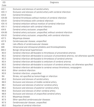
Cases were ascertained by using Inter-national Classification of Diseases (ICD) revisions 9 and 10 codes applied to discharge diagnoses (Table 1), on the basis of standardized methods rec-ommended by the International Pediat-ric Stroke Study. These cases were cross-referenced with the institution’s hematology database and stroke reg-istry (which commenced in 2002) to ensure that no patients were missed. AIS was defined as (1) an acute
neuro-logic deficit lastingⱖ24 hours and (2)
parenchymal infarction on neuroimag-ing scans. Inclusion criteria included a first presentation of AIS in patients from the newborn period (defined as
⬍28 days of age) up to 18 years of age,
with radiologic confirmation of AIS. Pa-tients with the following conditions were excluded: sinovenous thrombo-sis and hemorrhagic stroke subtypes, systemic vasculitides, birth before 36 weeks of gestation, other non-AIS dis-orders associated with focal deficits, and recurrent strokes. Subjects who did not have documentation of the times of clinical onset and neuroimag-ing were excluded.
Medical records were reviewed retro-spectively by Dr Srinivasan, and data on the pathways to stroke diagnosis Code
433.1 Occlusion and stenosis of carotid artery
433.11 Occlusion and stenosis of carotid artery with cerebral infarction 434.0 Cerebral thrombosis
434.00 Cerebral thrombosis without mention of cerebral infarction 434.01 Cerebral thrombosis with cerebral infarction
434.10 Cerebral embolism without mention of cerebral infarction 434.11 Cerebral embolism with cerebral infarction
434.9 Cerebral artery occlusion, unspecified
434.90 Cerebral artery occlusion, unspecified, without cerebral infarction 434.91 Cerebral artery occlusion, unspecified, with cerebral infarction 437.5 Moyamoya disease
437.9 Cerebrovascular disease, unspecified 438 Late effects of cerebrovascular disease
G08 Intracranial and intraspinal phlebitis and thrombophlebitis G93.2 Benign intracranial hypertension
I63.0 Cerebral infarction attributable to thrombosis of precerebral arteries
I63.2 Cerebral infarction attributable to occlusion of precerebral arteries, not otherwise specified I63.3 Cerebral infarction attributable to thrombosis of cerebral arteries
I63.4 Cerebral infarction attributable to embolism of cerebral arteries
I63.5 Cerebral infarction attributable to occlusion of cerebral arteries, not otherwise specified I63.6 Cerebral infarction attributable to cerebral venous thrombosis, nonpyogenic
I63.8 Other cerebral infarction I63.9 Cerebral infarction, unspecified
I64 Stroke, not specified as hemorrhage or infarction I65.2 Occlusion and stenosis of carotid artery I66.0 Occlusion and stenosis of middle cerebral artery I66.1 Occlusion and stenosis of anterior cerebral artery I66.2 Occlusion and stenosis of posterior cerebral artery I66.8 Occlusion and stenosis of other cerebral artery I66.9 Occlusion and stenosis of unspecified cerebral artery I67.5 Moyamoya disease
I67.8 Other specified cerebrovascular diseases I67.9 Cerebrovascular disease, unspecified I69.3 Sequelae of cerebral infarction
were collected. “Prehospital” times re-corded included the time of clinical on-set and the time to the Royal Children’s Hospital or another medical center. Whether the child presented to the ter-tiary center directly or indirectly (through a family physician or periph-eral hospital) also was ascertained.
“Posthospital” times recorded
in-cluded the time of physician assess-ment, the time of initial imaging, and the time of neuroimaging confirming AIS. The time of neuroimaging confirm-ing AIS was obtained from a time stamp recorded at the time of the pro-cedure and might not have been equiv-alent to the time of initial neuroimag-ing, if a diagnosis of AIS was not made immediately after the first imaging study (for example, if the initial study was a computed tomographic [CT] study that did not reveal the AIS). The time of clinical onset was deter-mined in a number of ways. The pre-ferred method was to use the exact
time of clinical onset (such as 9:00AM),
as documented in the medical chart by physicians or nurses. In the absence of such information, the clinical onset was extrapolated from other docu-mented information. For example, if the physicians documented that “hemiparesis began 2 hours ago” at
11:00 AM, then stroke onset was
esti-mated at 9:00AM. If the symptoms were
present at awakening, then the last time the patient was seen in normal condition was used. For the sedated population and for certain ICU pa-tients, the time of clinical onset was determined from the detailed regular neurologic observations performed by ICU nursing staff members, which yielded a time of clinical onset within 15 minutes. In cases with discrepan-cies between the physicians and nurses, the order of preference was as follows: earliest precise time documented, neurologic observations charted by nurses, and information
ex-trapolated from other documented data as described above. The following information also was collected: age at time of diagnosis, presence of localiz-ing signs, seizures at presentation, risk factors, documented differential diagnoses (including suspicion of AIS), and neuroimaging modality.
Focal neurologic deficits included fo-cal neurologic signs and fofo-cal sei-zures. A focal sign was defined as a motor (hemiplegia, monoplegia, or ataxia), sensory (paresthesia or dyses-thesia), visual (visual field deficit or eye deviation), or speech (dysarthria or dysphasia) deficit. Stroke subtypes were classified into 8 categories by us-ing the Pediatric Stroke Classification, a preliminary modification of the Trial of ORG 10172 in Acute Stroke Treat-ment system, that is, sickle cell dis-ease, cardioembolic stroke,
moya-moya syndrome, cervical arterial
dissection, stenoocclusive arteriopa-thy, other determined cause, multiple probable/possible causes, and
unde-termined cause.12
The associations between variables were analyzed by using Stata 9.2 (Stata, College Station, TX), with the 2-sample rank-sum (Mann-Whitney) test and Kruskal-Wallis test where
ap-propriate.Pvalues ofⱕ.05 were
con-sidered statistically significant.
RESULTS
Study Group
During this 12.5-year period, 375
pa-tients hadⱖ1 of the listed ICD codes
(Fig 1). A total of 107 patients, including 19 neonates and 88 children, met the
inclusion criteria. Most patients
(⬎70%) had a precise time of clinical
onset documented.
The overall median age at the time of diagnosis was 20 months (interquar-tile range [IQR]: 62 days to 112 months), and the male/female ratio was 1.27:1. The median age at the time
of diagnosis for children was 36 months (IQR: 6 –118 months). The me-dian age at the time of diagnosis for neonates was 6 days (IQR: 3–12 days). There were no significant differences between the excluded and included populations with respect to gender or age. Sixty percent of the study subjects were inpatients at the time of stroke onset. The remaining 40% were not in-patients at the time of AIS onset, with 13% presenting directly to the Royal Children’s Hospital emergency depart-ment after clinical onset and 27% pre-senting through a peripheral center.
Children (Nonneonatal Population) Times to AIS Confirmation
A median time of 24.8 hours (IQR: 10.2– 67.0 hours) from clinical onset to ra-diologic confirmation of AIS was noted for the population of children (Tables 2 and 3). For inpatients, the median time was 21.3 hours (IQR: 5.8 –54.1 hours); for outpatients, the median time was
27.4 hours (IQR: 18.9 –74.6 hours;P⬍
.001).
Six (6.8%) of 88 patients were diag-nosed as having AIS within 3 hours (Ta-ble 2); all were inpatients with cardio-embolic risk factors (Table 3). Five patients had structural heart disease. Five of the 6 children had focal signs. Five of the 6 children also had radio-logic evidence of hemorrhagic conver-sion or involvement of more than one third of the middle cerebral artery ter-ritory on initial imaging scans. Fifty percent of all 88 children with AIS did not have their stroke radiologically
confirmed untilⱖ24 hours after
clini-cal onset.
Times to Seeing a Physician and Initial Imaging
The time of first physician assessment was documented for 37 (42%) of the 88 children, and the median time from clinical onset to the first evaluation by a physician in this group was 1 hour
(IQR: 0 – 4.5 hours). Of all children, 75% were seen by a physician within 3 hours. For inpatients, the median time to physician assessment was 20 min-utes (IQR: 0 –180 minmin-utes); for outpa-tients, the median time was 120
min-utes (IQR: 57– 408 minmin-utes;P⫽.003)
(Table 2). The time to initial imaging was 11.4 hours (IQR: 4.6 –25.5 hours) for inpatients and 9.6 hours (IQR: 4.5–
20.6 hours) for outpatients (P⬎.05).
Initial Suspicion of AIS
A suspected diagnosis or differential diagnosis was documented by the first physician to see the patient for 44 (50%) of the 88 patients. Physicians documented suspicion of AIS for only 23 (26%) of the 88 children. Other
documented differential diagnoses
included seizures, central nervous system infections, sepsis, tumors, in-creased intracranial pressure, and, less commonly, migraines, musculo-skeletal conditions, and electrolyte disturbances. The small number of cli-nicians who documented a suspicion of AIS precluded analysis of whether suspicion of AIS influenced the time to neuroimaging.
Presence of Focal Signs or Focal Seizures
At the time of the first physician as-sessment, 76 (86%) of 88 patients had a focal sign (Table 4). The major-ity of focal signs were motor deficits except for the neonates, for whom focal seizures predominated. The presence of a focal sign was associ-ated with increased suspicion of AIS
(P⫽.02), with physicians suspecting
AIS for 23 (30%) of the 76 children with focal deficits, compared with no suspicion of AIS for the 12 children without focal deficits. However, the presence of a focal sign was not as-sociated with a shorter time to stroke confirmation.
179 patients excluded as not having had AIS
66 patients excluded as not first presentation of AIS
130 neonates and children with first presentation of AIS identified
23 patients excluded as having inadequate documentation of timing
Total of 107 patients: 19 neonates 88 children
19 neonates 88 children
FIGURE 1
Patients included in study.
TABLE 2 Distribution of Times From Clinical Onset to Confirmation of AIS Diagnosis (N⫽88)
Time n(%)
Within 3 h 6 (6.8)
⬎3 to 6 h 9 (10.2)
⬎6 to 9 h 5 (5.7)
⬎9 to 24 h 24 (27.3)
⬎24 h 44 (50.0)
There were no significant associations between seizures at presentation and suspicion of AIS or between seizures at presentation and time to confirmation. None of the cases reviewed in our ret-rospective study had documentation detailing cognitive deficits in children with acute stroke.
Risk Factors for AIS
By using the Pediatric Stroke Classifi-cation system, detailed stroke causes for the 88 nonneonates were catego-rized (Table 5). The 19 neonates were excluded from this analysis. Two of the 8 Pediatric Stroke Classification cate-gories, namely, moyamoya syndrome and sickle cell disease, were not rep-resented among the children.
Admission Status at Time of Stroke
Almost 58% of our children (n⫽ 51)
were inpatients at the time of AIS (Ta-ble 6). All inpatients with AIS had iden-tifiable risk factors, compared with 84% of the outpatients. Almost 69% of inpatient strokes resulted from a likely cardioembolic mechanism secondary to previously recognized cardiac risk factors, including structural cardiac defects, previous cardiac surgery and/or cardiac catheterization, or treatment with an extracorporeal cir-culation device (eg, extracorporeal membrane oxygenation treatment).
Imaging
The proportions of children who un-derwent brain MRI, brain CT imaging, or ultrasonography as initial imaging were 88.9%, 94.9%, and 30%, respec-tively. MRI and CT scanning were per-formed as initial imaging primarily in the older population, with more ultra-sound studies in the neonatal popula-tion. Initial imaging yielded negative results despite AIS for 62 of 74 children who underwent CT imaging and 3 of 6 children who underwent ultrasonog-raphy. MRI, when used as the initial im-aging modality, confirmed all strokes in 8 children.
Neonatal Population
The median time to confirmation of AIS for the neonates was 87.9 hours (IQR: 53.4 –166.4 hours), which was signifi-cantly longer than the time for the chil-dren, for whom the median time to AIS confirmation was 24.8 hours (IQR:
10.2– 67.0 hours;P⫽.0002). There was
significantly less documentation of AIS suspicion in the medical records of
ne-onates (P⫽ .04), and seizures were
more common at presentation for
ne-onates, compared with children (P⫽
.0001). Anterior circulation AIS pre-dominated for both age groups (68% for neonates and 63% for children).
DISCUSSION
Children
There is significant delay in the diagno-sis of stroke in children, with a median time of almost 25 hours from clinical onset to radiologic confirmation of AIS.
Gabis et al3demonstrated a large
pre-hospital delay (mean time: 28.5 hours) contributing to the overall delay in stroke diagnosis (mean time: 35.7 hours). In contrast, our study showed that the median time from clinical on-set to assessment by a physician was 1
TABLE 5 Etiologic Subtypes in Childhood Population, on the Basis of Modified Trial of ORG 10172 in Acute Stroke Treatment Classification
Classification n(%)
Total Inpatient at Time of AIS (N⫽51)
Noninpatient at Time of AIS (N⫽37)
Cardioembolic stroke 40 (45.4) 35 (68.6) 5 (13.5)
Dissection 2 (2.3) 0 (0) 2 (5.4)
Stenoocclusive arteriopathy 8 (9.1) 1 (2) 7 (18.9)
Other determined cause 12 (13.6) 3 (5.9) 9 (24.3)
Possible/probable risk factor 20 (22.7) 12 (23.5) 8 (21.6)
No identifiable risk factors 6 (6.8) 0 (0) 6 (16.2)
Total 88 51 37
No patients who met the inclusion criteria were identified within the categories of sickle cell disease or moyamoya syndrome. Cardioembolic causes include congenital heart disease, cardiac surgery, extracorporeal membrane oxygen-ation, and endocarditis. Other determined causes include fibromuscular dysplasia, primary angiitis of the central nervous system, radiation-related angiopathy, transtentorial herniation, chronic illnesses, and trauma. Possible/probable risk factors include migraine, central nervous system infection, sepsis, postprocedural status, and thrombophilia.
TABLE 6 Relationship of AIS to Age and Admission Status
Time P
Time to diagnosis, median (IQR), h
Neonate (n⫽19) 87.9 (53.4–166.4) .0002
Nonneonate (n⫽88) 24.8 (10.2–67)
Time to AIS confirmation, median (IQR), h
Inpatient (n⫽51) 21.3 (5.8–54.1) ⬍.001
Noninpatient (n⫽37) 27.4 (18.9–74.6)
Time to first physician assessment, median (IQR), min
Inpatient (n⫽37) 20 (0–180) .003
Noninpatient (n⫽26) 120 (57–408)
TABLE 4 Types of Focal Neurologic Deficits in Childhood Population (N⫽76)
Focal Neurologic Deficit n
Motor alone 30
Motor with other features
Seizures 2
Speech deficit 9
Visual deficit 8
Sensory deficit 1
ⱖ2 of above 5
Focal seizures alone 4
Focal seizures and visual deficit 6 Sensory (visual and/or other sensory) 11
Motor deficit indicates focal weakness or uncoordination; speech deficit, dysphasia or aphasia; visual deficit, visual field deficit or eye deviation.
hours after clinical onset. These find-ings are similar to those of a recent study from the United Kingdom, which found that three fourths of children were seen by a physician within 6
hours.4 Although prehospital delay
was demonstrated recently to be
pre-dictive of delayed AIS diagnosis,7 our
findings suggest that the delay in diag-nosis is more likely posthospital delay, largely related to the lack of aware-ness of stroke among medical staff members.
Eighty-six percent of our children had
ⱖ1 focal sign at the time of
presenta-tion, but one third of our patients had nonmotor (ie, nonhemiplegic) presen-tations. One half of the physicians doc-umented a differential diagnosis, and even fewer included stroke in the list of possibilities.
Our findings suggest that pediatric stroke is an underrecognized problem within the medical community, despite the significant associated long-term disability and the fact that more than one half of children have residual
func-tional deficits.1 Furthermore, almost
58% of our older subjects were inpa-tients at the time of their AIS, and al-most 69% had known cardiac risk fac-tors for stroke. This is important because cardiac disease is the most common, previously recognized risk
factor.13–15 Children with cardiac
dis-ease represent the one group that could potentially be targeted with pri-mary prevention strategies.
Although they met the theoretical 3-hour criterion for intravenous throm-bolysis, 5 of 6 children in our series
who presented within 3 hours
would not have been eligible for intra-venous thrombolysis because of exten-sive infarction or the presence of
hemorrhagic conversion.16,17 Cranial
ultrasonography and CT scanning are not as sensitive as MRI of the brain
showed that the stroke diagnosis was missed for 3 of 6 children who initially underwent ultrasonography and 62 of 74 children who initially underwent CT scanning. This is similar to the results of a United Kingdom study in which AIS was not detected on CT scans in 47% of
cases.4
Stroke-mimickers, such as hemiplegic migraine or Todd paresis, can cause
delay in AIS diagnosis.5Emergency
ra-diologic confirmation of acute infarc-tion and arterial occlusion will un-doubtedly be essential requirements in the first pilot safety trials of
throm-bolysis in childhood stroke.18
Hyperacute therapies have not been approved for children, and physicians may feel less urgency to perform rapid imaging. If such therapies were to be shown to be effective for children, then the time to diagnosis would likely de-crease as clinicians and radiologists prioritize neuroimaging studies.
Neonates
Pediatric stroke registry data suggest that 25% to 30% of all cases occur in neonates and almost 50% in children
⬍1 year of age.13Not surprisingly,
di-agnosis took longer for neonates with AIS than for the childhood population. This is because localizing signs often are absent, which makes stroke on-set difficult to determine accurately for neonates. Focal seizures can pre-dominate over motor deficits. Stroke should be suspected for any neonate presenting with seizures, lethargy, or apnea, the latter 2 features being seen in neonatal stroke in the setting
of encephalopathy.19
Study Population and Limitations Our study had a larger study popula-tion, compared with previous
stud-ies.3,4We defined our time of stroke
di-agnosis as the time of neuroimaging
there is close collaboration between pediatric neurologists and radiolo-gists, which makes use of the time of neuroimaging a more-reliable ap-proach to assess the time of stroke di-agnosis in our population. The results were not reviewed independently, and the timing of negative studies was not analyzed, because positive imaging re-sults represented an inclusion crite-rion. All scans were evaluated by at-tending radiologists who were familiar with neuroimaging. When assessment is performed with a suspicion of AIS, the radiologist’s interpretation of the neuroimaging scans at the Royal Chil-dren’s Hospital is conveyed immedi-ately to the referring doctor.
Other studies defined the time of AIS diagnosis to be the time the diagnosis
was entered in the medical chart.4We
chose radiologic confirmation be-cause a suspected diagnosis or differ-ential diagnosis was documented by the first physician to see the patient for only 50% of our population and a suspicion of AIS was documented for only 26% of children. A limitation of our definition for determining the time to stroke diagnosis is that radiologic con-firmation may not equal clinical confir-mation; however, because there often are delays or omissions in medical documentation, we think that the time to radiologic diagnosis is most likely to represent the true time to AIS diagno-sis in our population.
from a large tertiary pediatric hospi-tal, which houses a major cardiac unit. Therefore, cardioembolic stroke is more frequent in our population than in other studies, and the results are not entirely applicable to the general population. However, we would have anticipated that such a high-risk popu-lation would have a shorter time to stroke diagnosis than that in a gen-eral, community-based center. The fact that only 17% of our patients were neo-nates reflects the fact that we are not colocated with an obstetric unit and most neonatal cases are treated at other centers. In addition, sickle cell disease is uncommon in the Australian pediatric population, and patients with moyamoya syndrome might have been excluded for reasons such as previous AIS.
Patient-related factors influencing the time to diagnosis include the possible need for a general anesthetic for imag-ing for young children, and comorbidi-ties such as cardiac disease and criti-cal illness may mean that children are too ill to be transported to the scanner. In addition, admission to an ICU, and thus the need for sedation, may mask focal signs. Many inpatients may
have contraindications to hyperacute therapy, leading to delayed imaging because clinicians think that imag-ing results are unlikely to change management.
The delays associated with AIS are at-tributable to both decreased aware-ness in the medical community and problems with access to neuroimag-ing, including those associated with patient comorbidities precluding ur-gent scanning. The lack of available treatment options for the pediatric stroke population is the likely reason for delayed imaging for children, be-cause similar findings have been
ob-served for adult stroke patients.20
Cur-rent practice is the likely culprit in perpetuating delays, and this factor needs to be addressed. Although thrombolysis is an important factor, there are other measures for address-ing delays in diagnosis that can change long-term outcomes. Mea-sures that have been shown to im-prove outcomes in adults include control of fever and hypertension, maintenance of normal oxygenation, normalization of serum glucose levels,
and early access to rehabilitation.11
CONCLUSIONS
It is hoped that progress will de-crease the time lag to stroke diagno-sis through better understanding of risk factors in the pediatric
popula-tion.21The American College of Chest
Physicians has stated, “the infre-quency of stroke in children results in delayed recognition and an inabil-ity to intervene early with medica-tions that may reduce subsequent
neurologic deficits.”9 Clearly, there
are delays in childhood stroke diag-nosis that must be addressed with increased awareness among medi-cal professionals, particularly for high-risk groups such as children with cardiac disease. Ideally, devel-opment of hospital-based pediatric stroke services will improve early di-agnosis of stroke, to allow children the benefits of acute medical inter-ventions that have been shown to
improve outcomes in the adult
population.
ACKNOWLEDGMENTS
We thank Drs James H. Lee and Manali Chitre for their valuable contributions in editing the manuscript.
REFERENCES
1. Ganesan V, Hogan A, Shack N, Gordon A, Isaacs E, Kirkham FJ. Outcome after ischemic stroke in childhood.Dev Med Child Neurol.2000;42(7):455– 461
2. Saver JL. Time is brain: quantified.Stroke.2006;37(1):263–266
3. Gabis LV, Yangala R, Lenn NJ. Time lag to diagnosis of stroke in children.Pediatrics.2002;110(5): 924 –928
4. McGlennan C, Ganesan V. Delays in investigation and management of acute arterial ischaemic stroke in children.Dev Med Child Neurol.2008;50(7):537–540
5. Shellhaas RA, Smith SE, O’Tool E, Licht DJ, Ichord RN. Mimics of childhood stroke: characteristics of a prospective cohort.Pediatrics.2006;118(2):704 –709
6. Braun KP, Kappelle LJ, Kirkham FJ, Deveber G. Diagnostic pitfalls in pediatric ischemic stroke.Dev Med Child Neurol.2006;48(12):985–990
7. Rafay MF, Pontigon AM, Chiang J, et al. Delay to diagnosis in acute pediatric arterial ischemic stroke.Stroke.2009;40(1):58 – 64
8. Braun KP, Rafay MF, Uiterwaal CS, Pontigon AM, DeVeber G. Mode of onset predicts etiological diagnosis of arterial ischemic stroke in children.Stroke.2007;38(2):298 –302
9. Monagle P, Chan A, Massicotte P, Chalmers E, Michelson AD. Antithrombotic therapy in children: the Seventh ACCP Conference on Antithrombotic and Thrombolytic Therapy.Chest.2004;126(3 suppl):645S– 687S
10. Royal College of Physicians, Paediatric Stroke Working Group.Stroke in Childhood: Clinical Guide-lines for Diagnosis, Management and Rehabilitation. London, England: Royal College of Physicians;
11. Roach ES, Golomb MR, Adams R, et al. Management of stroke in infants and children: a scientific statement from a special writing group of the American Heart Association Stroke Council and the Council on Cardiovascular Disease in the Young.Stroke.2008;39(9):2644 –2691
12. Wraige E, Pohl KR, Ganesan V. A proposed classification for subtypes of arterial ischemic stroke in children.Dev Med Child Neurol.2005;47(4):252–256
13. deVeber G; Canadian Pediatric Ischemic Stroke Study Group. Canadian Pediatric Ischemic Stroke Study registry: analysis of children with arterial ischaemic stroke.Ann Neurol.2000;48(4):514 14. Ganesan V, Prengler M, McShane MA, Wade AM, Kirkham FJ. Investigation of risk factors in
children with arterial ischemic stroke.Ann Neurol.2003;53(2):167–173
15. Chabrier S, Husson B, Lasjaunias P, Landrieu P, Tardieu M. Stroke in childhood: outcome and recurrence risk by mechanism in 59 patients.J Child Neurol.2000;15(5):290 –294
16. Hacke W, Kaste M, Fieschi C, et al. Randomised double-blind placebo-controlled trial of thrombo-lytic therapy with intravenous alteplase in acute ischaemic stroke (ECASS II).Lancet.1998; 352(9136):1245–1251
17. National Institute of Neurological Disorders and Stroke rt-PA Stroke Study Group. Tissue plasmin-ogen activator for acute ischemic stroke.N Engl J Med.1995;333(24):1581–1587
18. Whelan HT, Cook JD, Amlie-Lefond CM, et al. Practical model-based dose finding in early-phase clinical trials: optimizing tissue plasminogen activator dose for treatment of ischemic stroke in children.Stroke.2008;39(9):2627–2636
19. Ramaswamy V, Miller SP, Barkovich AJ, Partridge JC, Ferriero DM. Perinatal stroke in term infants with neonatal encephalopathy.Neurology.2004;62(11):2088 –2091
20. Rose KM, Rosamond WD, Huston SL, Murphy CV, Tegeler CH. Predictors of time from hospital arrival to initial brain-imaging among suspected stroke patients: the North Carolina Collaborative Stroke Registry.Stroke.2008;39(12):3262–3267
DOI: 10.1542/peds.2008-3544 originally published online July 20, 2009;
2009;124;e227
Pediatrics
Jayasri Srinivasan, Steven P. Miller, Thanh G. Phan and Mark T. Mackay
Delayed Recognition of Initial Stroke in Children: Need for Increased Awareness
Services
Updated Information &
http://pediatrics.aappublications.org/content/124/2/e227
including high resolution figures, can be found at:
References
http://pediatrics.aappublications.org/content/124/2/e227#BIBL
This article cites 20 articles, 9 of which you can access for free at:
Subspecialty Collections
http://www.aappublications.org/cgi/collection/neurology_sub
Neurology
following collection(s):
This article, along with others on similar topics, appears in the
Permissions & Licensing
http://www.aappublications.org/site/misc/Permissions.xhtml
in its entirety can be found online at:
Information about reproducing this article in parts (figures, tables) or
Reprints
http://www.aappublications.org/site/misc/reprints.xhtml
DOI: 10.1542/peds.2008-3544 originally published online July 20, 2009;
2009;124;e227
Pediatrics
http://pediatrics.aappublications.org/content/124/2/e227
located on the World Wide Web at:
The online version of this article, along with updated information and services, is
by the American Academy of Pediatrics. All rights reserved. Print ISSN: 1073-0397.