• No results found

Invasive salivary duct carcinoma ex pleomorphic adenoma of the parotid gland: a teaching case giving rise to the genuine diagnostic difficulty on an inadequate cytology specimen

N/A
N/A
Protected

Academic year: 2020

Share "Invasive salivary duct carcinoma ex pleomorphic adenoma of the parotid gland: a teaching case giving rise to the genuine diagnostic difficulty on an inadequate cytology specimen"

Copied!
7
0
0

Loading.... (view fulltext now)

Full text

Loading

Figure

Figure 1 Cytomorphologic examination of the fine needle aspiration specimens. (A) The cytology specimens were inadequate, butconsisted of few small clusters of cohesive and three-dimensional pleomorphic tumor cells in a papillary-like fashion (rt
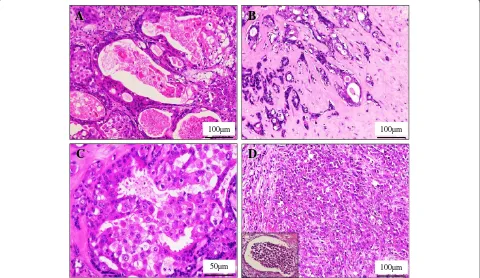
Figure 3 Microscopic examination of the invasive SDC ex PA. (A) In high power view, the ID components of SDC showed a proliferation ofhighly atypical epithelial cells having hyperchromatic pleomorphic nuclei and abundant eosinophilic cytoplasm, often arran
Figure 5B, inset). Also, the lost of myoepithelial cells
Figure 5 Myoepithelial markers analysis of the invasive SDC ex PA in immunohistochemistry.S-100 protein (side) (Original magnification × 200, Bar = 100mildly enlarged and hyperchromatic nuclei were sometimes negative for both of S-100 protein (A, inset) an

References

Related documents