International Journal of Medical Science and Current Research (IJMSCR)
Available online at: www.ijmscr.com
Volume1, Issue 2, Page No: 62-67 July-August 2018
62
Liver Function Tests in Burn Patients
Channabasavanna G. Halasagi, K F Kammar
1. Assistant Professor, Dept of Physiology, KMCT Medical College, Mukkam, Kerala. 2.
Professor and Head of the Dept, Dept of Physiology, KIMS Hubli, Karnataka
Corresponding Author: Channabasavanna G. Halasagi
Assistant Professor, Dept of Physiology, KMCT Medical College, Mukkam, Kerala
Type of Publication: Original Research Paper Conflicts of Interest: Nil
ABSTRACT
Background:Burn injury is associated with physiological, biochemical, immunological and anatomical alterations which require specialized care. Response to burn injury includes cellular protection mechanisms, inflammation, hypermetabolism, prolonged catabolism, organ dysfunction and immunosuppression. The present study is aimed at comparing the liver function tests in 30 female burn cases and 30 female healthy controls.
Aim and Objectives: The objectives of this study were to determine on the first day of admission, the Liver Function Tests (LFT)- Total Serum Protein, Serum Albumin, Total Serum Bilirubin, Direct Bilirubin, Aspertate Transaminase (AST), Alanine Transaminase (ALT) and Alkaline Phosphatase (ALP) and to compare the values with healthy controls.
Materials and Methods:Investigations were carried out in 30 female burn patients with burns of 21 – 50% on the first day of admission to burns ward and 30 healthy females were taken as controls.Statistical analysis was done by using unpaired‘t’ test. Analysis was done using SPSS 20 software.
Results:The LFT profile showed following values, the mean of total serum protein was 5.47 ± 0.63g/dL, serum albumin was 2.68 ± 0.58g/dL, total serum bilirubin 1.27 ± 0.28mg/dL, direct bilibubin was 0.68 ± 0.26mg/dL, AST was 70.80 ± 21.71U/L, ALT was 54.10 ± 12.35U/L and ALP was 57.66 ± 13.90U/L.The values of Total serum protein and Serum albumin were significantly lower as compared to healthy controls. Whereas Total serum bilirubin, direct bilirubin, AST, ALT and ALP were significantly increased as
compared to healthy controls.
Keywords: Burns;Albumin;Total protein;AST;ALT;ALP;Bilirubin.
INTRODUCTION
The skin is one of the largest and most versatile organs of the body. As the first line of defense, the skin is continuously subjected to potentially harmful environmental agents including solid matter, liquids, gases, sunlight and microorganisms. The skin serves
as immunological barrier1.
Burn trauma is caused by a wide variety of substances and external sources such as exposure to chemicals, friction, electricity, radiation and heat.
It is one of the most common injuries - mainly in developing countries. Two main factors define burn severity: depth of burn injury (which depends on the temperature and exposition time) and burnt body surface. Burn injury, affecting more than 20% of the
body surface area can lead to burn
disease.Immunodeficiency can develop because of the reduced immunoglobulin synthesis (down-regulation), with consequence of an increased
acquisition of infection2.
The immune system activates a high amount of inflammatory mediator release.
Macrophage and leukocyte activation triggers free radical, arachidonic acid and metabolites formation which play a role in early edema formation and cytokine (TNFα, IL-1,2, 6) production. The released metabolites have significant effect on both local
wound and systemic inflammatory reaction3.After
Pag
e
63
Pag
e
63
Pag
e
63
Pag
e
63
Pag
e
63
Pag
e
63
Pag
e
63
Pag
e
63
Pag
e
63
Pag
e
63
Pag
e
63
Pag
e
63
Pag
e
63
Pag
e
63
Pag
e
63
Pag
e
63
Pag
e
63
Pag
e
63
Pag
e
63
Pag
e
63
Pag
e
63
leukotriene, proinflammatory cytokines and free radicals play a role. i.e. more than the 20% of the BSA, generalized edema formation occurs; this fluid loss leads to hypovolaemia in the intravascular space and to hypoperfusion which subsequently results in the damage of cells and organs.Thermal injuries result in significant pathophysiological changes by interplay of various mediators in early stages. These changes further exacerbate the whole body inflammatory response into vicious cycle of
accelerating organ dysfunction4.The burn wounds
have much higher incidence of sepsis as compared to other forms of trauma due to disruption of skin barrier and alteration in cellular and humoral responses5.
In the view of the fact that numerous complications can occur in burn patients, a prospective study was undertaken to determine liver function tests in burn patients.Variations in biochemical parameters like total protein, albumin, bilirubin, AST, ALT, alkaline phosphatase were studied which reflects hepatic changes in response to inflammatory changes.
Aims and Objectives:
The present study was undertaken in the Department of Physiology and with laboratory assistance from the Department of Pathology and Biochemistry KIMS, Hubballi from January 2014 to June 2015.
Objectives:
1. To study Liver Function Tests in same
patients. (21 to 50% burns)
2. To compare Liver Function Tests with
control subjects.
Material and Methods:
This study was conducted in the Department of Physiology, KIMS Hubballi with the assistance of laboratory setup of the Department of Pathology and Biochemistry, KIMS, Hubli.Ethical approval was obtained from the ethical committee of KIMS, Hubli. This study was performed from January 2014 to September 2015 in the Department of Physiology.
Source of data
The present study included 30 burn victims all females, burns ranging from 21 to 50% TBSA (total body surface area). The sample of study group was taken from Burns Ward, KIMS, Hubli.
30 healthy females were taken for the study as controls.
The subjects were selected on the basis of inclusion and exclusion criteria.
Inclusion criteria:
All female burn patients with burns of 21 – 50 % in KIMS, Hubli.
Exclusion criteria:
1. Patients should not have blood
disorders and hepatic diseases.
2. Patients should not have h/o
medications which affect hematological parameters and liver function tests.
All values are expressed as mean ± standard deviation. The data is analyzed by using computer SPSS-20 software and p value < 0.05 as the lowest limit of significant. Unpaired “t” test is used analyze the data of cases and controls.
As all burn cases are admitted as medico legal cases, we have restricted access to these patients. Patient attenders were told about the study undertaken and the laboratory results would be helpful for the patient treatment as well. Patient attenders were explained about the procedure in their own local language. Informed consent was obtained from patient attenders. Blood from the patients whose relatives have denied this procedure was excluded from the studies.Complete clinical examination was done when the patient was selected as per our study protocol. The collection of blood sample was done at 9 AM on the first day of admission. Five ml of blood was drawn under aseptic precautions.
Following Liver function test were done.
1. Total protein
2. Serum Albumin
3. Serum Bilirubin
4. Serum Aspartate transaminase (AST)
and Alanine transaminase (ALT)
5. Serum Alkaline phosphatase.
Results:
Pag
e
64
Pag
e
64
Pag
e
64
Pag
e
64
Pag
e
64
Pag
e
64
Pag
e
64
Pag
e
64
Pag
e
64
Pag
e
64
Pag
e
64
Pag
e
64
Pag
e
64
Pag
e
64
Pag
e
64
Pag
e
64
Pag
e
64
Pag
e
64
Pag
e
64
Pag
e
64
Pag
e
64
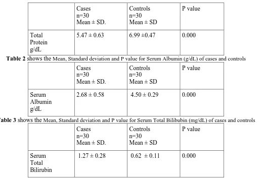
0.63 in patients whereas in controls the mean value is 6.99 with standard deviation as 0.47. Total Serum Protein in burn patients was reduced with highly significant values.
Table 2 shows mean value of Serum Albumin (g/dL) as 2.68 with Standard deviation of 0.58 in patients whereas in controls the mean value is 4.50 with standard deviation as 0.29. Serum Albumin in burn patients was reduced with highly significant values.
Table 3 shows highly significant increase in Total Serum Bilirubin in patients with a mean value of 1.27mg/dL with standard deviation of 0.28 whereas in controls it was 0.62 with SD of 0.11.
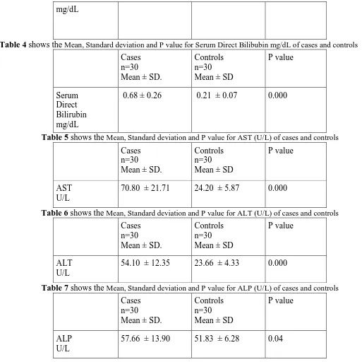
Table 4 shows a high significant increase in Direct Bilirubin of 0.68mg/dL with SD 0.26
Table 5 shows mean AST levels of 70.80 with SD of 21.71 in patients whereas controls showed 24.20 with SD of 5.87. There was a highly significant increase in AST level in patients.
Table 6 showed a highly increase in ALT (U/L) values with a mean of 54.10 and SD 12.35 in patients.
Table 7 showed an increase in the levels of ALP (U/L) with mean of 57.66 and SD 13.90 in patients. There was a significant increase in ALP values in patients.
Discussion
During the study period, a total of 30 burn cases (21 – 50% TBSA) all females and 30 controls all healthy females were evaluated for liver function tests.
Total Serum Protein and Serum Albumin.
Mean Total Serum Protein level (g/dL) in cases was 5.47 ± 0.63 and in control was 6.99 ± 0.47. The difference between the two groups was very highly significant
(‘p’ value < 0.0001).
Mean Serum Albumin level (g/dL) in cases was 2.68 ± 0.58 and in control was
4.50 ± 0.29. The difference between the two groups was very highly significant
(‘p’ value < 0.0001).
Similar observations were made by M Lehnhardt et
al6, protein levels in serum were significantly lower
as compared to physiological levels while wound fluid protein levels were elevated and remained high. This study qualifies and quantifies a significant protein loss in second degree burn wounds.
Study conducted by Aguayo-Becerra OA et al7
suggests that albumin levels showed highest sensitivity and specificity for mortality. At admission the albumin level could be used as a sensitive and specific marker of burns severity and indicator of mortality.
In a study conducted by Waxman et al8, considerable
protein losses were measured. These losses were greatest in the first 3 postburn days, being somewhat greater in full thickness burns compared to partial thickness burns. These data demonstrate significant protein losses through burn wounds greater than recent studies have considered. It is possible that inadequate nutritional replacement of these protein losses is partly responsible for the marked negative nitrogen balance of the early postburn period.
Similar observations were made by Vinha PP et al9,
the inflammatory stress caused by acute thermal injury was characterised by lower levels of total protein and albumin and higher levels of C-reactive protein and α-1 glycoprotein acid.
Similar observations were made by Ruot B et al10, a
reduction of serum albumin concentration in general consequences of various factors, including a change in its rate of synthesis, an increased catabolic rate, or a redistribution of albumin from to intestinal compartment. While in burn serum albumin is well known to decrease in response to inflammation.
Serum Total Bilirubin and Serum Direct Bilirubin
In our study, mean Serum Total Bilirubin level (mg/dL) in cases was 1.27 ± 0.28 and in control was 0.62 ± 0.11. The difference between the two groups was very highly significant
(‘p’ value < 0.0001).
Mean Serum Direct Bilirubin level (mg/dL) in cases was 0.68 ± 0.26 and in control was 0.21 ± 0.07. The difference between the two groups was very highly significant
(‘p’ value < 0.0001).
Similar observations were made by Czaja AJ et al11,
Pag
e
65
Pag
e
65
Pag
e
65
Pag
e
65
Pag
e
65
Pag
e
65
Pag
e
65
Pag
e
65
Pag
e
65
Pag
e
65
Pag
e
65
Pag
e
65
Pag
e
65
Pag
e
65
Pag
e
65
Pag
e
65
Pag
e
65
Pag
e
65
Pag
e
65
Pag
e
65
Pag
e
65
suggested that acute hemodynamic alterations were important etiologically. Intrahepatic cholestasis and jaundice developed later in septic or hypoxic patients and was accentuated by hemolysis and blood
transfusion. Late emergence of conjugated
hyperbilirubinemia suggested an underlying septic process.
AST, ALT and Alkaline Phosphatase (ALP)
Mean AST level (U/L) in cases was 70.80 ± 21.71 and in control was
24.20 ± 5.87. The difference between the two groups was very highly significant
(‘p’ value < 0.0001).
Mean ALT level (U/L) in cases was 54.10 ± 12.35 and in control was
23.66 ± 4.33. The difference between the two groups was very highly significant
(‘p’ value <0.0001).
Mean ALP level (U/L) in cases was 57.66 ± 13.90 and in control was
51.83 ± 6.28. The difference between the two groups was significant
(‘p’ value < 0.05).
Similar observations were made by Marc G Jeschke
et al12, AST and ALT significantly increased upon
burn trauma and ALP levels were significantly
altered in response to burn37.
Similar observations were made by Chiarelli et al13,
results showed an increase in alanine and aspertate
aminotransferases during recovery and after
discharge.
Study by MG Hala et al14 suggests an elevated AST,
ALT, ALP and gamma GT –because of impaired hepatic functions in burns patients.
Summary and Conclusion
The present study was done during January 2014 to September 2015, regarding Liver function tests in burn patients.Thirty female burn cases 21-50% TBSA and healthy females as controls were selected for the study. The cases were taken from burns ward KIMS, Hubli.Total serum protein, and Serum albumin levels were decreased significantly when compared with controls. AST, ALT, and ALP were increased
significantly.The total protein levels were
significantly decreased due to the well known explanation as capillary leak and protein loss lead to accumulation proteins in wound sera and ultimately to oedema formation. Serum albumin levels were decreased significantly due to acute hepatic injury leading to the decreased synthesis by hepatocytes. Hypoalbuminemia is associated with complications related to increased extravascular fluid, including edema, abnormal healing, and susceptibility to sepsis. The albumin levels could be used as sensitive and specific marker of burn severity in predicting mortality. Bilirubin levels were raised significantly due to hemolysis and morphologic changes in red blood cells with oxidative damage. Underlying septic process lead to hyperbilirubinemia as haemoglobin is broken down into bilirubin. AST and ALT levels were significantly raised because an increase in edema formation leads to liver cell damage, with release of hepatic enzymes. AST and ALT are the most sensitive indicators of hepatocyte injury. ALP levels were raised provides an elevation of the patency of bile channels at all levels, intrahepatic or extrahepatic. Intrahepatic cholestasis is developed due to septic or hypoxic consequences or accentuated hemolysis.
References
1. Muhammad Obaid Al-Muhammadi and HayderAbdulhusseinAzeez. Some physiological changes in burn patients.: Medical Journal of Babylon. 2011;8(3):303-17.
2. Babcock GF. Predictive medicine. Severe Trauma and Burns. Cytometry B ClinCytom 2003;50:48-53.
3. Robson MC. Burn sepsis, Crit Care Clin 1988;4:281-98.
Pag
e
66
Pag
e
66
Pag
e
66
Pag
e
66
Pag
e
66
Pag
e
66
Pag
e
66
Pag
e
66
Pag
e
66
Pag
e
66
Pag
e
66
Pag
e
66
Pag
e
66
Pag
e
66
Pag
e
66
Pag
e
66
Pag
e
66
Pag
e
66
Pag
e
66
Pag
e
66
Pag
e
66
7. Aguayo-Becerra OA, Torres-Garibay C,
Macias-Amezcua MD, Fuentes-Orozco C,
Chavez-Tostado Mde G, Andalon-Duenas E, Espinosa Partida A, Alvarez-Villasenor Adel S, Cortes-Flores AO, Gonzalez-Ojeda A. Serum albumin level as a risk factor for mortality in burn patients. 2013 Jul; 68(7): 940-5.
8.Waxman, Kenneth, Rebello, Tessio, Pinderski, Laura, O’Neal, Kelly, Khan, Nassir, Tourangeau, Steve, Himes, Edward, Cordill, Kelly RD. Journal of Trauma-Injury Infection & Critical Care. 1987 Feb; 27(2).
9. Vinha PP, Martinez EZ, Vannuchi H, Marchini JS, Farina JA Jr, Cunha SF. Effect of acute thermal injury in status of serum vitamins, inflammatory markers, and oxidative stress markers: preliminary data. 2013 Mar-Apr; 34(2):e87-91.
10. Rout B, Breuille D, Rambourdin F, Bayle G, Capitan P, Obled C. Synthesis rate of plasma
albumin is a good indicator of liver albumin synthesis in sepsis. 2000 Aug; 279(2): E244-51. 11.Czaja AJ, Rizzo TA, Smith WR Jr, Pruitt BA Jr. Acute liver disease after cutaneous thermal injury. 1975 Oct; 15(10): 887-94.
12. Marc G.Jeschke, Gerd G. Gauglitz, Garbiela A. Kulp, Celeste C, Finnerty, Felicia N. Williams, Robert Kraft, Oscar E. Suman, Ronald P. Mlcak, David N. Herndo. Published: July 18, 2011. DOI: 10.1371/journal.pone.0021245.
13.Chiarelli A, Casadei A, Pornaro E, Siliprandi L, Mazzoleni F. Alanine and Aspartate aminotransferases serum levels in burned patients: a long term study. 1987 Jul; 27(7):790-4. 14. MG Hala, MH Mahfouz. Biochemical alterations of amino acids, neurotransmitters and hepatic functions after thermal injury in rats. Egyptian Journal of Biochemistry and Molecular Biology. 2008; Vol.26(2):13-28.
Tables
Table 1 shows theMean, Standard deviation and P value for Total Serum Protein (g/dL) of cases and control
Cases n=30 Mean ± SD.
Controls n=30 Mean ± SD
P value
Total Protein g/dL
5.47 ± 0.63 6.99 ±0.47 0.000
Table 2 shows theMean, Standard deviation and P value for Serum Albumin (g/dL) of cases and controls
Cases n=30 Mean ± SD.
Controls n=30 Mean ± SD
P value
Serum Albumin g/dL
2.68 ± 0.58 4.50 ± 0.29 0.000
Table 3 shows theMean, Standard deviation and P value for Serum Total Bilibubin (mg/dL) of cases and controls
Cases n=30 Mean ± SD.
Controls n=30 Mean ± SD
P value
Serum Total Bilirubin
Pag
e
67
Pag
e
67
Pag
e
67
Pag
e
67
Pag
e
67
Pag
e
67
Pag
e
67
Pag
e
67
Pag
e
67
Pag
e
67
Pag
e
67
Pag
e
67
Pag
e
67
Pag
e
67
Pag
e
67
Pag
e
67
Pag
e
67
Pag
e
67
Pag
e
67
Pag
e
67
Pag
e
67
mg/dL
Table 4 shows theMean, Standard deviation and P value for Serum Direct Bilibubin mg/dL of cases and controls
Cases n=30 Mean ± SD.
Controls n=30 Mean ± SD
P value
Serum Direct Bilirubin mg/dL
0.68 ± 0.26 0.21 ± 0.07 0.000
Table 5 shows theMean, Standard deviation and P value for AST (U/L) of cases and controls
Cases n=30 Mean ± SD.
Controls n=30 Mean ± SD
P value
AST U/L
70.80 ± 21.71 24.20 ± 5.87 0.000
Table 6 shows theMean, Standard deviation and P value for ALT (U/L) of cases and controls
Cases n=30 Mean ± SD.
Controls n=30 Mean ± SD
P value
ALT U/L
54.10 ± 12.35 23.66 ± 4.33 0.000
Table 7 shows theMean, Standard deviation and P value for ALP (U/L) of cases and controls
Cases n=30 Mean ± SD.
Controls n=30 Mean ± SD
P value
ALP U/L