Management of Intra-articular Fracture of
Calcaneus by Combined Percutaneous and
Minimal Internal Fixation
Background: Fractures of the calcaneus are among the most challenging for the orthopaedic surgeon. The
treatment of the intra-articular calcaneum fracture remains controversial due to complications and complexity of
surgical anatomy. Treatment of calcaneal fracture ranges from non-operative treatment to operative. We present
intra-articular fracture of calcaneus treated by combined percutaneous and minimal internal fixation.
Methods: All cases evaluated either by X-ray or CT scan. All fractures were sanders two or three type evaluated
by CT scan and either joint depression or tongue type fracture by X-ray. Lateral approach was used, posterior facet
was exposed, reduced and fixed with one 4 mm canulated cancellous screws and 2 axial pins percutaneously from
tuberosity. Clinical evaluation of the outcomes was done by modified Rowe Score.
Results: Out of 22 patients, 14 were male and 8 cases were female. Average age of the patients was 30.5 yrs (15-63
yrs). Mode of the injury was RTA in 6 cases and fall from height in 16 cases. There was no soft tissue problem in any
patient. All fractures united without secondary displacement in an average of 8 weeks. Average duration of follow up
was 26 months (6-37 months). Average Modified Rowe Score was 80 (Range 55-95). Ten patients had excellent, 10
had good and 2 had satisfactory outcome.
Conclusions: Intra-articular fracture of the calcaneus can be well managed by minimal opening at the fracture
and fixation by single cancellous screw and 2 axial k-wires, so minimizes complications and results in comparable
outcomes.
Keywords: calcaneum; intra-articular fracture; internal fixation; Rowe Score.
Lamichhane A,
1Mahara D
11Department of Orthopaedics, Institute of Medicine, Tribhuwan University Teaching Hospital, Kathmandu, Nepal.
Correspondence:
Dr. Arjun Lamichanne, Department of Orthopaedics, Institute of
Medicine Tribhuwan University Teaching Hospital, Kathmandu, Nepal. Email: drajun@
ABSTRACT
INTRODUCTION
Calcaneal fractures have been described since the time of Hippocrates and throughout history as being associated with poor outcomes.1 Modern medicine has yet to come to a consensus opinion on the management of intra-articular calcaneal fractures.2 Management has ranged from no reduction to open reduction. Operative management has included primary arthrodesis, percutaneous reduction, and open reduction internal fixation.
Fractures of the calcaneus are among the most challenging for the orthopaedic surgeon. The treatment
of the intra-articular calcaneum fracture remains controversial due to complications and complexity of surgical anatomy. Treatment of calcaneal fracture ranges from non-operative treatment to operative methods using percutaneous pins, open reduction and grafting and internal or external fixations. Open reduction and internal fixation is becoming more widely accepted as the treatment of choice for displaced intra-articular fractures of the calcaneum.
Major surgical complications are wound healing and the peroneal tendon problems due to implants and inherent healing characteristics of the tissues around foot and
J Nepal Health Res Counc 2013 Jan;11(23):70-5
Original
ankle. We present intra-articular fracture of calcaneus treated by combined percutaneous and minimal internal fixation.
METHODS
There were 25 intra articular calcaneal fracture operated for the duration of 6 years. Clinical evaluation for the swelling, and possible compartment syndrome was done in all cases. Radiological evaluation of the patients done either by x-ray or computer tomogram. Cases included were sanders 2 and 3 or both joint depression and tongue type intra-articular fractures. Surgical fixation was planned once swelling was subsided. Average duration between injury and surgery was 10 days (5-21 days).
Surgical Technique
Patients are positioned on supine position with sand bag under the ipsilateral buttock.
Exsanguination is done with tourniquet applied in the thigh 250 mmHg. We use a straight incision on the lateral side of the foot approximately 1 to 1.5 cm distal to the tip of the fibula and roughly perpendicular to the long axis of the fibula. The incision starts approximately 1 cm posterior to the fibula and continues distally for a total length of 3 to 4 cm (Figure 1). This allows visualization of the posterior facet.
The peroneal tendons are identified and the distal portion of the retinaculum is released (Figure 2). The tendons are mobilized and retracted either anteriorly or posteriorly according to need for visualization of the facets. The fat pad in the sinus tarsi is mobilized proximal to distal and the capsule of the posterior talo-calcaneal joint is opened.
Figure 1. Superficial incision.
Figure 2. Deep incision.
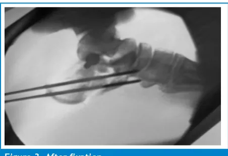
Posterior facet is elevated to align it with the medial sustantcular fragment and the depressed posterior facet is reduced to the talus. One K-wire is used to provisionally stabilize the facet and checked under image. One guide wire is passed from lateral to medial under image control, checked by lateral and axial views. The facet fragment is fixed with 4 mm cannulated cancellous screw and washer. Once this posterior facet fragment is fixed to the medial fragment and the joint has been successfully elevated, correction of the varus deformity of the posterior tuberosity is performed. The corrected tuberosity is fixed with 2 axial K-wires of 3 mm from posterior tuberosity percutaneously (Figure 3).
Figure 3. After fixation.
Evaluation of the patients was done with clinical and radiological means like, pain, infection, maintenance of reduction, union and functional outcomes. Functional outcomes were evaluated by using Modified Rowe Score3
Table 1. Modified Rowe score3 for clinical
evaluation.
Pain
None 30
Exercise induced 25
Mild on daily activity 20 Pain with weight-bearing 10
Pain at rest 0
Range of motion (per cent)
100 to 75 20
74 to 50 10
49 to 25 5
24 to 0 0
Gait
Normal 15
Mild limp (exercise) 10
Moderate limp 5
Severe limp 0
Activities
Normal 20
Restricted on rough ground
15
Moderate daily restrictions
10
Able to walk short distances only
5
Unable to walk 0
Work
No restrictions 15
Some restrictions on usual occupation
10
Change of job or substantial restrictions
5
Unable to work 0
Total 100
Grading was done by : Excellent: >85 Good: 70-85 Satisfactory: 55-70 Poor: <55
RESULTS
There were 25 cases operated for the fracture of calcaneus. Three cases lost to follow up, so 22 cases were evaluated for functional outcomes. Fourteen cases were male and 8 were female. Average age of
the patients was 30.5 yrs (Range 15-63 years). Common mode of injury was fall from height 16 (72.72%) cases followed by RTA in rest of the cases. There was no deep infection and soft tissue necrosis except superficial infection in 3 cases. All the fractures united at 8 weeks. All the patients were followed up for at least 2 years and maximum of 4 years. Average modified Rowe score was 80 (Range 55-95) with excellent in 8 (36.66%), good in 12 (54.54%) and satisfactory in 2 (9%) cases.
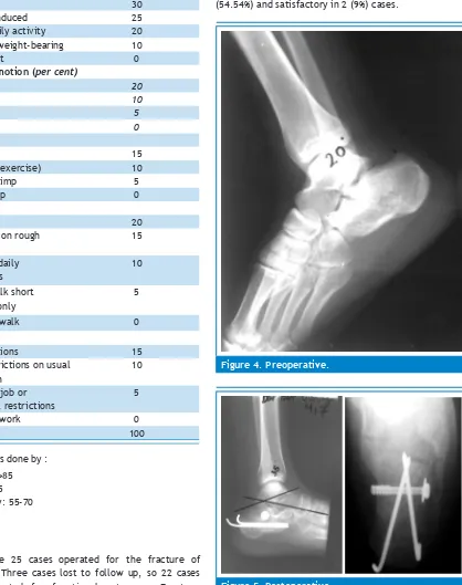
Figure 4. Preoperative.
Figure 6. At 3 months.
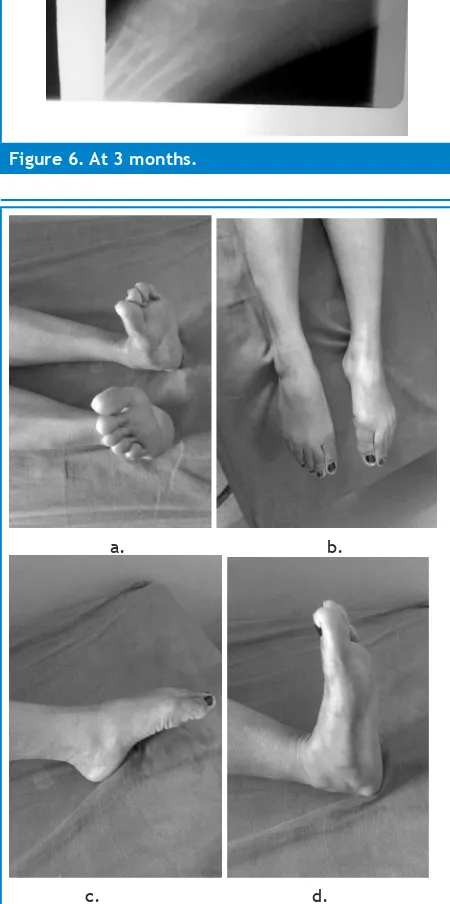
a. b.
c. d.
Figure 7(a-d). Movement at ankle and subtalar joint.
DISCUSSION
There is no consensus in the current literature regarding the optimal treatment of intra-articular fractures of the calcaneum. A meta-analysis, however, published in the year 2000,4 demonstrated that there was no
level-I evidence that enabled a surgeon to decide upon optimal treatment for a displaced intra-articular calcaneal fracture. Historically, displaced intra-articular calcaneal fractures were treated non-operatively as predictable operative reduction and fixation were not possible.5 Operative reduction became more popular as fracture care improved.6,7 Richard buckley et al,8 in a prospective, randomized, controlled multicenter trial demonstrated that operative treatment as a whole provides no improvement over non-operative treatment of displaced intra-articular calcaneal fractures. However, careful stratification of the patient population and clinical outcome information distinguishes certain features that support surgical care for displaced intra-articular calcaneal fractures.
Operative methods of treatment have included attempts at closed reduction or percutaneous manipulation, open reduction with either internal fixation, bone grafting or both, and primary arthrodesis of the subtalar joint.9
Mechanism of injury of the Calcaneum fracture causes a major soft tissue injury that includes heel pad, skin and other soft tissues. Also both lateral and medial walls are subcutaneous, any implants on the surfaces can lead to complications like wound necrosis, dehiscence, exposure of implants and infection. So minimal soft tissue dissection, and use of minimal implants is the viable option to avoid these complications.
seen roentgenographically within four to eight weeks after surgery.12 So we did not put bone graft in all cases.
Outcome is expressed in a variety of ways in the existing literature. The present trend is to evaluate subjectively so it represents the patient satisfaction. The subjective clinical systems used by P. S. Kerr’ et al,13 which include pain, work, walking and walking aids and VAS score by Kevin A et al,14 So we also did not include radiographic
evaluation (i.e., normalization of the Böhler angle) in our analysis, choosing instead to focus on patient’s functional outcomes.
CT scan could not be done in all cases due to cost and unavailability, so we were thus not able to evaluate the classification according to Sanders in all cases.
Zeman P et al,15 reported surgical treatment of intra-articular calcaneal fractures by open reduction and internal fixation with a calcaneal locking compression plate (LCP) from an extended lateral approach. Early post-operative complications were recorded in six patients (20.7 %). Wound dehiscence was found in two (6.9 %), necrosis of wound edges in two (6.9 %), and early superficial infection responding to antibiotic therapy also in two patients (6.9 %), Excellent Rowe scores were achieved in 10 patients (34.5 %), good in 15 (51.7 %) and satisfactory in two (6.9 %). Only two patients (6.9 %) reported poor outcome. We did not record any complications related to wound. The score obtained in our series is comparable to observed by Zeman et al.15
Open reduction and internal fixation of displaced intra articular calcaneal fracture with plate and screws needs removal in later date so need 2nd operation to be done. Harvey et al,16 in 183 calcaneal fracture
patients, removed 43.5% of hardware because of patient complaints. This problem was not seen in our patients because single screw put deep within the soft tissue does not cause any irritation and pain to the patients. David S Levine et al,17 treated intra-articular fracture of the calcaneum by minimally invasive technique and found almost complete preservation of subtalar motion despite the nearly anatomical articular surface reconstruction. The minimum dissection required during procedure result in less post operative swelling, less periarticular scaring, and greater tolerance of early postoperative motion.
Intra-articular fracture of the calcaneum treated in time by an experienced surgeon can also lead to some amount of subtalar stiffness despite skillfully treated with perfect articular reconstruction. Articular congruency is not the only factor to influence the outcome. There
are other factors as well. So Michael Q. Potter et al,18 stated that, an entire year may be required to fully assess patients for residual disability that occurs after a fracture of the calcaneus.
CONCLUSION
Intra articular fracture of the calcaneus can be well managed by minimal opening at the fracture and fixation by single cancellous screw and 2 axial k-wires, so minimizes complications and results in comparable outcomes.
REFERENCES
1. Eastwood DM, Phipp L. Intra-articular fractures of the calcaneum: why such controversy? Injury1997;28:247–59.
2. Lowery RBW, Calhoun JH. Fractures of the calcaneus: Part II. Foot Ankle Int. 1996;17:360–6.
3. Buckley RE, Meek RN. Comparison of open versus closed reduction of intraarticular calcaneal fractures: a matched cohort in workmen. J Orthop Trauma. 1992;6:216-22.
4. Randle JA, Kreder HJ, Stephen D, Williams J, Jaglal S, Hu R. Should calcaneal fractures be treated surgically? A meta-analysis. Clin Orthop Relat Res. 2000; 377:217-27.
5. Pozo JL, Kirwan EO, Jackson AM. The long-term results of conservative management of severely displaced fractures of the calcaneus. J Bone Joint Surg Br. 1984;66:386-90.
6. Sanders R. Intra-articular fractures of the calcaneus: present state of the art. J Orthop Trauma. 1992;6:252-65.
7. Leung KS, Yuen KM, Chan WS. Operative treatment of displaced intraarticular fractures of the calcaneum. Medium-term results. J Bone Joint Surg Br. 1993;75:196-201.
8. Richard Buckly, Suzanne Tough, Robert Mccormack et al. Operative Compared with Nonoperative Treatment of Displaced Intra-Articular Calcaneal Fractures: a prospective randomised controlled multicenter trial. J Bone Joint Surg Am. 2002;84:1733-44.
9. Sanders R, Fortin P, DiPasquale T, Walling A. Operative treatment in 120 displaced intraarticular calcaneal fractures. Results using a prognostic computed tomography scan classification. Clin Orthop Relat Res. 1993 May; 290:87-95.
10. Zeman P, Zeman J, Matejka J, Koudela K. Long-term results of calcaneal fracture treatment by open reduction and internal fixation using a calcaneal locking compression plate from an extended lateral approach. Acta Chir Orthop Traumatol Cech. 2008 Dec;75(6):457-64.
12. McRaynold IS. Trauma to os calcis and heel cord. In Jahss, M.H (ed.): Disorder of the foot. Philadelphia, W.B Saunders; 1982. p. 1497-542.
13. Kerr PS, Prothero DL, Atkins RM. Assessing outcome following calcaneal fracture: a rational scoring system. Injury. 1996 Jan;27(1):35-8.
13. Kevin A et al. Functional oucome measures after dispalced intra-articular calcaneal fractures. J Bone Joint Surg Br. 1996;78-B:119-23.
14. Zeman P, Zeman J, Matejka J, Koudela K. Long-term results of calcaneal fracture treatment by open reduction and internal fixation using a calcaneal locking compression plate from an extended lateral approach. Acta Chir Orthop Traumatol Cech. 2008 Dec;75(6):457-64.
15. Harvey EJ, Grujic L, Early JS, et al. Morbidity associated with ORIF of intra articular calcaneus fractures using a lateral approach. Foot Ankle Int.2001;22:868–73.
16. Levine DS, Helfet DL. An introduction to the minimally invasive osteosynthesis of intra-articular calcaneal fractures. Injury. 2001 May;32 Suppl 1:SA51-4.