R E S E A R C H A R T I C L E
Open Access
Image quality to estimate ventricular
ejection fraction by last year medical
students improves after short courses
of training
Tobias Hüppe
1*, Heinrich Volker Groesdonk
2, Thomas Volk
1, Stefan Wagenpfeil
3and Benedict Wallrich
1Abstract
Background:Transthoracic echocardiography is the primary imaging modality for diagnosing cardiac conditions but medical education in this field is limited. We tested the hypothesis that a structured theoretical and supervised practical course of training in focused echocardiography in last year medical students results in a more accurate assessment and more precise calculation of left ventricular ejection fraction after ten patient examinations.
Methods:After a theoretical introduction course 25 last year medical students performed ten transthoracic
echocardiographic examination blocks in postsurgical patients. Left ventricular function was evaluated both with an
eye-balling methodand with thecalculated ejection fractionusing diameter and area of left ventricles. Each examination block was controlled by a certified and blinded tutor. Bias and precision of measurements were assessed with Bland and Altman method.
Results:Using theeye-balling methodstudents agreed with the tutor’s findings both at the beginning (88%) but more at the end of the course (95.7%). The variation between student and tutor forcalculationof area, diameter and ejection fraction, respectively, was significantly lower in examination block 10 than in examination block 1 (eachp< 0.001). Students underestimated both the length and the area of the left ventricle at the outset, as complete imaging of the left heart in the ultrasound sector was initially unsuccessful.
Conclusions:A structured theoretical and practical transthoracic echocardiography course of training for last year medical students provides a clear and measurable learning experience in assessing and measuring left ventricular function. At least 14 examination blocks are necessary to achieve 90% agreement of correct determination of the ejection fraction.
Keywords:Echocardiography, Training program, Students, Left ventricular ejection fraction, Ultrasound
Background
Transthoracic echocardiography is the primary imaging modality for diagnosing cardiac conditions. Although cardiac ultrasound has become standard imaging in many disciplines today, the standards and goals to teach remain still undefined. The inclusion of cardiac ultra-sound training in undergraduate medical training may
be advantageous for many disciplines because cardiac risk stratification is becoming more important in in-creasingly aging patients.
Cardiac ultrasound has been used successfully to teach car-diac anatomy and physiology to medical students [1–4]. Moreover, structured echocardiographic education in stu-dents provides measurable increasing skills in image acquisi-tion [5–7], volume assessment [8], cardiomyopathies [9–13], valve dysfunction [11, 13–15], and pericardial effusion [12, 13]. Furthermore, although inferior to teaching by an expert cardiographer, hands-on training by student tutors led to a significant gain in echocardiography
© The Author(s). 2019Open AccessThis article is distributed under the terms of the Creative Commons Attribution 4.0 International License (http://creativecommons.org/licenses/by/4.0/), which permits unrestricted use, distribution, and reproduction in any medium, provided you give appropriate credit to the original author(s) and the source, provide a link to the Creative Commons license, and indicate if changes were made. The Creative Commons Public Domain Dedication waiver (http://creativecommons.org/publicdomain/zero/1.0/) applies to the data made available in this article, unless otherwise stated.
* Correspondence:[email protected]
1Department of Anesthesiology, Intensive Care and Pain Therapy, Saarland
University, Medical Center, Kirrberger Straße 100, 66421 Homburg, Saar, Germany
skills in 4th and 5th year students [16]. Even the use of simulations as a supplement to traditional educational approaches improves learning among medical students [17]. However, it is still unclear whether such trainings improve the correct determination of the ejection frac-tion as, in addifrac-tion to the velocity time integral (VTI) [18] or the global longitudinal strain (GLS) [19], one of the most commonly used surrogate parameters of the left ventricular systolic pump function [19].
We, therefore, specifically tested the hypothesis that a structured theoretical and supervised practical course of training in focused echocardiography for last year med-ical students leads to significant more accurate assess-ments and more precise calculations of the left ventricular ejection fraction.
Methods
Student selection
We enrolled 25 last year medical students during their one-year internship without any previous theoretical and practical experience in transthoracic echocardiography into the study. After consultation with the local ethics committee, an ethics approval was not necessary.
Protocol
All students received a 3 h introduction course in focused transthoracic echocardiography, consisting of 1.5 h lecture and 1.5 h hands-on exercises on healthy volunteers. Teaching included the physical basics of ultrasound, the
various cutting planes with focus on the apical 4-chamber view, the morphology and function of the left ventricle, normal and pathological left ventricular pump functions as well as the calculation of the ejection fraction. There was a maximum of four weeks between the introduction course and patient examinations.
All patient examinations were carried out in the recov-ery room in patients after general, trauma or heart sur-gery with a portable echocardiography device (VScan, GE, Solingen, Germany). Patient inclusion criteria were consent to echocardiography, ASA status I-IV, spontan-eous breathing as well as the possible left lateral position and elevation of the left arm.
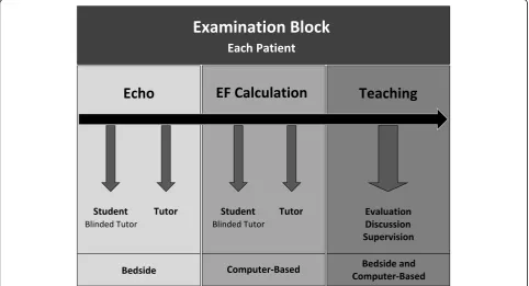
Each student performed a total of 10 examination blocks, one per patient (Fig.1). Each examination block
consisted of bedside echocardiography, where first the student (with blinded tutor) and then the tutor recorded three best possible loops of the apical four-chamber view to assess the left ventricular function. This was followed by the computer-based calculation of the ejection frac-tion (VScan Gateway Software, GE Healthcare, Solingen, Germany), where first the student (with blinded tutor) and then the tutor carried out the calculation. Finally, the bedside and computer-based teaching was held, which included the evaluation of imaging and calcula-tion, discussion and supervision. Specifically, feedback was provided by evaluating position and angle of ultra-sound transducer, determining the correct endocardial edge as well as defining end-diastole and end-systole.
Using this formative approach, students were able to learn from their own mistakes. Both, the introduction course and all control examinations were carried out by the same tutor (TH).
Measurements
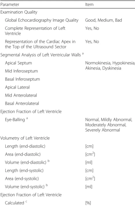
The evaluation of the left ventricular function was per-formed as aneye-balling methodand as acalculated ejec-tion fracejec-tion. For the latter, both the diameter and the area of the left ventricle were measured, each end-diastolic and end-systolic. The volume was determined using the area-length formula (volume = 0.85 x Area2 / Diameter). Ejection fraction was calculated from the ratio of the stroke volume to end-diastolic volume. The eye-bal-ling method and the ordinally scaled calculated ejection fractionused the categoriesnormal(≥55%),mildly abnor-mal(45–55%),moderately abnormal(30–44%) orseverely abnormal (< 30%) [20]. All measurements were carried out using a protocol shown in Table1.
Statistical analysis
For ordinal scaled variables (global image quality, complete representation of left ventricle, representation of the cardiac apex in the top of the ultrasound sector, segmental analysis of left ventricular walls, ejection frac-tion of left ventricle witheye-balling method and calcu-lated, ordinally scaled) the differences between student and tutor were expressed as percent matches given over the course of ten examination blocks. In order to esti-mate the bias of the measurements, additional positive or negative deviations were indicated. The increase in agreements between students and tutor in the course of the examination blocks was examined on the basis of the Spearman correlation. The two-sided significance level was defined at 5%. All data were tested for normal distribution (Shapiro-Wilk). We defined saturation of agreement when the number of examination blocks cor-responded to at least 90% of linear fitted agreement.
Precision and bias for metric calculated diameter, area and ejection fraction of the left ventricle were assessed with Bland and Altman plots. The limit of agreement was defined as 1.96 times standard deviation. Addition-ally, we compared variances of student-tutor-differences for area, diameter and ejection-fraction between examin-ation block 1 and 10 using the t-test statistics for two dependent samples, however, replacing means with vari-ances. Statistical evaluation was performed with R software, version 3.2.3 (R Foundation for Statistical Computing).
Results
25 students examined 250 patients, resulting in 500 transthoracic echocardiographies with 1.500 loops. No student and no patient were excluded from the study.
Ten examination blocks of a single student were com-pleted within one week.
During the course of training, the student-tutor-agreement for global echocardiography image quality
significantly increased. All other items showed an im-proved agreement as well, without reaching significance. Using the eye-balling method to assess left ventricular function, students agreed with the tutor‘s findings both at the beginning (88%) but more at the end of training (95.7%). When the ejection fraction was calculated, the agreement between student and tutor was lower at the beginning (60%) than at the end of the course of training (91.3%). Both,eye-balling methodandcalculation of ejec-tion fracejec-tionshowed a trend towards a more precise de-termination, but they did not show any statistical significance. When the ejection fraction was calculated,
Table 1Documentation parameters by students and tutor
Parameter Item
Examination Quality
Global Echocardiography Image Quality Good, Medium, Bad
Complete Representation of Left Ventricle
Yes, No
Representation of the Cardiac Apex in the Top of the Ultrasound Sector
Yes, No
Segmental Analysis of Left Ventricular Wallsa
Apical Septum Normokinesia, Hypokinesia,
Akinesia, Dyskinesia Mid Inferoseptum
Basal Inferoseptum
Apical Lateral
Mid Anterolateral
Basal Anterolateral
Ejection Fraction of Left Ventricle
Eye-Ballinga Normal, Mildly Abnormal,
Moderately Abnormal, Severely Abnormal
Volumetry of Left Ventricle
Length (end-diastolic) [cm]
Area (end-diastolic) [cm2]
Volume (end-diastolic)b [ml]
Length (end-systolic) [cm]
Area (end-systolic) [cm2]
Volume (end-systolic)b [ml]
Ejection Fraction of Left Ventricle
Calculatedc [%]
Documented parameters by the student and tutor with corresponding items for ordinal and dichotomous scaled variables (examination quality, segmental analysis of left ventricular walls,eye-balling methodfor determination of ejection fraction).aAccording to the recommendations for chamber quantification from the American Society of Echocardiography [20]. Length and area of left ventricle were measured.b
Volume was calculated using the formula [volume = 0.85 x Area2
/ Diameter].c
Ejection fraction was calculated using the formula [[end-diastolic volume - end-systolic volume] /
the students tended to overestimate the pump function (bias + 11.7%). Using the linear fitted approach global echocardiography image quality reached saturation after at least 10, calculated ejection fraction of left ventricle
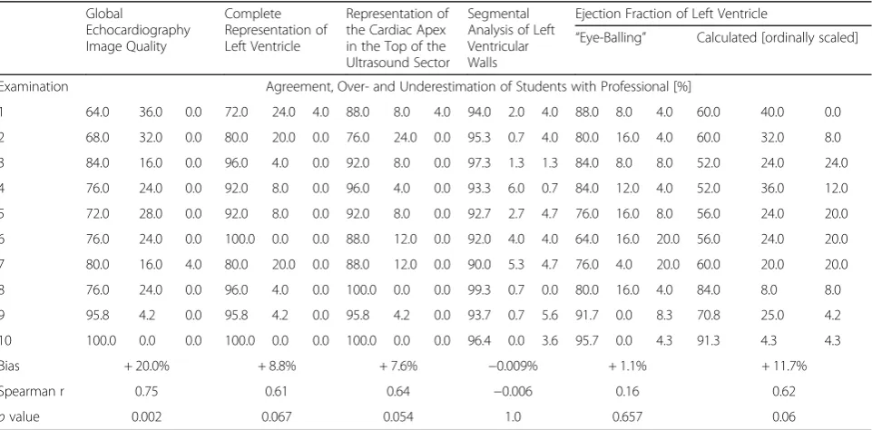
after at least 14 examination blocks. Agreement, over-and underestimation of students with tutor in the assess-ment of the left ventricular function are presented in Table2.
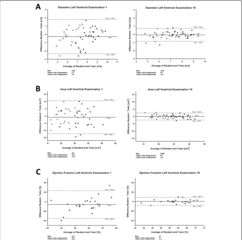
The variation of student-tutor-differences for calcula-tion of area, diameter and ejeccalcula-tion fraccalcula-tion, respectively, was significantly lower in examination block 10 than in examination block 1 (each of the two-sided p-values < 0.001). Bland and Altman plots in Fig. 2 show the im-proved precision and the respective bias with limits of agreement of the students in the tenth compared to the first examination block for left ventricular diameters, areas, and calculated ejection fractions.
Discussion
A structured supervised course of training in focused echocardiography for last year medical students results in an improved global image quality and a more accurate assessment of left ventricular function. Latter applies to both theeye-balling method and in particular to the ac-curatecalculation of the left ventricular ejection function. Although significance levels were not reached in all items, the clinical and educational effects are visible and relevant.
We obtained these results from 500 echocardiography studies. High agreement rates between tutor and stu-dents already at the beginning of the course using the
eye-balling method suggests that even a theoretical lesson allows a rough estimation of the left ventricular function. This is different for the actual calculation. At the beginning, the student often fails to fully visualize the left ventricle and edge the endocard in the echocar-diographic sector leading to underestimation of the ven-tricular diameter and area. This becomes clear from the negative bias in both the determination of the diameter and the area in the first examination block (Fig.2). As a result, the student fails to accurately calculate the ejec-tion fracejec-tion. However, this measurement error was much less in the tenth examination block, leading to a more precise calculated ejection fraction.
We used a linear approach with at least 90% of agree-ments to detect saturation. We are aware of the high variability especially in the last examination blocks. However, more precise methods like three-dimensional echocardiography or three-dimensional cardiac magnetic resonance imaging show frequently worse agreements with two-dimensional transthoracic echocardiography compared to our student-tutor-differences [21]. Malm and colleagues demonstrated both, volume and ejection underestimation by transthoracic echocardiography compared to the gold standard cardiac magnetic reson-ance imaging. They reported a bias of −56 ml (± 48 ml
Table 2Agreement, over- and underestimation of students with tutor in the assessment of the left ventricle
Global
Echocardiography Image Quality
Complete Representation of Left Ventricle
Representation of the Cardiac Apex in the Top of the Ultrasound Sector
Segmental Analysis of Left Ventricular Walls
Ejection Fraction of Left Ventricle
“Eye-Balling” Calculated [ordinally scaled]
Examination Agreement, Over- and Underestimation of Students with Professional [%]
1 64.0 36.0 0.0 72.0 24.0 4.0 88.0 8.0 4.0 94.0 2.0 4.0 88.0 8.0 4.0 60.0 40.0 0.0
2 68.0 32.0 0.0 80.0 20.0 0.0 76.0 24.0 0.0 95.3 0.7 4.0 80.0 16.0 4.0 60.0 32.0 8.0
3 84.0 16.0 0.0 96.0 4.0 0.0 92.0 8.0 0.0 97.3 1.3 1.3 84.0 8.0 8.0 52.0 24.0 24.0
4 76.0 24.0 0.0 92.0 8.0 0.0 96.0 4.0 0.0 93.3 6.0 0.7 84.0 12.0 4.0 52.0 36.0 12.0
5 72.0 28.0 0.0 92.0 8.0 0.0 92.0 8.0 0.0 92.7 2.7 4.7 76.0 16.0 8.0 56.0 24.0 20.0
6 76.0 24.0 0.0 100.0 0.0 0.0 88.0 12.0 0.0 92.0 4.0 4.0 64.0 16.0 20.0 56.0 24.0 20.0
7 80.0 16.0 4.0 80.0 20.0 0.0 88.0 12.0 0.0 90.0 5.3 4.7 76.0 4.0 20.0 60.0 20.0 20.0
8 76.0 24.0 0.0 96.0 4.0 0.0 100.0 0.0 0.0 99.3 0.7 0.0 80.0 16.0 4.0 84.0 8.0 8.0
9 95.8 4.2 0.0 95.8 4.2 0.0 95.8 4.2 0.0 93.7 0.7 5.6 91.7 0.0 8.3 70.8 25.0 4.2
10 100.0 0.0 0.0 100.0 0.0 0.0 100.0 0.0 0.0 96.4 0.0 3.6 95.7 0.0 4.3 91.3 4.3 4.3
Bias + 20.0% + 8.8% + 7.6% −0.009% + 1.1% + 11.7%
Spearman r 0.75 0.61 0.64 −0.006 0.16 0.62
pvalue 0.002 0.067 0.054 1.0 0.657 0.06
for the limits of agreement) for calculation of end-diastolic volume, −16 ml (± 32) for end-systolic volume and−6% (±14) for calculation oft the ejection fraction. Moreover, mean inter-observer variability for calculation of ejection fraction was 13.9%, mean intra-observer vari-ability 5.4% [22]. Regarding our differences between stu-dents and tutor in calculation of ejection fraction
especially in the tenth examination block, we report limits of agreement between –7.7 and 9.1% comparable to the previous mentioned findings. Again, this residual error might be attributed of course to worse skills of stu-dents compared to the tutor but also to the inaccuracy of transthoracic echocardiography itself as well as inter-and intra-observer variability.
Numerous studies have already shown that struc-tured echocardiography training for students and resi-dents improves theoretical knowledge and practical skills [6, 23]. In particular, clear learning success by a structured training program could be shown for rough assessment of pump function [9, 12, 14]. Hope et al. showed that a visual approach using template matching led to a sufficient categorical assessment of left ventricular function with minimal training in stu-dents [10]. Nevertheless, the didactics were very dif-ferent between all studies.
A theoretical introduction is indispensable for the basic understanding of anatomy and physiology. How-ever, the practical training on patients seems to deter-mine the learning success. We demonstrated that a structured course of training significantly improves the precision of metric determination of left ventricular diameter and area, both needed for exact calculation of left ventricular ejection fraction. This effect is noticeable already after ten supervised echocardiographic examin-ation blocks. Our data suggest that on average at least 14 examination blocks are necessary to achieve 90% agreement of correct calculation of left ventricular ejec-tion fracejec-tion. Nevertheless, it must be mentioned that re-petitive and long-term training is required to keep up to the same level of performance and maintain high quality in transthoracic echocardiography.
We consciously selected students with neither prior theoretical knowledge nor practical experience in trans-thoracic echocardiography. And even for this study population a learning success could be shown. Probably the correct measurement of left ventricular diameter and area is one of the most demanding methods in transtho-racic echocardiography especially at the beginning. How-ever, this method allows measuring the learning success quantitatively by comparing diameter and area between student and tutor.
Our cardiac ultrasound course of training followed a formative approach. In contrast to a summative proced-ure, teaching was combined with direct assessment of the knowledge and skills in each individual examination block. As a result, the students received feedback not just at the end of the course of training, so that errors could be corrected directly and assistance could be im-plemented immediately. Thus, the greatest possible learning success could be ensured.
Perhaps conventional ultrasound imaging devices would have led to a better image quality and thus to a simpler understanding of anatomy and physiology. The use of pocket-sized ultrasound devices allows more flex-ible and mobile use. Furthermore, numerous studies have demonstrated that hand-held devices can be easily used in clinical routine and especially in student educa-tion [11–13,24–26].
Limitations
Our study has several limitations. First, determination of the left ventricular ejection fraction may be an inappro-priate outcome parameter. For example, both underesti-mated end-diastolic and end-systolic volumes by students might result in a correct ejection fraction. Nevertheless, the improvement of precision is especially detectable for the ventricular diameter and the area. Thus, correct determination of ejection fraction is not a coincidence, but the result of a more precise measure-ment of area and diameter during the course of training. Second, the ejection fraction was measured only mono-but not biplane. However, this would have necessitated the inclusion of another transthoracic plane and possibly overwhelmed the students in this setting. Third, the training focused only on the determination of the left ventricular ejection fraction. The detection of heart valve defects or other pathologies has not been considered, but could be closely related. Fourth, the majority of pa-tients had no major cardiac pre-existing conditions with mostly normal pump function. Nevertheless, the principle of measurements should not be affected. It also has to be considered critically that the students only car-ried out ten examination blocks. An even higher number could possibly have shown an even stronger effect with a lower variability. In case number planning, we have based our own clinical experience in education as well as previously published work in this field [11, 12, 27]. And finally, only one tutor supervised all students. Thus, he might have been aware of the design and might have expected outcomes and results. Moreover, we are aware that a period of up to four weeks between theoretical introduction and examination blocks might have an individual impact on learning success. Due to the small number of students we were not able to adjust the learning success to this confounder.
We did not evaluate any basic knowledge prior to the the-oretical three-hour lesson, nor did we retrospectively review the theoretical learning success. Moreover, we did not know if and to what extent the theoretical training improved the practical implementation. Maybe this normalization would have affected the results. However, we hypothesized that the theoretical knowledge and practical skills in transthoracic echocardiography were initially marginal in last year medical students, so the initial conditions were similar. Beside that, long-term retention was not assessed and thus the durability of the improvement in precise measurement of left ventricu-lar ejection fraction cannot be assessed in this study. There-fore, the improvement shown in our present data may be lost over time [28].
Conclusions
A structured theoretical and practical transthoracic echocardiography course of training for last year medical students provides a measurable learning experience for the assessment and calculation of left ventricular pump function. Incorporating training of transthoracic echo-cardiography in medical student education may be one step further towards a more widespread use of ultra-sound for many specialties.
Abbreviations
EF:Ejection fraction; GLS: Global longitudinal strain; VTI: Velocity time integral
Acknowledgements
We acknowledge support by the Deutsche Forschungsgemeinschaft (DFG, German Research Foundation) and Saarland University within the funding programme Open Access Publishing.
Authors’contributions
Conception: TH, HVG, BW. Design: TH, BW. Acquisition, Analysis: SW. Interpretation of Data: TH, TV, BW. Drafted the Work: TH, HVG, SW, BW. All authors (TH, HVG, TV, SW, BW) approved the submitted version. All authors (TH, HVG, TV, SW, BW) agreed both to be personally accountable for the author’s own contributions and to ensure that questions related to the accuracy or integrity of any part of the work, even ones in which the author was not personally involved, are appropriately investigated, resolved, and the resolution documented in the literature.
Funding
Not applicable, no funding.
Availability of data and materials
The datasets used and/or analysed during the current study are available from the corresponding author on reasonable request.
Ethics approval and consent to participate
An approval by the ethics committee as well as a patient consent was not necessary after consultation with the authority (Ärztekammer des Saarlandes, Germany).
Consent for publication
Not applicable.
Competing interests
The authors declare that they have no competing interests.
Author details
1Department of Anesthesiology, Intensive Care and Pain Therapy, Saarland
University, Medical Center, Kirrberger Straße 100, 66421 Homburg, Saar, Germany.2Department of Intensive Care Medicine, Helios Klinikum Erfurt, Erfurt, Germany.3Institute for Medical Biometry, Epidemiology and Medical
Informatics, Saarland University Medical Center, Homburg, Saar, Germany.
Received: 21 March 2019 Accepted: 13 September 2019
References
1. Paganini XM, Rubini A. Ultrasound-based lectures on cardiovascular physiology and reflexes for medical students. Adv Physiol Educ. 2016;40(2): 243–7.https://doi.org/10.1152/advan.00010.2016Ultrasound.
2. Brunner M, Moeslinger T, Spieckermann PG. Echocardiography for teaching cardiac physiology in practical student courses. Am J Phys. 1995;268(6):S2–9.
https://doi.org/10.1097/01.CCM.0000260673.66681.AF.
3. Bell FE, Wilson LB, Hoppmann RA. Using ultrasound to teach medical students cardiac physiology. Adv Physiol Educ. 2015;39(4):392–6.https://doi. org/10.1152/advan.00123.2015Ultrasound.
4. Hammoudi N, Arangalage D, Boubrit L, et al. Ultrasound-based teaching of cardiac anatomy and physiology to undergraduate medical students.
Arch Cardiovasc Dis. 2013;106(10):487–91.https://doi.org/10.1016/j.acvd. 2013.06.002.
5. Ray JJ, Meizoso JP, Hart V, et al. Effectiveness of a perioperative
transthoracic ultrasound training program for students and residents. J Surg Educ. 2017;74(5):805–10.https://doi.org/10.1016/j.jsurg.2017.02.005. 6. Felipe AA, García JD, Arcos ISL, et al. Teaching the basics of echocardiography
in the undergraduate: students as mentors. Rev Clin Esp. 2017;217(5):245–51
http://www.revclinesp.es/es/pdf/S0014256517300619/S300/.
7. Heiberg J, Hansen LS, Wemmelund K, et al. Point-of-care clinical ultrasound for medical students. Ultrasound Int Open. 2015;1(2):58–66.https://doi.org/ 10.1055/s-0035-1565173.
8. Kukulski P, Ward M, Carter K. Ultrasound for volume assessment in patients with shock: effectiveness of an educational intervention for fourth-year medical students. Cureus. 2018;10(1):e2129.https://doi.org/10.7759/cureus.2129. 9. Kobal SL, Lior Y, Ben-Sasson A, et al. The feasibility and efficacy of
implementing a focused cardiac ultrasound course into a medical school curriculum. BMC Med Educ. 2017;17(1):94. https://doi.org/10.1186/s12909-017-0928-x.
10. Hope MD, De la Pena E, Yang PC, et al. A visual approach for the accurate determination of echocardiographic left ventricular ejection fraction by medical students. J Am Soc Echocardiogr. 2003;16(8):824–31.https://doi.org/ 10.1067/S0894-7317(03)00400-0.
11. Ho AM, Critchley LA, Leung JY, et al. Introducing final-year medical students to pocket-sized ultrasound imaging: teaching transthoracic
echocardiography on a 2-week anesthesia rotation. Teach Learn Med. 2015; 27(3):307–13.https://doi.org/10.1080/10401334.2015.1044657.
12. Andersen GN, Viset A, Mjølstad OC, et al. Feasibility and accuracy of point-of-care pocket-size ultrasonography performed by medical students. BMC Med Educ. 2014;14:156.https://doi.org/10.1186/1472-6920-14-156. 13. FilipiakStrzecka D, John B, Kasprzak JD, et al. Pocketsize echocardiograph
-a v-alu-able tool for non-experts or just -a port-able device for
echocardiographers? Adv Med Sci. 2013;58(1):67–72.https://doi.org/10.2478/ v10039-012-0054-2.
14. Cawthorn TR, Nickel C, O’Reilly M, et al. Development and evaluation of methodologies for teaching focused cardiac ultrasound skills to medical students. J Am Soc Echocardiogr. 2014;27(3):302–9.https://doi.org/10.1016/j. echo.2013.12.006.
15. Yan BP, Fok JCY, Wong THY, et al. Junior medical student performed focused cardiac ultrasound after brief training to detect significant valvular heart disease. IJC Hear Vasc. 2018;19:41–5.https://doi.org/10.1016/j.ijcha. 2018.03.007.
16. Kühl M, Wagner R, Bauder M, et al. Student tutors for hands-on training in focused emergency echocardiography - a randomized controlled trial. BMC Med Educ. 2012;12:101.https://doi.org/10.1186/1472-6920-12-101. 17. Kusunose K, Yamada H, Suzukawa R, et al. Effects of transthoracic
echocardiographic simulator training on performance and satisfaction in medical students. J Am Soc Echocardiogr. 2016;29(4):375–7.https://doi.org/ 10.1016/j.echo.2015.12.002.
18. Chinen D, Fujino M, Anzai T, et al. Left ventricular outflow tract velocity time integral correlates with low cardiac output syndrome in patients with acute decompensated heart failure. Eur Heart J. 2013;34:4249.https://doi.org/10. 1093/eurheartj/eht309.P4249.
19. Chengode S. Left ventricular global systolic function assessment by echocardiography. Ann Card Anaesth. 2016;19(5):26.https://doi.org/10.4103/ 0971-9784.192617.
20. Lang RM, Bierig M, Devereux RB, et al. Recommendations for chamber quantification: a report from the American Society of Echocardiography’s guidelines and standards committee and the chamber quantification writing group, developed in conjunction with the European Association of Echocardiograph. J Am Soc Echocardiogr. 2005;18(12):1440–63.https://doi. org/10.1016/j.echo.2005.10.005.
21. Gurunathan S, Karogiannis N, Senior R. Imaging the heart failure patient -need for accurate measurements of left ventricular volumes and ejection fraction: the role of three-dimensional and contrast echocardiography. Curr Opin Cardiol. 2016;31(5):459–68.https://doi.org/10.1097/HCO.
0000000000000312.
23. Schall CT, Marouki M, Kangas JR, et al. Anesthesia Resident Transthoracic Echocardiogram Trial (ARTET). Annual Meeting of the American Society of Anesthesiologists San Francisco, USA 2018:4320.
24. Panoulas VF, Daigeler AL, Malaweera AS, et al. Pocket-size hand-held cardiac ultrasound as an adjunct to clinical examination in the hands of medical students and junior doctors. Eur Heart J Cardiovasc Imaging. 2013;14(4): 323–30.https://doi.org/10.1093/ehjci/jes140.
25. Mirabel M, Celermajer D, Beraud AS, et al. Pocket-sized focused cardiac ultrasound: strengths and limitations. Arch Cardiovasc Dis. 2015;108(3):197– 205.https://doi.org/10.1016/j.acvd.2015.01.002.
26. Chamsi-Pasha MA, Sengupta PP, Zoghbi WA. Handheld echocardiography: current state and future perspectives. Circulation. 2017;136(22):2178–88.
https://doi.org/10.1161/CIRCULATIONAHA.117.026622.
27. DeCara J, Kirkpatrick M, Spencer JN, et al. Use of hand-carried ultrasound devices to augment the accuracy of medical student bedside cardiac diagnoses. J Am Soc Echocardiogr. 2005;18(3):257–63.https://doi.org/10. 1016/j.echo.2004.11.015.
28. Lee Y, Shin H, Kim C, et al. Learning curve-cumulative summation analysis of visual estimation of left ventricular function in novice practitioners. Medicine. 2019;98(14):e15191.https://doi.org/10.1097/MD. 0000000000015191.
Publisher’s Note