INTRODUCTION
Liver dysfunction and endoplasmic reticulum (ER) stress are two major con-sequences of burn injury (1,2). It is well established that burn injury induces he-patic acute-phase response and decreases albumin production and that these con-ditions persist over a prolonged period of time (1–5). In addition, burn injury upregulates hepatic ER stress (6,7), caus-ing hepatic damage that can be detri-mental to recovery. We previously
pub-lished rodent studies that showed that insulin treatment after burn attenuates these processes and enhances hepatic function (6). We expect that insulin treat-ment may improve postburn morbidity and mortality in patients; however, the mechanism of action of insulin in the context of burn injury is unclear.
To determine whether the beneficial effects of insulin treatment are due to insulin-specific signaling or glucose mod-ulation, we attenuated hyperglycemia
with metformin, an antidiabetic drug. Recently, it was reported that metformin reduces burn-induced hyperglycemia and insulin resistance, indicating an im-provement in postburn patient outcome (8). A similar effect was also observed in metformin-treated burn rats (9). Met-formin activates multiple physiological pathways, including energy sensor AMP-activated protein kinase (AMPK) (10,11). The activation of AMPK leads to the in-hibition of hepatic gluconeogenesis (12). Whereas many studies claim the capacity of metformin to suppress hepatic gluco-neogenesis (10,13–15), some report that metformin primarily decreases plasma glucose by activating glucose utilization (16). In addition, metformin is known to mobilize fat in the liver (17). However, it is important to this study that metformin decreases blood glucose levels by non–insulin-mediated mechanisms, which allows us to distinguish between insulin- and glucose-mediated effects.
Reticulum Stress in Male Rats
Yaeko Hiyama,
1,2*Alexandra H Marshall,
1,2*Robert Kraft,
3*Nour Qa’aty,
1,2Anna Arno,
1,2David N Herndon,
4,5and Marc G Jeschke
1,21Ross Tilley Burn Centre, Sunnybrook Health Sciences Centre, Sunnybrook Research Institute, Toronto, Ontario, Canada; 2Department
of Surgery, Division of Plastic Surgery, Department of Immunology, University of Toronto, Toronto, Ontario, Canada; 3Department of Trauma, Klinikum Memmingen, Memmingen, Germany; and 4Shriners Hospitals for Children, Galveston, Texas, United States of
America; 5Department of Surgery, University of Texas Medical Branch, Galveston, Texas, United States of America
Severe burn injury causes hepatic dysfunction that results in major metabolic derangements including insulin resistance and hy-perglycemia and is associated with hepatic endoplasmic reticulum (ER) stress. We have recently shown that insulin reduces ER stress and improves liver function and morphology; however, it is not clear whether these changes are directly insulin mediated or are due to glucose alterations. Metformin is an antidiabetic agent that decreases hyperglycemia by different pathways than insulin; therefore, we asked whether metformin affects postburn ER stress and hepatic metabolism. The aim of the present study is to determine the effects of metformin on postburn hepatic ER stress and metabolic markers. Male rats were randomized to sham, burn injury and burn injury plus metformin and were sacrificed at various time points. Outcomes measured were hepatic dam-age, function, metabolism and ER stress. Burn-induced decrease in albumin mRNA and increase in alanine transaminase (p < 0.01 versus sham) were not normalized by metformin treatment. In addition, ER stress markers were similarly increased in burn injury with or without metformin compared with sham (p < 0.05). We also found that gluconeogenesis and fatty acid metabolism gene ex-pressions were upregulated with or without metformin compared with sham (p < 0.05). Our results indicate that, whereas thermal injury results in hepatic ER stress, metformin does not ameliorate postburn stress responses by correcting hepatic ER stress.
Online address: http://www.molmed.org doi: 10.2119/molmed.2012.00330
*YH, AHM, and RK contributed equally to this work.
Address correspondence toMarc G Jeschke, Sunnybrook Health Sciences Centre, 2075 Bayview Avenue, Room D704, Toronto, ON, Canada M4N 3M5. Phone: 416-480-6703; Fax: 416-480-6763; E-mail: marc.jeschke@sunnybrook.ca.
The aim of the present study was to determine whether postburn metformin-mediated glucose modulation reduces hepatic ER stress, alters hepatic meta-bolic marker expressions and improves function. We hypothesized that met-formin does not alleviate postburn ER stress and that the beneficial effects of in-sulin are due to inin-sulin-specific effects.
MATERIALS AND METHODS
Animals and Treatments
All procedures were in accordance with current National Institutes of Health guidelines and were approved by the University of Texas Medical Branch Institutional Animal Care and Use Com-mittee. All animals were housed in wire-bottom cages with a 12-h light–dark cycle and received food and water ad libi-tumfor the entire study period.
A well-established method (18) was used to induce a 60% total body surface area scald burn in adult male Sprague Dawley rats (approximately 350 g). Rats received analgesia (buprenorphine, 0.05 mg/kg; intramuscularly) and were anesthetized with ketamine (40 mg/kg body weight; intraperitoneally) and xy-lazine (5 mg/kg body weight; intraperi-toneally) before burn. These animals were secured in a protective mold with an opening of approximately 60% of total body surface area. The exposed skin was immersed in 96–100°C water (10 s back, 1.5 s front) to induce third-degree burn. After receiving thermal injury, rats were resuscitated with Ringer lactate
(60 mL/kg; intraperitoneally). Analgesia was administered every 12 h. Sham ani-mals received the same treatment except for the scald burn. Metformin (450 mg/kg
per os[PO]; Sigma-Aldrich, St. Louis, MO, USA) was administered immediately after burn. Animals were sacrificed at 24-or 48-h postburn, and livers were rapidly removed and frozen in liquid nitrogen.
Western Blotting
Livers were homogenized in lysis buffer (150 mmol/L NaCl, 50 mmol/L Tris-HCl, pH 7.8, 1% [w/v] Triton X-100,
1 mmol/L EDTA, 0.5 mmol/L phenyl-methanesulfonyl fluoride) with a pro-tease inhibitor (Roche Molecular Bio-chemicals, Indianapolis, IN, USA) and phosphatase inhibitor (Sigma-Aldrich). Samples were centrifuged, and protein concentrations were measured by BCA assay (Thermo Scientific, Waltham, MA, USA). A total of 25 μg denatured protein from tissues were separated on sodium dodecyl sulfate–polyacrylamide gel elec-trophoresis (SDS-PAGE) gel. After trans-fer to nitrocellulose membrane, the mem-brane was blocked with 5% nonfat milk solution and washed with buffer (Tris-buffered saline, 0.05% Tween). Primary antibodies for p-AMPK and β-actin, and anti-rabbit IgG was purchased from Cell Signaling Technology (Danvers, MA, USA) and used at the recommended dilu-tions. The bound antibody was detected by SuperSignal West Pico Chemilumines-cent Substrate (Thermo Scientific).
RNA Isolation and Gene Expression Analysis
Total RNA was extracted from the liv-ers by using an RNeasy Mini Kit (Qiagen) and was reverse-transcribed by using a High-Capacity cDNA Reverse Transcrip-tion Kit (Applied Biosystems; Life Tech-nologies, Carlsbad, CA, USA) following the manufacturer’s instructions. Quantita-tive real-time polymerase chain reaction (PCR) was performed on the ABI PRISM 7000 Sequence Detection System with SYBR Green PCR Master Mix (Applied Biosystems; Life Technologies). The 18S rRNA was used as the housekeeping gene to normalize expression. PCR primer se-quences are available upon request.
Liver Injury Measurement
Alanine transaminase (ALT) activity levels were determined in rat livers using enzymatic kits (Cayman Chemical, Ann Arbor, MI, USA). Activity was nor-malized to protein concentration mea-sured by BCA Assay (Thermo Scientific).
Liver Triglyceride Content
Liver lipids were extracted based on the method described by Folch et al.(19).
Briefly, liver homogenates were mixed with a chloroform-methanol (2:1) mix-ture. After centrifugation, the bottom or-ganic layer was removed and evapo-rated under a stream of nitrogen. Triglyceride concentration was mea-sured enzymatically (INFINITY; Thermo Scientific).
Statistical Analysis
All data are presented as the mean ± standard error of the mean. Statistical significance was examined with one-way analysis of variance with a Tukey post hoc
test, unless noted otherwise. All data were analyzed by using GraphPad Prism software.
RESULTS
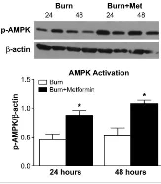
Hepatic AMPK Activation
To determine AMPK activation, a downstream target of metformin, we measured phosphorylated-AMPK (p-AMPK) levels in the livers by Western blot. As expected, there was a significant increase (p< 0.05) in the phosphorylation of AMPK (at Ser108) in metformin-treated livers compared with nonmetformin-treated livers (Figure 1). This result confirmed the adequate administration of the drug,
since phosphorylation at this site is a re-quirement for the activation of the AMPK enzyme (20).
Hepatic Function
We investigated the effects of met-formin on albumin expression and ALT levels. At 24-h postburn, there was a nonsignificant decrease in albumin ex-pression in burn livers; however, there was a further decrease with metformin treatment (Figure 2A). At 48-h postburn, there were no significant differences be-tween the groups.
In addition, we measured ALT enzyme activity. At 24-h postburn, there was a significant increase in ALT levels in both burn groups (Figure 2B; p < 0.01 for both burn versus sham, and burn + met-formin versus sham.) These results indi-cate that burn-induced liver dysfunction but metformin did not correct it.
Hepatic ER Stress
As expected, ER stress markers were significantly increased in burn injury (Figure 3). We measured x-box protein 1
(Xbp-1) spliced, DnaJ homolog subfamily B member 9 (Dnajb9), protein disulfide isomerase family A member 3 (Pdia3), and CCAAT/enhancer binding protein (Chop) mRNA levels and found that all of these ER stress–related gene expres-sions were significantly upregulated (p < 0.05 versus sham) at 24-h postburn. Met-formin did not have any effects on ER stress at this time point. At 48-h post-burn, ER stress was induced in burn but less so than at 24-h postburn. Metformin further upregulated expression (p < 0.05 versus sham). Our results confirm that ER stress is increased in burn; however, metformin did not reduce ER stress ei-ther at 24- or 48-h postburn.
Hepatic Gluconeogenesis
To examine the effects of metformin on hepatic gluconeogenesis, we measured peroxisome proliferator–activated recep-tor γcoactivator 1-α(Pgc1), glucose 6-phosphatase (G6pc) and
phospho-enolpyruvate carboxykinase 1 (Pck1) gene expressions (Figure 4). Although burn induces hepatic gluconeogenesis, only at 24-h postburn were the expres-sions increased in burn compared with sham (p < 0.05). Moreover, metformin did not reduce these expressions.
At 48-h postburn, there were similar ex-pression levels of G6pcand Pck1among the treatment groups. Pgc1was increased ~40-fold with metformin treatment (p < 0.001 versus all other groups and time points). These results show that there is a time-dependent modification in gluco-neogenic gene expression and that, with the exception of Pgc1expression at 48-h postburn, metformin does not alter gluco-neogenic gene expression in burn livers.
Hepatic Fatty Acid and Glucose Metabolism
Because metformin is known to mobi-lize energy within the liver, we assessed the expression levels of acyl-CoA oxidase
Figure 2.Metformin does not alleviate he-patic damage after burn. Albumin con-centration (A) and ALT levels (B) were measured in rat livers. **p < 0.01 versus
sham. Figure 3.at the mRNA level. Chop is a downstream target of PERK, whereas Bip, Dnajb9 and Pdia3Metformin augments hepatic ER stress after burn. ER stress markers were assessed
(Acox1), fatty acid translocase (Cd36), fatty acid synthase (Fasn), stearoyl-CoA desaturase 1 (Scd1), glucose transporter 1 (Glut1) and glucose transporter 4 (Glut4). Many of these gene expressions had a trend to increase in burned animals com-pared with sham at both time points and were significantly increased in burn + metformin compared to sham (Figure 5). In addition, at 48-h postburn, expres-sions were significantly upregulated in
Acox1(100-fold), Cd36(50-fold), Scd1 (15-fold), Glut1(250-fold) and Glut4(20-fold) (p< 0.01 versus all other groups and time points). Our results signify a vast energy mobilization after burn, espe-cially with metformin treatment.
Hepatic Triglyceride Content
Because nutrient metabolism gene ex-pressions were altered in the burn livers, we measured triglyceride content to de-termine whether there was a physiologi-cal effect (Figure 6). We found that triglyc-eride content was slightly increased in burn livers and had a trend to normalize with metformin treatment. These data suggest that metformin potentially re-duces burn-induced hepatic steatosis by increasing fatty acid metabolism.
DISCUSSION
The aim of the current study was to determine the effects of metformin on burn-induced ER stress and hepatic metabolic markers. We found that while metformin did not reduce burn-induced liver injury and ER stress, it increased
energy metabolism, thereby potentially normalizing burn-induced hepatic steatosis. These results indicate that the beneficial effects observed with insulin
Figure 4.Gluconeogenesis gene expres-sion in rat livers after burn. mRNA levels of Pgc1 (A), G6pc (B) and Pck1 (C) were evaluated. *p < 0.05 versus sham. †p < 0.01 versus all other groups at both time points.
Figure 5.Gene expression of markers for fatty acid metabolism (A–D) and for glucose up-take (E, F). *p < 0.05, **p < 0.01, ***p < 0.001 versus sham. †p < 0.01 versus all other groups at both time points.
administration are most likely associated with insulin-specific modulations and not via glucose alterations. Metformin has been used as an antidiabetic drug for nearly a century. Previously, others have shown that metformin reduces burn-in-duced hyperglycemia by increasing glu-cose tolerance (8). In addition to patient data, there are rat data that also indicate that metformin decreases blood glucose levels in burn-injured rats (9). Recently, we demonstrated that, with insulin treat-ment, burn-induced hyperglycemia as well as liver damage is corrected, sug-gesting an association between glucose levels and hepatic ER stress (6,21). How-ever, it was not clear whether the im-proved outcomes were due to insulin ac-tion directly or due to reduced glucose levels.
Although metformin reduces blood glucose, it is unknown what its effects are on burn livers. We hypothesized that metformin does not alleviate postburn ER stress and that the beneficial effects of insulin previously published were due to insulin-specific effects. In concurrence with previous data, burn caused ER stress and liver dysfunction; these condi-tions, however, were not normalized with metformin treatment. Albumin lev-els were decreased and ALT levlev-els were increased similarly in burn rats with or without metformin. Elevated ALT is a marker for liver damage. Although met-formin is known to decrease elevated ALT levels back to baseline in diabetic patients, this does not seem to be the case after burn. It is possible that there are other liver function markers that are altered with metformin; however, it is more likely that metformin cannot cor-rect liver dysfunction itself. Metformin has been contraindicated in individuals with preexisting conditions that could in-crease the risk of lactic acidosis, such as lung disease and liver disease (22).
The ER, a cell organelle, is a major site of protein folding and trafficking. When there is an accumulation of unfolded or misfolded proteins in the lumen of the ER, uncoupled protein responses and ER stress occur. These responses inhibit
translation and activate the three branches of ER stress: activating tran-scription factor 6 (ATF6), insulin re-sponse element-1 (IRE-1), and protein ki-nase RNA-like endoplasmic reticulum kinase (PERK), each of which are trans-membrane proteins that are phosphory-lated and activated in response to in-creased ER stress (23). In the present study, we assessed downstream targets of each of these branches, including
Chop, Bip, Dnajb9, Pdia3and Xbp-1
spliced. We showed that ER stress is in-creased after burn, as we have previ-ously demonstrated in rodents and pa-tients (6,7). We did not observe a correction in ER stress markers with metformin. It was shown that metformin reduces some, but not all, branches of ER stress in cardiomyocytes as well as cultured hepatocytes (24–26). However, there are little data that illustrate the ef-fect of metformin on hepatic ER stress in vivo.Possibly, ER stress cannot be cor-rected by metformin in the context of burn, especially at the time points (24-and 48-h postburn) we studied.
Next, we assessed the expression of genes involved in hepatic gluconeogene-sis. Metformin is known to lower glucose by inhibiting hepatic gluconeogenesis (15). We measured three of the most common gluconeogenic genes: Pgc1,
G6pcand Pck1(Figure 4). At 24-h post-burn, there was an increase in these glu-coneogenic gene expressions with burn injury; however, at 48 h, there were no differences. Moreover, metformin did not reduce these gene expressions. It is possi-ble that metformin treatment after burn injury does not reduce glucose by in-hibiting gluconeogenesis but rather by increasing glucose utilization. We mea-sured Glut1and Glut4expression and found that, at 48-h postburn, metformin increased these glucose transporter ex-pressions significantly (Figures 5E, F).
Because metformin is also known to alleviate hepatic steatosis (17), we quan-tified the expression of several fatty acid metabolism genes, specifically Acox1,
Cd36, Fasnand Scd1(Figures 5A–D). It is important to note that in all of the
meta-bolic gene expressions we evaluated, the burn injury groups had higher expres-sion than shams, regardless of whether the gene was responsible for synthesis or breakdown of fatty acids. We postulate that this is due to the hypermetabolism that occurs after burn. Metformin further upregulated these expressions, especially after 48 h; Cd36and Scd1genes, which are associated with the uptake and syn-thesis of fat, were further upregulated (50- and 15-fold versus sham, respec-tively). However, Acox1, a gene responsi-ble for the oxidation of fatty acids, had an even greater increase (100-fold), sug-gesting that numerous pathways involv-ing fat metabolism were altered with metformin treatment. The discrepancies in the gene expression pattern may affect hepatic phenotype such as triglyceride content (Figure 6). Because hepatic steatosis is known to occur in burn, we expected the increased triglyceride levels in the burn livers. Although this was not statistically significant, we observed a trend toward increased triglyceride in burn livers. Rats that received metformin had decreased triglyceride compared with nontreated rats. These results are in agreement with our gene expression data that Acox1is significantly increased in metformin-treated burn livers.
What do we think are the mechanisms of metformin? There are several results in this study that showed even detrimen-tal effects of metformin in terms of ER stress and gluconeogenesis at some time points. The possible explanations are not easy. Both ER stress and gluconeogenic gene expressions are increased with met-formin. It could be that the observed changes with metformin are a time effect, or it could be that many of the gluco-neogenic gene expressions are upregu-lated when they “sense” high
not reach statistical significance. We pos-tulated that metformin possibly exerts its effects not through the liver, but through other organs such as the adipose or the muscle. The main physiological and mo-lecular changes could occur in other or-gans, and it will be important in future studies to examine extrahepatic tissues. It may also be valuable in the future to study time points beyond 48 h, since the rats sacrificed at 48-h postburn were given an additional dose of metformin, which may be a confounding factor.
CONCLUSION
We have shown that metformin does not reverse burn-induced hepatic de-rangements such as ER stress and dys-function and that there is a possibility that it may improve patient outcome by upregulating hepatic fatty acid metabo-lism. We also conclude that insulin and metformin act very differently after burn on liver and liver-associated metabolic and morphologic changes.
ACKNOWLEDGMENTS
This research was supported by the National Institutes of Health (R01-GM087285-01 to MG Jeschke), Canadian Institutes of Health Research (123336), the CFI’s Leader’s Opportunity Fund (25407 to MG Jeschke) and the Health Research Grant Program from the Physi-cians’ Services Incorporated Foundation (to MG Jeschke). We thank the SRI Ge-nomics Core Facility for genotyping the samples.
DISCLOSURE
The authors declare that they have no competing interests as defined by Molec-ular Medicine, or other interests that might be perceived to influence the re-sults and discussion reported in this paper.
REFERENCES
1. Jeschke MG, et al.(2011) Long-term persistence of the pathophysiologic response to severe burn in-jury. PLoS One.6:e21245.
2. Jeschke MG, et al.(2008) Pathophysiologic response to severe burn injury. Ann. Surg.248:387–401. 3. Jeschke MG, Micak RP, Finnerty CC, Herndon
DN. (2007) Changes in liver function and size after a severe thermal injury. Shock. 28:172–7. 4. Jeschke MG. (2009) The hepatic response to
ther-mal injury: is the liver important for post-burn outcomes? Mol. Med.15:337–51.
5. Jeschke MG, Barrow RE, Herndon DN. (2004) Ex-tended hypermetabolic response of the liver in severely burned pediatric patients. Arch. Surg.
139:641–7.
6. Jeschke MG, et al.(2011) Insulin protects against hepatic damage post-burn. Mol. Med.17:516–22. 7. Song J, Finnerty CC, Herndon DN, Boehning D, Jeschke MG. (2009) Severe burn-induced endo-plasmic reticulum stress and hepatic damage in mice. Mol. Med.15:316–20.
8. Gore DC, Wolf SE, Herndon DN, Wolfe RR. (2003) Metformin blunts stress-induced hyperglycemia after thermal injury. J. Trauma. 54:555–61. 9. Frayn KN. (1976) Effects of metformin on insulin
resistance after injury in the rat. Diabetologia. 12:53–60.
10. Zhou G, et al.(2001) Role of AMP-activated pro-tein kinase in mechanism of metformin action.
J. Clin. Invest.108:1167–74.
11. Shaw RJ, et al.(2005) The kinase LKB1 mediates glucose homeostasis in liver and therapeutic ef-fects of metformin. Science. 310:1642–6. 12. Towler MC, Hardie DG. (2007) AMP-activated
protein kinase in metabolic control and insulin signaling. Circ. Res.100:328–41.
13. Argaud D, Roth H, Wiernsperger N, Leverve XM. (1993) Metformin decreases gluconeogenesis by enhancing the pyruvate kinase flux in isolated rat hepatocytes. Eur. J. Biochem.213:1341–8. 14. Otto M, Breinholt J, Westergaard N. (2003)
Met-formin inhibits glycogen synthesis and gluconeo-genesis in cultured rat hepatocytes. Diabetes Obes. Metab. 5:189–94.
15. Kim YD, et al.(2008) Metformin inhibits hepatic gluconeogenesis through AMP-activated protein kinase-dependent regulation of the orphan nu-clear receptor SHP. Diabetes. 57:306–14. 16. Yoshida T, et al.(2009) Metformin primarily
de-creases plasma glucose not by gluconeogenesis suppression but by activating glucose utilization in a non-obese type 2 diabetes Goto-Kakizaki rats. Eur. J. Pharmacol.623:141–7.
17. Lin HZ, et al.(2000) Metformin reverses fatty liver disease in obese, leptin-deficient mice. Nat. Med.6:998–1003.
18. Herndon DN, Wilmore DW, Mason AD Jr. (1978) Development and analysis of a small animal model simulating the human post-burn hyper-metabolic response. J. Surg. Res.25:394–403. 19. Folch J, Lees M, Sloane Stanley GH. (1957) A
sim-ple method for the isolation and purification of total lipides from animal tissues. J. Biol. Chem.
226:497–509.
20. Warden SM, et al.(2001) Post-translational modi-fications of the beta-1 subunit of AMP-activated protein kinase affect enzyme activity and cellular localization. Biochem. J.354:275–83.
21. Leffler M, et al.(2007) Insulin attenuates
apo-ptosis and exerts anti-inflammatory effects in endotoxemic human macrophages. J. Surg. Res.
143:398–406.
22. Eurich DT, et al.(2007) Benefits and harms of an-tidiabetic agents in patients with diabetes and heart failure: systematic review. BMJ.335:497. 23. Ron D, Walter P. (2007) Signal integration in the
endoplasmic reticulum unfolded protein re-sponse. Nat. Rev. Mol. Cell. Biol.8:519–29. 24. Liu J, et al.(2010) Endoplasmic reticulum stress
involved in the course of lipogenesis in fatty acids-induced hepatic steatosis. J. Gastroenterol. Hepatol.25:613–8.
25. Yeh CH, Chen TP, Wang YC, Lin YM, Fang SW. (2010) AMP-activated protein kinase activation during cardioplegia-induced hypoxia/reoxy-genation injury attenuates cardiomyocytic apo-ptosis via reduction of endoplasmic reticulum stress. Mediators Inflamm.2010:130636. 26. Quentin T, Steinmetz M, Poppe A, Thoms S.