INTRODUCTION
The insulinlike growth factor-I (IGF-I), also called somatomedin C, is a cellular and secreted growth factor which is criti-cal for normal body growth, develop-ment and maintenance, and has impor-tant roles in multiple biological systems (1–3). A variety of cellular responses are induced by IGF-I, including cell prolifer-ation, differentiprolifer-ation, migration and sur-vival (4–8). These cellular responses have implicated IGF-I in several conditions such as the pathophysiology of several
cancers (9–11), or the mitogenic and myogenic processes during muscle de-velopment, regeneration or hypertrophy, since, unlike other growth factors, IGF-I acts as both a mitogen and a differentia-tion factor (12,13).
IGF-I is produced by many tissues, in-dicating that a significant component of IGF-I action is due to its autocrine and paracrine mode of function, although it also acts as a classical circulating hor-mone. In the endocrine mode of action, IGF-I acts as a mediator (somatomedin
C) of the growth-promoting effects of pi-tuitary growth hormone (GH, soma-totropin), which induces the synthesis and release of IGF-I by the liver (2,14,15). Circulating IGF-I is mainly derived from the liver, but also from skeletal muscle (3,16–18), and is mostly bound to high affinity IGF-binding proteins, which pro-tect it from proteolytic degradation and modulate its bioavailability to the IGF-I receptors (2,19).
Different IGF-I mRNA transcripts are produced as a result of the alternative splicing of the IGF1gene, encoding for several IGF-I precursor proteins. These IGF-I protein isoforms differ by the structure of their extension peptides, or E-peptides, on the carboxy-terminal end and by the length of their terminal signal peptides. However, they share the same mature peptide, which is the common part of all the IGF-I precur-sors (20–23). IGF-I mediates its actions through the binding and activation of several receptors and the IGF-I domain
Modification and Bioactivity
Anastassios Philippou,
1Maria Maridaki,
2Spiros Pneumaticos,
3and Michael Koutsilieris
11
Department of Experimental Physiology, Medical School, 2Department of Sports Medicine and Biology of Physical Activity, Faculty of Physical Education and Sport Science, and 3Third Department of Orthopaedic Surgery, Medical School, National and Kapodistrian University of Athens, Athens, Greece
The insulinlike growth factor-I (IGF-I) is an important factor which regulates a variety of cellular responses in multiple biological systems. The IGF1 gene comprises a highly conserved sequence and contains six exons, which give rise to heterogeneous mRNA transcripts by a combination of multiple transcription initiation sites and alternative splicing. These multiple transcripts code for dif-ferent precursor IGF-I polypeptides, namely the IGF-IEa, IGF-IEb and IGF-IEc isoforms in humans, which also undergo posttransla-tional modifications, such as proteolytic processing and glycosylation. IGF-I actions are mediated through its binding to several cell-membrane receptors and the IGF-I domain responsible for the receptor binding is the bioactive mature IGF-I peptide, which is derived after the posttranslational cleavage of the pro-IGF-I isoforms and the removal of their carboxy-terminal E-peptides (that is, the Ea, Eb and Ec). Interestingly, differential biological activities have been reported for the different IGF-I isoforms, or for their E-peptides, implying that IGF-I peptides other than the IGF-I ligand also possess bioactivity and, thus, both common and unique or complementary pathways exist for the IGF-I isoforms to promote biological effects. The multiple peptides derived from IGF-I and the differential expression of its various transcripts in different conditions and pathologies appear to be compatible with the dis-tinct cellular responses observed to the different IGF-I peptides and with the concept of a complex and possibly isoform-specific IGF-I bioactivity. This concept is discussed in the present review, in the context of the broad range of modifications that this growth factor undergoes which might regulate its mechanism(s) of action.
Online address: http://www.molmed.org doi: 10.2119/molmed.2014.00011
Address correspondence to Anastassios Philippou, Department of Experimental Physiol-ogy, Medical School, National and Kapodistrian University of Athens, 75 Micras Asias, Goudi-Athens, 115 27, Greece. Phone: + 30210 7462690; Fax: + 30210 7462690; E-mail: [email protected]
which is responsible for the receptor binding is the biologically active mature peptide. It is derived after the posttrans-lational cleavage of the pro-IGF-I (iso)forms and the removal of the E-peptides (23–26). Interestingly, it has been proposed that the E-peptides also possess bioactivity that is distinct from that of mature IGF-I (20,27).
Thus, during the last decade, many in vitroand in vivostudies have investigated the aspect of the differential IGF-I iso-forms or their E-peptides actions in vari-ous conditions and pathologies (28–38). This concept was further supported by recent findings which revealed differen-tial, E-peptide– or IGF-I isoform–specific signaling (31,33,34,38–40).
In the present review, focus has been on the propounded concept of the differ-ential roles and bioactivity of the IGF-I isoforms or peptides, in the context of the complexity that characterizes the al-ternative splicing, posttranscriptional regulation and posttranslational modifi-cations of this growth factor and which might modulate its mechanism(s) of action.
HUMAN IGF1 GENE STRUCTURE AND ALTERNATIVE SPLICING
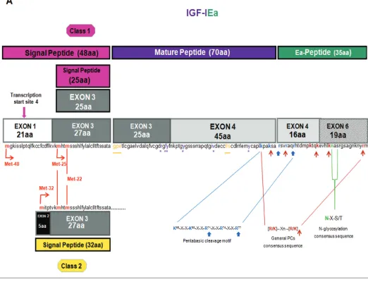
The IGF1gene spans a region of over 80 kb of genomic DNA located on the long arm of chromosome 12 in humans, it is a highly conserved sequence in mammals and primates (23), and con-tains six exons, which give rise to hetero-geneous mRNA transcripts by a combi-nation of multiple transcription initiation sites (that is, alternative leader se-quences), alternative splicing and differ-ent polyadenylation signals (41,42). These multiple IGF-I transcripts code dif-ferent precursor polypeptides, which also undergo posttranslational modifica-tions (26,43,44), (Figures 1A–C).
More specifically, the different leader sequences result in two different classes of IGF-I mRNA variants: class 1 tran-scripts have their initiation sites on exon 1 (promoter 1), whereas class 2 tran-scripts use exon 2 as leader exon (pro-moter 2), and class 1 (exon 1 to exon 3)
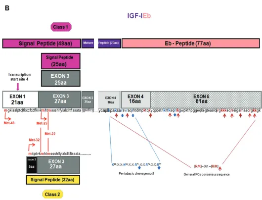
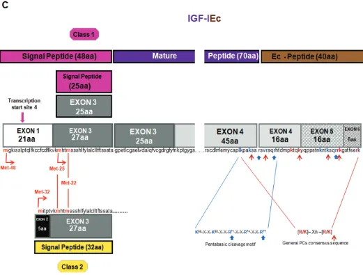
or class 2 (exon 2 to the exon 3) mRNA transcripts are produced by differential splicing of exons 1 and 2 to the common exon 3. Alternative splicing of exon 5 also results in different mRNA variants containing exon 5, generally defined as class B (IGF-IEb), or containing exon 6 (and excluding exon 5) defined as class A (IGF-IEa) (26,45,46), (see Figures 1A, B). A third variant, the IGF-IEc, which corre-sponds to IGF-IEb in rodents, also is gen-erated by alternative splicing in the human IGF1gene and contains both exon 5 and 6 (22), (see Figure 1C). Simi-lar to the human IGF1gene, multiple forms of pro-IGF-I mRNA have been de-scribed in other species in which IGF-I genomic sequences have been deter-mined, such as the designated pro-IGF-I Ea-1, Ea-2, Ea-3 and Ea-4 in teleosts (23,47–50).
All possible combinations between promoter usage and terminal exon (5 or 6) can occur in different IGF-I transcripts (45,51,52). It has been proposed that the use of promoter 1 could be associated with the synthesis of paracrine IGF-I and may influence interactions with insulin-like growth factor binding proteins (IGF-BPs), or promote the formation of the truncated IGF-I peptide (46), (see below: IGF-I Processing, Secretion and Glycosy-lation). Transcripts initiating at promoter 1 are widely expressed in many tissues, whereas transcripts initiating at pro-moter 2 are expressed mainly in the liver (circulating forms) and kidney (53) and are thought to be more GH-dependent (23,54–58), or equally GH-responsive (59,60). However, the two promoters are probably not mutually exclusive, and GH can also stimulate the expression of tissue-specific (local) transcripts, al-though the existent evidence is still equivocal (48,61–65).
IGF-IEa transcript derives from the splicing pattern exon 1 or 2–3–4–6 of the IGF1gene, which represents the main pro-IGF-I mRNA produced in liver (sys-temic IGF-IEa) but also in other tissues with similar exon sequence (22,66), (see Figure 1A). IGF-IEb transcript is a splice variant of exon 1 or 2–3–4–5. Its
expres-sion was firstly detected in the human liver (67), while it was also found to be expressed in lung carcinoma cells (20), in skeletal muscle (31,62,68) and more re-cently in various tissues and cells such as prostate, endometrium and lens epithe-lial cells (34,35,65,69), (see Figure 1B). Whether there is equivalent to human IGF-IEb splice pattern in nonhuman pri-mates is not known (23,43,70). IGF-IEc mRNA transcript is an exon 1 or 2–3–4–5–6 splice variant (see Figure 1C), which was initially identified also in human liver, where, however, it is ex-pressed approximately at 10% relative to the main IGF-IEa transcript (22). Struc-turally, its cDNA differs from the IGF-IEa variant by the presence of the first 49 base pairs from exon 5 (52 bp in rodents), It results from a splice acceptor site in the intron preceding exon 6 and, due to a reading frame shift, it gives rise to a dif-ferent carboxy-terminal peptide sequence and a premature stop codon in exon 6. This transcript was named mechano-growth factor (MGF) since it was found to be upregulated in response to muscle stretch and/or damage (71); for review see (72). However, its expression also has been identified in various tissues such as endometrium (35), normal and cancerous prostatic cells (34), as well as in
osteoblast-like osteosarcoma cells (73). The biological significance of IGF-I splice variants is currently unknown and the physiological and molecular mecha-nisms that regulate their expression are unclear; however, the presence of distinct transcripts is indicative of diverse re-sponses of cells to different stimuli (74) and they probably reflect the complexity of IGF-I actions mediated via its various isoforms (75,76).
(64,72). The differential expression of the IGF-I splice variants observed in various pathologies is of particular interest, as it could indicate distinct regulatory mecha-nisms and biological roles of the different IGF-I isoforms; however, their particular functions remain as yet unclear.
The different IGF-I mRNA transcripts encode the corresponding precursor pro-teins IGF-IEa, IGF-IEb and IGF-IEc (23,44). The 5′end, by alternative splic-ing of exons 1, 2 and 3, encodes for the
signal peptide of the IGF-I prohormone. Four different transcription start sites are present in exon 1 and their positions rel-ative to the translational initiation codons (that is, Met-48 located in exon 1, and Met-25 and Met-22 located in exon 3) can give rise to three distinct IGF-I signal peptides from class 1 mRNAs (exon 1 to exon 3), (23,43). However, translation of mRNAs initiated at the four transcription start sites mentioned above is expected to produce signal sequences of 48
(tran-scription start sites 1, 2 and 3) and 25 (transcription start site 4) amino acids, since between two translation start sites contained in an mRNA, (for example, Met-25 and Met-22, located downstream of the transcription start site 4), the pref-erence is given to the upstream site (42,44,55,66), (see Figures 1A–C). It was suggested that when translation initiates at Met-25, the nucleotide sequence corre-sponding to the first 21 amino acids (en-coded by exon 1) of the sequence of the
Figure 1.Gene structure, alternative splicing, amino acid sequence and posttranslational modifications of human IGF-I. Posttranslational cleavage of pro-IGF-I polypeptides can occur at a unique pentabasic motif and at sites containing the general PC consensus se-quence (panels 1A–C; potential processing sites are indicated by arrows; see text for details). Only the Ea-peptide of human IGF-I con-tains an N-linked glycosylation site. Mature IGF-I peptide residues that interact with IGFBPs are shown in orange color and underlined whereas residues that interact with IGF-IR are in purple color and marked with an asterisk (*) (panel 1A).
48 amino acids (aa) signal peptide may have a specific function as part of the promoter 1, or may play a role in post-transcriptional regulation of IGF-I mRNA by GH (23,55,58).
From class 2 (exon 2 to exon 3) tran-scripts, three transcription start sites and their upstream position relative to the translational initiation codon Met-32 (lo-cated in exon 2) give rise to an IGF-I pre-cursor polypeptide with 32 aa long signal sequence (23,42,44), (see Figures 1A–C).
The mature IGF-I peptide is coded by exons 3 (25 aa) and 4 (45 aa). The first 16 amino acids of the amino-terminal por-tion of the IGF-I E-peptide are coded by
exon 4. Exons 5 and 6 encode, by alter-native splicing, distinct portions of the E-peptide with alternative terminal sequences that contain also dis-tinct termination codons (22,44). Three different E-peptides have been identified in humans, encoded by three mRNA variants produced by alternative splic-ing of the 3′end of the pre-IGF-I mRNA. Exon 4 to exon 6 mRNA splicing en-codes the Ea-peptide, which contains 35 aa. The first 16 aa, which are common in all the E-peptides, are encoded by the exon 4 and the remaining 19 are en-coded by exon 6 (44,66). Splice variant of exon 4 to exon 5 yields the Eb-peptide
which, apart from the 16 common aa en-coded by the exon 4, contains 61 addi-tional aa encoded by exon 5, resulting in the 77 aa long Eb-peptide (44,67). The third mRNA splice variant, which con-tains exon 4, only 49 bp from exon 5, and then exon 6, produces the peptide with a predicted length of 40 aa, that is, 16 aa from the exon 4, 16 aa from the exon 5 and 8 aa from the exon 6 (22,44), (see Figures 1A–C). It is noted, that the last 8 aa of Ec-peptide are en-coded by exon 6, however they differ from the corresponding Ea-peptide se-quence, because of a frameshift at the splice point. Ec-peptide is thought to
Figure 1. Continued.
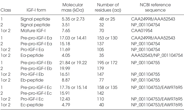
occur by use of a cryptic IGF633donor splice site, which is located 49 bp down-stream from the 5′end of the exon 5. When this cryptic IGF633donor splice site is not used, the alternative splicing of exon 4–5 occurs, that is, the Eb pep-tide (22,43). The predicted molecular mass and the residues of the three differ-ent isoforms of the human IGF-I precur-sor polypeptide as well as of their vari-ous forms and partial peptides (based on the amino acid sequence derived from the various IGF-I mRNA transcripts) are summarized in Table 1.
In general, the complexity introduced by the transcriptional and splicing vari-ants, posttranscriptional regulation and
posttranslational modifications of the IGF1gene (77), giving rise to various IGF-I isoforms, probably indicate their different biological roles under various conditions or pathologies and following different stimuli (78). The development of epitope-specific antibodies for distin-guishing the different IGF-I E-peptides (20,38,79–82) can contribute to a more definitive analysis of IGF-I isoforms ex-pression in various tissues and physio-logical or pathophysiophysio-logical conditions.
IGF-I PROCESSING, SECRETION AND GLYCOSYLATION
Post translational processing of IGF-I precursor protein may be a regulatory
mechanism of the IGF-I activity, as indi-cated by the unique processing features of IGF-I precursor polypeptides that have been described (25,78). Posttransla-tional endoproteolysis of those polypep-tides produces the signal, the mature and the E-peptides (E domains). It is thought that the signal peptide at the start of a precursor is removed after facilitating the passage of the polypeptide into the en-doplasmic reticulum and the secretory pathway, with possibly no further bio-logical significance (23,83). The se-quences of the signal peptides and the E-peptides are less strongly conserved compared with mature IGF-I peptide, though to a variable extent (23).
The mature peptide comprises four domains, that is, the B amino-terminal domain, C and A domain and D carboxy-terminal domain, of IGF-I polypeptides (25,84). In addition, two other protein products have been identified in the human brain; the tripeptide prolyl-glutamate (GPE) corresponding to the NH2-terminal of the B domain of ma-ture IGF-I and a truncated IGF-I form (–3N:IGF-I) that lacks the first three amino acids of the amino terminal end of mature peptide, probably due to alter-nate signal peptides or the combined ac-tion of some peptidases (78,85,86). Re-moval of the NH2-terminal tripeptide could be a mechanism for increasing the biological potency and availability of IGF-I, since the truncated –3N:IGF-I has less affinity for IGF-binding proteins than mature IGF-I, thus, increasing its bioactivity (78), (see Figure 1A) (see below: IGF-I Receptors and Binding Proteins).
The mature IGF-I is a 70 aa long single-chain peptide and a highly con-served sequence among primate species (23,24). Cleavage of pro-IGF-I removes the carboxyl-terminal E domain and can occur at the highly conserved, unique pentabasic motif K65
-X-X-K68-X-X-R71 -X-X-R74-X-X-R77. More specifically, the
Arg71-Ser72bond is cleaved followed by the removal of the Arg residue by the ac-tion a carboxypeptidase (25), (see Fig-ures 1A–C). In general, proproteins can be processed at this specific motif, usu-ally residing at the end of their pro re-gions, by proprotein convertases (PCs) such as furin (25,87,88). Furin belongs to the subtilisin-related PCs (SPCs), a major family of endoproteolytic processing en-zymes of the secretory pathway in mam-mals (89,90). Seven mammalian PCs have been identified, namely PC1, PC2, furin, PC4, PC5, paired basic amino acid cleav-ing enzyme 4 (PACE4) and PC7, and a method of prediction of the general PC-specific or furin-PC-specific cleavage sites has been proposed (88). PCs process pre-cursors at sites usually containing the specific consensus sequence [R/K]-Xn-[R/K], where X indicates any amino acid residue, and n, the number of spacer amino acid residues, which is 0, 2, 4 or 6 (91), (see Figures 1A–C). However, furin appears to have a more stringent speci-ficity and preferentially recognizes sites that contain the sequence motif R-X-[R/K]-R (87), while R-X-X-R is its mini-mal cleavage sequence (88). Thus, apart from the cleavage site Arg71-Ser72for ma-ture IGF-I, another furin-mediated pro-cessing at Arg77has been observed in
overexpression studies, which produces an extended, 76 aa long mature IGF-I (25,78), (see Figures 1A–C).
PC-mediated processing of pro-IGF-I to mature peptide has been shown to occur intracellularly (92), as expected for intracellular convertases such as furin, which are located in the secretory path-way (92,93). Nevertheless, evidence has been provided that the E domains are not cleaved intracellularly (94), and the secretion of unprocessed pro-IGF-IEa iso-form, both glycosylated and nonglycosy-lated, has been reported (25,30,95–97), while there are potential proprotein con-vertases that could process pro-IGF-I ex-tracellularly (97,98).
Conversion of pro-IGF-I to mature peptide cleaves off the E domains of the IGF-I precursors and one peptide is pro-duced from the human Ea domain after posttranslational processing of pro-IGF-IEa isoform (23,80), (see Figure 1A). However, the Eb domain contains po-tential processing sites (see Figure 1B), and at least two putative peptide prod-ucts of the human Eb peptide have been identified: the EB1peptide (residues 103–124 aa) with a C-terminal amide and the EB2peptide amide (residues 129–142) (20). The Ec domain also con-tains potential processing sites, from which one corresponds to the EB1 pep-tide cleavage site and cleaves off the last eight residues of the Ec domain encoded by the exon 6 (26,82), (see Figures 1B, C). However, so far only the proform of IGF-IEc isoform and not the Ec peptide or other cleavage products have been detected (31,33–35,73,82).
The Ea-peptide of human IGF-I con-tains an N-linked glycosylation site at Asn92based on the consensus sequence Asn-X-Ser/Thr, where X represents any encoded amino acid except proline (99,100), (see Figure 1A). However, it has been found that the human IGF-IEa pre-cursor with a signal sequence of 48 aa (Met-48) is not glycosylated, implying that the greater the number of the amino acids contained in the signal peptide the lesser the extent of a glycosylation process (101). The human Eb- and Ec-peptides
Table 1.Predicted molecular mass and residues of the three different isoforms (i.e., IGF-IEa, IGF-IEb, IGF-IEc), as well as of their various forms and partial peptides, of the human IGF-I precursor polypeptide.
Molecular Number of NCBI reference Class IGF-I form mass (kDa) residues (aa) sequence
1 Signal peptide 5.35 or 2.73 48 or 25 CAA24998/AAA52543 2 Signal peptide 3.51 32 NP_001104754 1or 2 Mature IGF-I 7.65 70 CAA01954
1 Pre-pro-IGF-I Ea 17.03 or 14.41 153 or 130 CAA24998/AAA52543 2 Pre-pro-IGF-I Ea 15.18 137 NP_001104754 1or 2 Pro-IGF-I Ea 11.69 105 NP_001104754
1or 2 Ea-peptide 4.05 35 AAA52543/NP_001104754 1 Pre-pro-IGF-I Eb 21.84 or 19.22 195 or 172 NP_001104755
2 Pre-pro-IGF-I Eb 19.99 179
1or 2 Pro-IGF-I Eb 16.51 147 NP_001104755 1or 2 Eb-peptide 8.87 77 NP_001104755
1 Pre-pro-IGF-I Ec 17.76 or 15.14 158 or 135 NP_001104753/EAW97695 2 Pre-pro-IGF-I Ec 15.91 142
lack this cotranslational modification due to the reading frame shift resulted from the inclusion of the exon 5 in these two IGF-I isoforms (see Figures 1B, C). N-linked glycosylation involves the trans-fer of a lipid-linked tetradecasaccharide (GlcNAc2-Man9-Glc3) to an asparagine side chain (100,102), (see Figure 1A). It oc-curs in endoplasmic reticulum and the subsequent diversification of the conju-gates occurs both in the endoplasmic reticulum and Golgi apparatus (100,102). Recently, it has been found that the gly-cosylation status of pro-IGF-IEa does not affect its processing (97), while the secre-tion of a peptide similar or identical to Ea from a human B-lymphocyte cell line, in a partially glycosylated form has been reported (80). Although the deglycosyla-tion process is expected to occur intracel-lularly, whether the deglycosylation of pro-IGF-IEa or the Ea-peptide also could be clipped extracellularly is not known.
Considering the unique role of glycosy-lation in the protein biosynthesis process (100), it is possible that the Ea-peptide glycosylation might play a role in interac-tions with chaperones in the endoplasmic reticulum (78), or in regulation of the bioavailability of the different species of this IGF-I isoform (that is, pro-IGF-I Ea, mature IGF-I or Ea-peptide) (78,97,103). Thus, the existence of an N-linked glyco-sylation site in the Ea-peptide, which is absent in the Ec- and Eb-peptide, might reflect a differential and specific biologi-cal action of the IGF-IEa isoform medi-ated by this posttranslational modifica-tion of its Ea-peptide (25,43,97). Recently, it has been shown that the species of IGF-I produced by the IGF-IEa isoform has a differential ability to activate IGF-IR. Glycosylated pro-IGF-IEa is less effi-cient at receptor activation than pro-IGF-I and mature IGF-I (97), resembling the de-creased receptor- binding affinity of pegy-lated IGF-I forms (104,105) and implying that glycosylated pro-IGF-IEa may serve as a reservoir for IGF-I that can be stored until needed (97). The strong conserva-tion observed in the sequence of the Ea domain also suggests a specific biological function for the Ea-peptide (23).
Nevertheless, whether the E-peptides are more stable and/or bioactive within their pro-IGF-I forms, or they are processed to act directly on their targets remains to be elucidated (see further dis-cussion in next sections). Moreover, it would be essential to verify whether the IGF-I isoforms could be released in the circulation as different proforms or E-peptides (3,79), or the final peptide that enters the circulation after extracellular endoproteolysis of the IGF-I prohormone is only the mature peptide (103,106).
IGF-I RECEPTORS AND BINDING PROTEINS
IGF-I actions are mediated through its binding to several receptors, such as IGF-IR (or type I IGF receptor) and IGF-IIR (or type II IGF receptor), insulin receptor (IR), and some atypical receptors such as the hybrid IR/IGF-IR (107–109).
More specifically, the mature IGF-I peptide, which is responsible for binding to the receptors, binds IGF-IR with the highest affinity, IGF-IIR with low affinity and is also able to interact with IR. The IGF-IR exhibits a high degree of homol-ogy to IR (110) and, given the significant structural similarity between IGF-I and insulin, these ligands can cross-activate both receptors, while the IGF-IR signal-ing pathways share multiple intracellular mediators with the insulin signaling cas-cade (19,52). The IGF-IR/IR hybrid re-ceptor is thought to function predomi-nantly as an IGF-I receptor, since its binding affinity for insulin is lower than that for IGF-I, however the functional importance of IGF-IR/IR hybrid receptor remains poorly understood (74,109,111).
It is widely recognized that most of the observed IGF-I biological effects on cell growth, differentiation, invasion and sur-vival depend on the binding and activa-tion of IGF-IR, which is a ligand-activated receptor tyrosine kinase (4,112). Specifi-cally, functional epitope mapping of IGF-I has revealed that the IGF-IR interacts with the residues 21, 23, 24, 44 as well as the ty-rosines 31 and 60, which are located in the C and A domains (113), (see Figure 1A). IGF-IR is a transmembrane protein
con-sisting of two extracellular α-subunits, which contain the cysteine-rich ligand binding site, and two transmembrane β-subunits that have a cluster of three ty-rosine residues, which undergo phospho-rylation and activation upon IGF-I bind-ing (2,110,114). A structural rearrangement in the transmembrane βsubunits of the receptor is caused by binding of IGF-I to IGF-IR, resulting in transautophosphory-lation of the cytoplasmic tyrosine kinase domain of the receptor, as one kinase do-main phosphorylates the other, and thus destabilizing the autoinhibitory conforma-tion within the kinase domain (115,116). This conformational change permits unre-stricted access to the binding sites for pro-tein substrates (117), thus recruiting spe-cific cytoplasmic molecules, such as insulin receptor substrate (IRS) proteins, and activating specific intracellular path-ways including Ras/mitogen-activated protein kinase (MAPK)/extracellular signal-regulated kinases 1 and 2 (ERK1/2) and phosphatidylinositol 3-kinase (PI3K)/ Akt (118).
However, some IGFBPs can exhibit IGF-I potentiating effects and their IGF-I-inhibitory or stimulatory activities are dictated by factors such as the tissue-specific distribution of particular IGFBPs and the ratio between free (active) IGF-I and IGFBP-IGF-I bound (4,11). Moreover, it has been shown that certain IGFBPs have IGF-independent activities, imply-ing that they can modulate cell survival and apoptosis, or inhibit tumor growth in the absence of the ligand (11,127). In addition, proteolytic fragments of IGFBP-3 were reported to possess mito-genic activity in the peritoneal fluid of women with endometriosis (128).
There is also a group of cysteine-rich proteins, known as IGFBP-related pro-teins (IGFBP-rPs), that share important structural similarities with the IGFBPs but they have low binding affinity to IGFs. It has been proposed that these proteins and the IGFBPs constitute an IGFBP superfamily (129,130), however the functions of the IGFBP-rPs regarding the IGFs actions are as yet unclear (131).
IGF-I PEPTIDES ACTIONS AND SIGNALING
Although, by the general consensus, IGF-I is thought to exert its biological actions predominantly through mature peptide, differential biological activities have been reported for the different IGF-I isoforms (propeptides), or for their E-peptides, exogenously adminis-trated or overexpressed in various in vivo(28,29,36,132–134) and in vitro mod-els (20,27,31–33,135,136), implying that there are peptides other than the IGF-I ligand that also possess bioactivity and, thus, both common and unique or com-plementary pathways exist for the IGF-I isoforms to promote biological effects (31,36).
A differential expression profile of the IGF-I isoforms have been shown in vari-ous conditions or pathologies in humans, such as skeletal muscle damage (31,68), endometriosis (35) or prostate (34), cervi-cal (69) and colorectal cancer (137). The diversity and the patterns of differential expression have been proposed to reflect
potential biological activities associated with the E domain peptides (27,52). Di-vergent actions and signaling of the dif-ferent pro-IGF-I forms or mature IGF-I lacking any E-peptide have been re-ported after viral-mediated expression of the IGF-I isoforms (IGF-IA and IGF-IB) in mouse skeletal muscle (36,132). Specifi-cally, it has been shown that overexpres-sion of mature IGF-I in skeletal muscle did not promote muscle hypertrophy in young mice, suggesting that the pro-IGF-I forms are required for this effect and that E-peptides, either as a part of pro-IGF-I or independent of mature pro-IGF-I, may be necessary for IGF-I-mediated muscle hypertrophy (36). Moreover, by using the MKR transgenic mouse model where functional IGF-IRs are lacking in muscle fibers (as a dominant negative IGF-IR is expressed specifically in skele-tal muscle), it was shown that, regardless of which isoform is overexpressed, IGF-I receptors on muscle fibers are required for IGF-I-mediated hypertrophy (36). In-terestingly, however, after viral-mediated delivery of murine IGF-I isoforms into skeletal muscle, even though both iso-forms caused increased phosphorylation of the IGF-IR, increased expression of IGF-IB (murine IGF-IEb) drove both main pathways downstream of IGF-IR, that is, the PI3K/Akt pathway and the MAPK pathway, whereas IGF-IA
(murine IGF-IEa) overexpression resulted in increased Akt phosphorylation only (132). These findings imply distinguish-ing IGF-I isoform–specific actions, re-gardless of any potential receptor(s) acti-vated (see also discussion in following sections). In addition, constitutively overexpression of the IGF-IEa isoform specifically in cardiac muscle was shown to protect the heart from oxidative stress via Sirtuin 1 (SirT1)⁄c-Jun N-terminal ki-nase 1 (JNK1) activity while, conversely, mature IGF-I triggered oxidative stress in the heart and did not affect SirT1 activity (138,139). Moreover, it has been shown that although both mature IGF-I and IGF-IEa propeptide triggered the phos-phorylation of IGF-IR, its downstream canonical PI3K/Akt/mTOR signaling
pathway was not induced in the trans-genic mice overexpressing IGF-IEa (138,140). Instead, this specific IGF-I propeptide activated alternate signaling intermediates dependent protein kinase-1 (PDK1) and serum/glucocorticoid regulated kinase 1 (SGK1), as well as SirT1. Thus, it was suggested that this downstream of IGF-I receptor(s) signaling activated by IGF-IEa employs novel pathways and that the divergent signaling mechanisms be-tween the two IGF-I forms (that is, ma-ture IGF-I and IGF-IEa propeptide) may account for their opposing effects on the heart (136,138,140).
Considering particularly the bioactiv-ity of the E-peptides, in vitrostudies have suggested that the E-peptides of the human IGF-I precursors may act as inde-pendent growth factors, since their syn-thetic analogs, generated from unique re-gions within the E domains, were demonstrated to possess mitogenic (20,27,34,35,73,141), angiogenic (142) and migratory activity (28,141,143), and regu-late cell differentiation (27,28) in various human cells or cell lines. Antitumor ac-tivity of human Eb-peptide also has been reported in some cancer cells (144).
The differential biological effects of the synthetic Ec-peptide compared with ma-ture IGF-I peptide, such as cell prolifera-tion versus differentiaprolifera-tion, and the lack of suppression of the synthetic E-peptide bioactivity after blocking (mature) IGF-I signaling with IGF-IR neutralizing anti-bodies, makes it tempting to postulate that the Ec-peptide acts via a different re-ceptor (28,135,145). However, concerns have been raised about the effectiveness of the IGF-IR neutralizing antibodies to block IGF-I signaling, since they could internalize and, in this way, even activate the receptor, or change its localization, thus facilitating an E-peptide action (38). Nevertheless, the mature IGF-I bioactiv-ity appeared to be suppressed in those cells where IGF-IR neutralizing antibod-ies were used.
Interestingly, an autonomous, IGF-IR-independent bioactivity has been re-ported for specific regions of the human Eb domain of IGF-I; the synthetic analog of the EB1peptide (see above: IGF-I Pro-cessing, Secretion and Glycosylation) was found not only to possess mitogenic ac-tivity in human bronchial epithelial cells but also to bind to specific high-affinity receptors on those cells. Furthermore, neither ligand binding was inhibited by recombinant IGF-I or recombinant in-sulin, nor did a monoclonal antibody an-tagonist to the IGF-IR suppress the prolif-erative response induced by the synthetic EB1peptide. It was suggested that IB1is a growth factor that mediates its effect through a specific receptor (20). Similarly, in accordance with the well established notion that the molecular action of a bioactive peptide initiates through its specific binding to a cell surface receptor, it was demonstrated that the human Eb-peptide binds to common cell surface molecules on human neuroblastoma cells (27). Again, the saturation of mature IGF-I or insulin did not displace the bind-ing of the human Eb-peptide, suggestbind-ing the existence of distinct putative receptor components on those cells (148).
Further evidence for distinct, independent bioactivity of the human Ec domain was provided from its divergent
signaling compared with mature IGF-I. Our group (31,33,34,65) and others (39) have shown that a synthetic analog of the human Ec peptide possesses distinct sig-naling compared with IGF-IR ligand. Spe-cifically, to distinguish any unique biolog-ical effect of the Ec domain from a potential bioactivity possessed by the common part (that is, the first 16 residues) of the IGF-I E domains, we utilized a syn-thetic Ec-peptide corresponding to the region beyond that common sequence, that is, a peptide similar to the C-terminal 24-residues, of which 16 are encoded by exon 5 and the last 8 by exon 6. It was documented that this sequence of the human Ec domain possesses distinct sig-naling since, in contrast to mature IGF-I, it activates only ERK1/2 and not Akt (31,33,34,65). Moreover, the selective acti-vation of only one of the two main signal-ing pathways downstream of IGF-I/IGF-IR and the IGF-IGF-I/IGF-IR- and IGF-I/IGF-IR- independent bioactivity of this synthetic part of the Ec domain was further documented by using the siRNA knock-out model in various human cell lines (34,35,73).
More recently, synthetic E-peptides corresponding to the rodent Ea and Eb domain sequences were utilized in a ki-nase receptor activation (KIRA) assay to test IGF-I-dependent and -independent activation of IGF-IR by these E-peptides, showing that they do not directly induce IGF-IR phosphorylation in mouse fibrob-lasts (P6 cells) (38). Interestingly, how-ever, the presence of either of those E-peptides increased IGF-IR activation by IGF-I in the murine C2C12 cell line, sug-gesting that they may modulate IGF-I ac-tivity (38). Moreover, from the findings of that model, it was concluded that E-peptides signaling, as well as mitogenic and motogenic effects are dependent upon a functional IGF-IR, and their activ-ity plausibly reflects actions of pro-IGF-I (38). Further evidence has been provided recently, suggesting that IGF-I splice variants may exert their actions through mature IGF-I and not the E-peptides (149), or by supporting the bioactivity of pro-IGF-I forms (97). Furthermore, there is controversial evidence regarding
the role of proforms (103,136,149) or E-peptides (38) of the murine IGF-I iso-forms, particularly in cell differentiation. Whether it reflects a differential mode of action of the free E-peptides compared with the pro-IGF-I isoforms, or potential differences between their exogenous ad-ministration and overexpression (or nat-ural endogenous production), possibly should be addressed.
Collectively, the in vitromodels utiliz-ing the sequences of the rodent IGF-I E domains have provided specific infor-mation regarding the bioactivity and the mode of action of those E-peptides in murine cell lines. The findings of these studies suggest that the E-peptides have little or no independent activity and, in-stead, they modulate mature IGF-I (lig-and) activity and signaling (see review in [150]). Nevertheless, since these do-mains are very variable and much less conserved, it has been indicated that the species specificity must clearly be taken into account when assessing the activity of the human IGF-I Eb and Ec domains, from which peptides with important bi-ological activities have been reported (23). Thus, it remains to be further eluci-dated whether the autonomous, independent and IRindependent bio -activity, and the specific high-affinity receptor binding observed for human Eb- and Ec-peptides in various human cell lines (20,34,35,73,148) reflect an alter-native, species-specific ligand/ receptor mechanism of action for these human E domains. Species-specific models, in terms of utilizing peptide sequences that correspond to the IGF-I E domains of the species or the cells used in the model, are of particular importance to investigate physiological mechanisms of actions, such as a ligand/receptor mechanism.
which would act in favor of the MAPK arm of the IGF-IR downstream signaling (38). However, since the possibility that the E-peptides also activate ERK1/2 through an IGF-IR independent mecha-nism cannot be excluded, it remains to be further elucidated whether they af-fect the ERK1/2 pathway only at the level of the IGF-IR per se(that is, possi-bly by increasing its internalization, or by affecting its conformational change after its binding to IGF-I and, thus, acti-vating exclusively a specific pathway) or if the E-peptide–induced activation of ERK1/2 occurs at a level downstream of the IGF-IR, or even upstream, in that the E-peptide utilizes another “receptor” or cellular uptake mechanism, given the activation of ERK1/2 after exogenous administration of the E-peptide. The in-tracellular signaling pathways initiated by IGF-IR ligation may interact with signaling via G protein-coupled recep-tors or other mediarecep-tors, modulating some responses (151). However, the ex-istence of such a putative, canonical or noncanonical receptor or internalization mechanism for the E-peptide remains to be determined and characterized. Alter-natively, the exclusive phosphorylation of ERK1/2 has been proposed to be a re-sult of a possible tuning of the IGF-IR signaling cascade by the E-peptide to-ward MAPK (38).
The identification of the signaling pathways and effectors upstream and downstream of the E-peptide–induced activation of the extracellular-regulated kinases ERK1 and ERK2, the characterized members of the MAP ki-nase family (152), would provide infor-mation of particular importance regarding the mechanisms of E-peptide bioactivity. Furthermore, it is tempting to speculate that the selective activation of MAPK/ERK1/2 pathway up to a critical level by the E-peptide might further exert an inhibitory regulation of its com-petitive PI3K/Akt pathway, since it has been shown that the activity of one path-way might inactivate portions of the other (153,154), thus consisting of a regu-latory mechanism of competitive
biologi-cal actions such as cell proliferation and differentiation.
It should be also mentioned that, given the subcellular as well as differential local-ization of the E-peptides (94,141,155,156), an intracrine signaling mechanism that mediates E-peptides bioactivity, as pro-posed for some peptide growth factors (157), cannot be excluded.
CONCLUSION
IGF-I regulation occurs at multiple lev-els due to the alternative splicing of its pre-mRNA both at the 5′and 3′end, and the isoform-specific co- and posttransla-tional modifications, which appear to play key roles in modulating the bio -availabilty and bioactivity of this growth factor. New evidence supports the con-cept of IGF-I isoform– or IGF-I various peptides–specific functions, possibly via specific and divergent signaling. Never-theless, it remains to be determined where the distinct E-peptide signaling diverges from that of mature IGF-I, or whether this signaling is induced uniquely and au-tonomously by the E-peptides or syner-gistically with the mature IGF-I bioactive molecule, and in the form of processed or pro-IGF-I molecules. Thus, the regulatory mechanism(s) of a potentially specific and apportioned bioactivity of the various IGF-I peptides is of particular interest within the context of a revisited character-ization of the multiple IGF-I actions.
DISCLOSURE
The authors declare that they have no competing interests as defined by Molecu-lar Medicine, or other interests that might be perceived to influence the results and discussion reported in this paper.
REFERENCES
1. Liu JP, Baker J, Perkins AS, Robertson EJ, Efstratiadis A. (1993) Mice carrying null muta-tions of the genes encoding insulin-like growth factor I (Igf-1) and type 1 IGF receptor (Igf1r). Cell. 75:59–72.
2. Laviola L, Natalicchio A, Giorgino F. (2007) The IGF-I signaling pathway. Curr. Pharm. Des. 13:663–9. 3. Barton ER, et al. (2012) Deletion of muscle GRP94 impairs both muscle and body growth by inhibit-ing local IGF production. FASEB J. 26:3691–702. 4. Jones JI, Clemmons DR. (1995) Insulin-like
growth factors and their binding proteins: bio-logical actions. Endocr. Rev. 16:3–34.
5. Koutsilieris M, Mitsiades C, Sourla A. (2000) In-sulin-like growth factor I and urokinase-type plasminogen activator bioregulation system as a survival mechanism of prostate cancer cells in osteoblastic metastases: development of survival factor therapy for hormone-refractory prostate cancer. Mol. Med. 6:251–67.
6. Siddle K, et al. (2001) Specificity in ligand bind-ing and intracellular signallbind-ing by insulin and insulin-like growth factor receptors. Biochem. Soc. Trans. 29:513–25.
7. LeRoith D, Roberts CT, Jr. (2003) The insulin-like growth factor system and cancer. Cancer Lett. 195:127–37.
8. Kooijman R. (2006) Regulation of apoptosis by insulin-like growth factor (IGF)-I. Cytokine Growth Factor Rev. 17:305–23.
9. Werner H, LeRoith D. (1996) The role of the in-sulin-like growth factor system in human cancer. Adv. Cancer Res. 68:183–223.
10. Reyes-Moreno C, Sourla A, Choki I, Doillon C, Koutsilieris M. (1998) Osteoblast-derived survival factors protect PC-3 human prostate cancer cells from adriamycin apoptosis. Urology. 52:341–7. 11. Werner H, Bruchim I. (2009) The insulin-like
growth factor-I receptor as an oncogene. Arch. Physiol. Biochem. 115:58–71.
12. Ewton DZ, Roof SL, Magri KA, McWade FJ, Florini JR. (1994) IGF-II is more active than IGF-I in stimulating L6A1 myogenesis: greater mito-genic actions of IGF-I delay differentiation. J. Cell Physiol. 161:277–84.
13. Florini JR, Ewton DZ, Coolican SA. (1996) Growth hormone and the insulin-like growth factor sys-tem in myogenesis. Endocr. Rev. 17:481–517. 14. Daughaday WH, Rotwein P. (1989) Insulin-like
growth factors I and II. Peptide, messenger ri-bonucleic acid and gene structures, serum, and tissue concentrations. Endocr. Rev. 10:68–91. 15. Ohlsson C, et al. (2009) The role of liver-derived
in-sulin-like growth factor-I. Endocr. Rev. 30:494–535. 16. Naranjo WM, et al. (2002) Protein calorie restric-tion affects nonhepatic IGF-I producrestric-tion and the lymphoid system: studies using the liver-specific IGF-I gene-deleted mouse model. Endocrinology. 143:2233–41.
17. Klover P, Hennighausen L. (2007) Postnatal body growth is dependent on the transcription factors signal transducers and activators of transcrip-tion 5a/b in muscle: a role for autocrine/ paracrine insulin-like growth factor I. Endocrinol-ogy. 148:1489–97.
18. Stratikopoulos E, Szabolcs M, Dragatsis I, Kli-nakis A, Efstratiadis A. (2008) The hormonal ac-tion of IGF1 in postnatal mouse growth. Proc. Natl. Acad. Sci. U. S. A. 105:19378–83.
19. Duan C, Ren H, Gao S. (2010) Insulin-like growth factors (IGFs), IGF receptors, and IGF-binding proteins: roles in skeletal muscle growth and dif-ferentiation. Gen. Comp. Endocrinol. 167:344–51. 20. Siegfried JM, et al. (1992) A mitogenic peptide
the insulin-like growth factor IB prohormone. Proc. Natl. Acad. Sci. U. S. A. 89:8107–11. 21. Gilmour RS. (1994) The implications of
insulin-like growth factor mRNA heterogeneity. J. En-docrinol. 140:1–3.
22. Chew SL, Lavender P, Clark AJ, Ross RJ. (1995) An alternatively spliced human insulin-like growth factor-I transcript with hepatic tissue ex-pression that diverts away from the mitogenic IBE1 peptide. Endocrinology. 136:1939–44. 23. Wallis M. (2009) New insulin-like growth factor
(IGF)-precursor sequences from mammalian genomes: the molecular evolution of IGFs and associated peptides in primates. Growth Horm. IGF. Res. 19:12–23.
24. Rotwein P, Pollock KM, Didier DK, Krivi GG. (1986) Organization and sequence of the human insulin-like growth factor I gene. Alternative RNA processing produces two insulin-like growth factor I precursor peptides. J. Biol. Chem. 261:4828–32.
25. Duguay SJ, Lai-Zhang J, Steiner DF. (1995) Mu-tational analysis of the insulin-like growth fac-tor I prohormone processing site. J. Biol. Chem. 270:17566–74.
26. Barton ER. (2006) The ABCs of IGF-I isoforms: impact on muscle hypertrophy and implications for repair. Appl. Physiol. Nutr. Metab. 31:791–7. 27. Kuo YH, Chen TT. (2002) Novel activities of pro-IGF-I E peptides: regulation of morphological dif-ferentiation and anchorage-independent growth in human neuroblastoma cells. Exp. Cell Res. 280:75–89. 28. Mills P, Dominique JC, Lafreniere JF,
Bouchen-touf M, Tremblay JP. (2007) A synthetic mechano growth factor E Peptide enhances myogenic pre-cursor cell transplantation success. Am. J. Trans-plant. 7:2247–59.
29. Dluzniewska J, et al. (2005) A strong neuroprotec-tive effect of the autonomous C-terminal peptide of IGF-1 Ec (MGF) in brain ischemia. FASEB J. 19:1896–8.
30. Pfeffer LA, Brisson BK, Lei H, Barton ER. (2009) The insulin-like growth factor (IGF)-I E-peptides modulate cell entry of the mature IGF-I protein. Mol. Biol. Cell. 20:3810–7.
31. Philippou A, et al. (2009) Expression of IGF-1 iso-forms after exercise-induced muscle damage in humans: characterization of the MGF E peptide actions in vitro. In Vivo. 23:567–75.
32. Quesada A, Ogi J, Schultz J, Handforth A. (2011) C-terminal mechano-growth factor induces heme oxygenase-1-mediated neuroprotection of SH-SY5Y cells via the protein kinase C/Nrf2 path-way. J. Neurosci. Res. 89:394–405.
33. Stavropoulou A, et al. (2009) IGF-1 expression in infarcted myocardium and MGF E peptide actions in rat cardiomyocytes in vitro. Mol. Med. 15:127–35. 34. Armakolas A, et al. (2010) Preferential expression of
IGF-1Ec (MGF) transcript in cancerous tissues of human prostate: evidence for a novel and au-tonomous growth factor activity of MGF E peptide in human prostate cancer cells. Prostate. 70:1233–42. 35. Milingos DS, et al. (2010) Insulinlike growth factor-1Ec (MGF) expression in eutopic and ectopic
en-dometrium: characterization of the MGF E-peptide actions in vitro. Mol. Med. 17:21–8.
36. Barton ER, DeMeo J, Lei H. (2010) The insulin-like growth factor (IGF)-I E-peptides are required for isoform-specific gene expression and muscle hypertrophy after local IGF-I production. J. Appl. Physiol. (1985). 108:1069–76.
37. Gentile MA, et al. (2010) Androgen-mediated improvement of body composition and muscle function involves a novel early transcriptional program including IGF1, mechano growth fac-tor, and induction of {beta}-catenin. J. Mol. En-docrinol. 44:55–73.
38. Brisson BK, Barton ER. (2012) Insulin-like growth factor-I E-peptide activity is dependent on the IGF-I receptor. PLoS One. 7:e45588.
39. Quesada A, Micevych P, Handforth A. (2009) C-terminal mechano growth factor protects dopamine neurons: a novel peptide that induces heme oxygenase-1. Exp. Neurol. 220:255–66. 40. Deng M, et al. (2011) Mechano growth factor E
pep-tide promotes osteoblasts proliferation and bone-defect healing in rabbits. Int. Orthop. 35:1099–106. 41. Adamo ML, et al. (1993) Distinct promoters in the
rat insulin-like growth factor-I (IGF-I) gene are active in CHO cells. Endocrinology. 132:935–937. 42. Yang H, et al. (1995) Alternative leader sequences
in insulin-like growth factor I mRNAs modulate translational efficiency and encode multiple sig-nal peptides. Mol. Endocrinol. 9:1380–95. 43. Shavlakadze T, Winn N, Rosenthal N, Grounds
MD. (2005) Reconciling data from transgenic mice that overexpress IGF-I specifically in skele-tal muscle. Growth Horm. IGF. Res. 15:4–18. 44. Philippou A, Maridaki M, Halapas A, Koutsilieris
M. (2007) The role of the insulin-like growth factor 1 (IGF-1) in skeletal muscle physiology. In Vivo. 21:45–54.
45. Okazaki R, Durham SK, Riggs BL, Conover CA. (1995) Transforming growth factor-beta and forskolin increase all classes of insulin-like growth factor-I transcripts in normal human osteoblast-like cells. Biochem. Biophys. Res. Commun. 207:963–70. 46. Bloor CA, Knight RA, Kedia RK, Spiteri MA,
Allen JT. (2001) Differential mRNA expression of insulin-like growth factor-1 splice variants in pa-tients with idiopathic pulmonary fibrosis and pulmonary sarcoidosis. Am. J. Respir. Crit. Care Med. 164:265–72.
47. Shamblott MJ, Chen TT. (1993) Age-related and tissue-specific levels of five forms of insulin-like growth factor mRNA in a teleost. Mol. Mar. Biol. Biotechnol. 2:351–61.
48. Duguay SJ, Swanson P, Dickhoff WW. (1994) Dif-ferential expression and hormonal regulation of alternatively spliced IGF-I mRNA transcripts in salmon. J Mol. Endocrinol. 12:25–37.
49. Faulk CK, Perez-Dominguez R, Webb KA, Jr., Holt GJ. (2010) The novel finding of four distinct prepro-IGF-I E domains in a perciform fish, Sci-aenops ocellatus, during ontogeny. Gen. Comp. Endocrinol. 169:75–81.
50. Zhang J, et al. (2013) Cloning and characteriza-tion of new transcript variants of insulin-like
growth factor-I in Sika deer (Cervus elaphus). Growth Horm. IGF. Res. 23:120–7.
51. Jansen E, Steenbergh PH, LeRoith D, Roberts CT, Jr., Sussenbach JS. (1991) Identification of multiple tran-scription start sites in the human insulin-like growth factor-I gene. Mol. Cell. Endocrinol. 78:115–25. 52. Philippou A, Armakolas A, Koutsilieris M. (2013) Evidence for the Possible Biological Significance of the igf-1 Gene Alternative Splicing in Prostate Cancer. Front Endocrinol. (Lausanne). 4:31. 53. Adamo ML, Ben-Hur H, LeRoith D, Roberts CT,
Jr. (1991) Transcription initiation in the two leader exons of the rat IGF-I gene occurs from disperse versus localized sites. Biochem. Biophys. Res. Commun. 176:887–93.
54. D’Ercole AJ, Stiles AD, Underwood LE. (1984) Tissue concentrations of somatomedin C: further evidence for multiple sites of synthesis and paracrine or autocrine mechanisms of action. Proc. Natl. Acad. Sci. U. S. A. 81:935–9.
55. Rotwein P, Pollock KM, Watson M, Milbrandt JD. (1987) Insulin-like growth factor gene expression during rat embryonic development. Endocrinol-ogy. 121:2141–4.
56. Adamo M, Lowe WL, Jr., LeRoith D, Roberts CT, Jr. (1989) Insulin-like growth factor I messenger ribonucleic acids with alternative 5′-untranslated regions are differentially expressed during devel-opment of the rat. Endocrinology. 124:2737–44. 57. Wang X, Yang Y, Adamo ML. (1997)
Characteri-zation of the rat insulin-like growth factor I gene promoters and identification of a minimal exon 2 promoter. Endocrinology. 138:1528–36.
58. O’Sullivan DC, Szestak TA, Pell JM. (2002) Regu-lation of IGF-I mRNA by GH: putative functions for class 1 and 2 message. Am. J. Physiol. En-docrinol. Metab. 283:E251–8.
59. Woelfle J, Chia DJ, Rotwein P. (2003) Mechanisms of growth hormone (GH) action. Identification of conserved Stat5 binding sites that mediate GH-induced insulin-like growth factor-I gene activa-tion. J. Biol. Chem. 278:51261–6.
60. Chia DJ, Young JJ, Mertens AR, Rotwein P. (2010) Distinct alterations in chromatin organization of the two IGF-I promoters precede growth hor-mone-induced activation of IGF-I gene transcrip-tion. Mol. Endocrinol. 24:779–89.
61. Lund PK. (1994) Insulin-like growth factor I: molec-ular biology and relevance to tissue-specific expres-sion and action. Recent Prog. Horm. Res. 49:125–48. 62. Hameed M, et al. (2004) The effect of
recombi-nant human growth hormone and resistance training on IGF-I mRNA expression in the mus-cles of elderly men. J. Physiol. 555:231–40. 63. Imanaka M, et al. (2008) Growth hormone
stimu-lates mechano growth factor expression and acti-vates myoblast transformation in C2C12 cells. Kobe J. Med. Sci. 54:E46–54.
64. Aperghis M, et al. (2009) Serum IGF-I levels and IGF-I gene splicing in muscle of healthy young males receiving rhGH. Growth Horm. IGF. Res. 19:61–7.
receptor mRNAs in human HLE-B3 lens epithe-lial cells. In Vivo. 25:179–84.
66. Jansen M, et al. (1983) Sequence of cDNA encod-ing human insulin-like growth factor I precursor. Nature. 306:609–11.
67. Rotwein P. (1986) Two insulin-like growth factor I messenger RNAs are expressed in human liver. Proc. Natl. Acad. Sci. U. S. A. 83:77–81.
68. McKay BR, O’Reilly CE, Phillips SM, Tarnopolsky MA, Parise G. (2008) Co-expression of IGF-1 fam-ily members with myogenic regulatory factors following acute damaging muscle-lengthening contractions in humans. J. Physiol. 586:5549–60. 69. Koczorowska MM, Kwasniewska A,
Gozdzicka-Jozefiak A. (2011) IGF1 mRNA isoform expres-sion in the cervix of HPV-positive women with pre-cancerous and cancer lesions. Exp. Ther. Med. 2:149–56.
70. Shimatsu A, Rotwein P. (1987) Sequence of two rat insulin-like growth factor I mRNAs differing within the 5′untranslated region. Nucleic Acids Res. 15:7196. 71. Goldspink G. (2005) Mechanical signals, IGF-I
gene splicing, and muscle adaptation. Physiology (Bethesda). 20:232–8.
72. Matheny RW, Jr., Nindl BC, Adamo ML. (2010) Minireview: Mechano-growth factor: a putative product of IGF-I gene expression involved in tissue repair and regeneration. Endocrinology. 151:865–875. 73. Philippou A, et al. (2011) IGF1Ec expression in
MG-63 human osteoblast-like osteosarcoma cells. Anticancer Res. 31:4259–65.
74. Yu H, Rohan T. (2000) Role of the insulin-like growth factor family in cancer development and progression. J. Natl. Cancer Inst. 92:1472–89. 75. Mourkioti F, Rosenthal N. (2005) IGF-1,
inflam-mation and stem cells: interactions during mus-cle regeneration. Trends Immunol. 26:535–42. 76. Temmerman L, Slonimsky E, Rosenthal N. (2010)
Class 2 IGF-1 isoforms are dispensable for viabil-ity, growth and maintenance of IGF-1 serum lev-els. Growth Horm. IGF. Res. 20:255–63.
77. Zhang J, Whitehead RE, Jr., Underwood LE. (1997) Effect of fasting on insulin-like growth fac-tor (IGF)-IA and IGF-IB messenger ribonucleic acids and prehormones in rat liver. Endocrinology. 138:3112–8.
78. Duguay SJ. (1999) Post-translational processing of insulin-like growth factors. Horm. Metab. Res. 31:43–9.
79. Powell DR, Lee PD, Chang D, Liu F, Hintz RL. (1987) Antiserum developed for the E peptide region of insulin-like growth factor IA prohormone recog-nizes a serum protein by both immunoblot and ra-dioimmunoassay. J. Clin. Endocrinol. Metab65:868–75. 80. Wilson HE, Westwood M, White A, Clayton PE.
(2001) Monoclonal antibodies to the terminal Ea sequence of pro-insulin-like growth fac-tor-IA (proIGF-IA) recognize proIGF-IA secreted by IM9 B-lymphocytes. Growth Horm. IGF. Res. 11:10–7. 81. Kravchenko IV, Furalyov VA, Khotchenkov VP,
Popov VO. (2006) Monoclonal antibodies to mechano-growth factor. Hybridoma (Larchmt). 25:300–5.
82. Philippou A, et al. (2008) Characterization of a
rabbit antihuman mechano growth factor (MGF) polyclonal antibody against the last 24 amino acids of the E domain. In Vivo. 22:27–35. 83. Lingappa VR, Lingappa JR, Blobel G. (1980)
Sig-nal sequences for early events in protein secre-tion and membrane assembly. Ann. N. Y. Acad. Sci. 343:356–61.
84. Denley A, Cosgrove LJ, Booker GW, Wallace JC, Forbes BE. (2005) Molecular interactions of the IGF system. Cytokine Growth Factor Rev. 16:421–39. 85. Carlsson-Skwirut C, et al. (1986) Isolation and
characterization of variant IGF-1 as well as IGF-2 from adult human brain. FEBS Lett201:46–50. 86. Sara VR, et al. (1993) The biological role of
trun-cated insulin-like growth factor-1 and the tripep-tide GPE in the central nervous system. Ann. N. Y. Acad. Sci. 692:183–91.
87. Nakayama K. (1997) Furin: a mammalian subtil-isin/Kex2p-like endoprotease involved in pro-cessing of a wide variety of precursor proteins. Biochem. J. 327 ( Pt 3): 625–35.
88. Duckert P, Brunak S, Blom N. (2004) Prediction of proprotein convertase cleavage sites. Protein Eng. Des. Sel. 17:107–12.
89. Rouille Y, et al. (1995) Proteolytic processing mechanisms in the biosynthesis of neuroen-docrine peptides: the subtilisin-like proprotein convertases. Front Neuroendocrinol. 16:322–61. 90. Steiner DF. (1998) The proprotein convertases.
Curr. Opin. Chem. Biol. 2:31–9.
91. Seidah NG, Chretien M. (1999) Proprotein and prohormone convertases: a family of subtilases generating diverse bioactive polypeptides. Brain Res. 848:45–62.
92. Duguay SJ, Milewski WM, Young BD, Nakayama K, Steiner DF. (1997) Processing of wild-type and mutant proinsulin-like growth factor-IA by sub-tilisin-related proprotein convertases. J. Biol. Chem. 272:6663–70.
93. Thomas G. (2002) Furin at the cutting edge: from protein traffic to embryogenesis and disease. Nat. Rev. Mol. Cell. Biol. 3:753–66.
94. Tan NS, Ho B, Ding JL. (2002) Engineering a novel secretion signal for cross-host recombinant protein expression. Protein Eng. 15:337–45. 95. Conover CA, Baker BK, Hintz RL. (1989)
Cul-tured human fibroblasts secrete insulin-like growth factor IA prohormone. J. Clin. Endocrinol. Metab. 69:25–30.
96. Conover CA, et al. (1993) Human hepatoma cells synthesize and secrete insulin-like growth factor Ia prohormone under growth hormone control. Regul. Pept. 48:1–8.
97. Durzynska J, Philippou A, Brisson BK, Nguyen-McCarty M, Barton ER. (2013) The pro-forms of insulin-like growth factor I (IGF-I) are predomi-nant in skeletal muscle and alter IGF-I receptor activation. Endocrinology. 154:1215–24. 98. Tsuji A, et al. (2003) Secretory proprotein
conver-tases PACE4 and PC6A are heparin-binding pro-teins which are localized in the extracellular ma-trix. Potential role of PACE4 in the activation of proproteins in the extracellular matrix. Biochim. Biophys. Acta. 1645:95–104.
99. Marshall RD. (1972) Glycoproteins. Annu. Rev. Biochem. 41:673–702.
100. Imperiali B, O’Connor SE, Hendrickson T, Kel-lenberger C. (1999) Chemistry and biology of asparagine-linked glycosylation. Pure Appl. Chem. 71:777–87.
101. Simons G, et al. (1993) Overproduction of bovine beta-casein in Escherichia coli and engi-neering of its main chymosin cleavage site. Pro-tein Eng. 6:763–70.
102. Freeze HH. (2001) Endoglycosidase and gly-coamidase release of N-linked oligosaccharides. Curr. Protoc. Mol. Biol. Chapter 17:Unit17 13A. 103. Hede MS, et al. (2012) E-peptides control
bioavailability of IGF-1. PLoS One. 7:e51152. 104. Metzger F, et al. (2011) Separation of fast from
slow anabolism by site-specific PEGylation of insulin-like growth factor I (IGF-I). J. Biol. Chem. 286:19501–10.
105. Sivaramakrishnan M, et al. (2013) PEGylation of lysine residues reduces the pro-migratory activ-ity of IGF-I. Biochim. Biophys. Acta. 1830:4734–42. 106. Rinderknecht E, Humbel RE. (1978) The amino
acid sequence of human insulin-like growth fac-tor I and its structural homology with proin-sulin. J. Biol. Chem. 253:2769–76.
107. Federici M, et al. (1997) Increased abundance of insulin/IGF-I hybrid receptors in adipose tissue from NIDDM patients. Mol. Cell. Endocrinol. 135:41–7.
108. Nakae J, Kido Y, Accili D. (2001) Distinct and overlapping functions of insulin and IGF-I re-ceptors. Endocr. Rev. 22:818–35.
109. Taguchi A, White MF. (2008) Insulin-like signal-ing, nutrient homeostasis, and life span. Annu. Rev. Physiol. 70:191–212.
110. De Meyts P, Whittaker J. (2002) Structural biol-ogy of insulin and IGF1 receptors: implications for drug design. Nat. Rev. Drug. Discov. 1:769–83. 111. Kristensen C, Wiberg FC, Andersen AS. (1999)
Specificity of insulin and insulin-like growth factor I receptors investigated using chimeric mini-receptors. Role of C-terminal of receptor alpha subunit. J. Biol. Chem. 274:37351–6. 112. Baserga R, Peruzzi F, Reiss K. (2003) The IGF-1
receptor in cancer biology. Int. J. Cancer107:873–7. 113. Manes S, et al. (1997) Functional epitope
map-ping of insulin-like growth factor I (IGF-I) by anti-IGF-I monoclonal antibodies. Endocrinology. 138:905–15.
114. Philippou A, Halapas A, Maridaki M, Koutsilieris M. (2007) Type I insulin-like growth factor recep-tor signaling in skeletal muscle regeneration and hypertrophy. J. Musculoskelet. Neuronal Interact. 7:208–18.
115. Wu J, et al. (2008) Small-molecule inhibition and activation-loop trans-phosphorylation of the IGF1 receptor. EMBO. J. 27:1985–94. 116. Ozkan EE. (2011) Plasma and tissue insulin-like
acti-vated insulin receptor tyrosine kinase in com-plex with peptide substrate and ATP analog. EMBO J. 16:5572–81.
118. Baserga R. (1999) The IGF-I receptor in cancer research. Exp. Cell. Res. 253:1–6.
119. Baxter RC. (2000) Insulin-like growth factor (IGF)-binding proteins: interactions with IGFs and intrinsic bioactivities. Am. J. Physiol. En-docrinol. Metab. 278:E967–76.
120. Duan C. (2002) Specifying the cellular re-sponses to IGF signals: roles of IGF-binding proteins. J. Endocrinol. 175:41–54.
121. Cohen P. (2006) Overview of the IGF-I system. Horm. Res. 65 Suppl 1:3–8.
122. Baxter RC, Martin JL, Beniac VA. (1989) High molecular weight insulin-like growth factor binding protein complex. Purification and prop-erties of the acid-labile subunit from human serum. J. Biol. Chem. 264:11843–8.
123. Baxter RC, Martin JL. (1989) Structure of the Mr 140,000 growth hormone-dependent insulin-like growth factor binding protein complex: deter-mination by reconstitution and affinity-labeling. Proc. Natl. Acad. Sci. U. S. A. 86:6898–902. 124. Kelley KM, et al. (1996) Insulin-like growth
factor-binding proteins (IGFBPs) and their regulatory dynamics. Int. J. Biochem. Cell Biol. 28:619–37. 125. Clemmons DR. (1997) Insulin-like growth factor
binding proteins and their role in controlling IGF actions. Cytokine Growth Factor Rev. 8:45–62. 126. Firth SM, Baxter RC. (2002) Cellular actions of
the insulin-like growth factor binding proteins. Endocr. Rev. 23:824–54.
127. Oh Y. (1998) IGF-independent regulation of breast cancer growth by IGF binding proteins. Breast Cancer Res. Treat. 47:283–93.
128. Koutsilieris M, Lavergne E, Lemay A. (1997) As-sociation of protease activity against IGFBP-3 with peritoneal fluid mitogens: possible impli-cations for the ectopic growth of endometrial cells in women with endometriosis. Anticancer Res. 17:1239–44.
129. Hwa V, Oh Y, Rosenfeld RG. (1999) The insulin-like growth factor-binding protein (IGFBP) su-perfamily. Endocr. Rev. 20:761–87.
130. Hwa V, Oh Y, Rosenfeld RG. (1999) Insulin-like growth factor binding proteins: a proposed su-perfamily. Acta. Paediatr. Suppl. 88:37–45. 131. Lopez-Bermejo A, et al. (2006) Insulin resistance
is associated with increased serum concentra-tion of IGF-binding protein-related protein 1 (IGFBP-rP1/MAC25). Diabetes. 55:2333–9. 132. Barton ER. (2006) Viral expression of
insulin-like growth factor-I isoforms promotes different responses in skeletal muscle. J. Appl. Physiol. (1985) 100:1778–84.
133. Carpenter V, et al. (2008) Mechano-growth fac-tor reduces loss of cardiac function in acute my-ocardial infarction. Heart Lung Circ. 17:33–9. 134. Rae FK, et al. (2012) Proximal tubule
overex-pression of a locally acting IGF isoform, Igf-1Ea, increases inflammation after ischemic injury. Growth Horm. IGF. Res. 22:6–16.
135. Ates K, et al. (2007) The IGF-I splice variant MGF
increases progenitor cells in ALS, dystrophic, and normal muscle. FEBS Lett581:2727–32. 136. Poudel B, et al. (2011) Increased cardiogenesis in
P19-GFP teratocarcinoma cells expressing the propeptide IGF-1Ea. Biochem. Biophys. Res. Com-mun. 416:293–9.
137. Kasprzak A, et al. (2012) Differential expression of IGF-1 mRNA isoforms in colorectal carci-noma and normal colon tissue. Int. J. Oncol. 42:305–16.
138. Vinciguerra M, Musaro A, Rosenthal N. (2010) Regulation of muscle atrophy in aging and dis-ease. Adv. Exp. Med. Biol. 694:211–33.
139. Vinciguerra M, et al. (2012) mIGF-1/JNK1/SirT1 signaling confers protection against oxidative stress in the heart. Aging Cell. 11:139–49. 140. Santini MP, et al. (2007) Enhancing repair of the
mammalian heart. Circ. Res. 100:1732–40. 141. Durzynska J, Wardzinski A, Koczorowska M,
Gozdzicka-Jozefiak A, Barton ER. (2013) Human Eb peptide: not just a by-product of pre-pro-IGF1b processing? Horm. Metab. Res. 45:415–22. 142. Deng M, et al. (2012) New proangiogenic
activ-ity on vascular endothelial cells for C-terminal mechano growth factor. Acta. Biochim. Biophys. Sin. (Shanghai). 44:316–22.
143. Collins JM, Goldspink PH, Russell B. (2010) Migration and proliferation of human mes-enchymal stem cells is stimulated by different regions of the mechano-growth factor prohor-mone. J. Mol. Cell Cardiol. 49:1042–5. 144. Chen MJ, et al. (2007) Suppression of growth
and cancer-induced angiogenesis of aggressive human breast cancer cells (MDA-MB-231) on the chorioallantoic membrane of developing chicken embryos by E-peptide of pro-IGF-I. J. Cell. Biochem. 101:1316–27.
145. Yang SY, Goldspink G. (2002) Different roles of the IGF-I Ec peptide (MGF) and mature IGF-I in myoblast proliferation and differentiation. FEBS Lett522:156–60.
146. Qin LL, et al. (2013) Mechano growth factor (MGF) promotes proliferation and inhibits dif-ferentiation of porcine satellite cells (PSCs) by down-regulation of key myogenic transcrip-tional factors. Mol. Cell. Biochem. 370:221–30. 147. Tian XC, Chen MJ, Pantschenko AG, Yang TJ,
Chen TT. (1999) Recombinant E-peptides of pro-IGF-I have mitogenic activity. Endocrinology. 140:3387–90.
148. Kuo YH, Chen TT. (2003) Specific cell surface binding sites shared by human Pro-IGF-I Eb-peptides and rainbow trout Pro-IGF-I peptide. Gen. Comp. Endocrinol. 132:231–40. 149. Matheny RW, Jr., Nindl BC. (2011) Loss of
IGF-IEa or IGF-IEb impairs myogenic differentia-tion. Endocrinology. 152:1923–34.
150. Brisson BK, Barton ER. (2013) New Modulators for IGF-I Activity within IGF-I Processing Prod-ucts. Front Endocrinol. (Lausanne)4:42. 151. Malbon CC, Karoor V. (1998) G-protein-linked
receptors as tyrosine kinase substrates: new paradigms in signal integration. Cell Signal. 10:523–7.
152. Blenis J. (1993) Signal transduction via the MAP kinases: proceed at your own RSK. Proc. Natl. Acad. Sci. U. S. A. 90:5889–92.
153. Samuel DS, et al. (1999) Raf-1 activation stimu-lates proliferation and inhibits IGF-stimulated differentiation in L6A1 myoblasts. Horm. Metab. Res. 31:55–64.
154. Rommel C, et al. (1999) Differentiation stage-specific inhibition of the Raf-MEK-ERK path-way by Akt. Science. 286:1738–41.
155. Zhang B, Xian C, Luo Y, Wang Y. (2009) Expres-sion and subcellular localization of mechano-growth factor in osteoblasts under mechanical stretch. Sci. China C. Life Sci. 52:928–34. 156. Peng Q, et al. (2012) The nuclear localization of
MGF receptor in osteoblasts under mechanical stimulation. Mol. Cell Biochem. 369:147–56. 157. Re RN. (2003) The intracrine hypothesis and