Open Access
Review
Ion channel diversity, channel expression and function in the
choroid plexuses
Ian D Millar, Jason IE Bruce and Peter D Brown*
Address: Faculty of Life Sciences, Core Technology Facility, University of Manchester, Manchester M13 9NT, UK
Email: Ian D Millar - [email protected]; Jason IE Bruce - [email protected]; Peter D Brown* - [email protected]
* Corresponding author
Abstract
Knowledge of the diversity of ion channel form and function has increased enormously over the last 25 years. The initial impetus in channel discovery came with the introduction of the patch clamp method in 1981. Functional data from patch clamp experiments have subsequently been augmented by molecular studies which have determined channel structures. Thus the introduction of patch clamp methods to study ion channel expression in the choroid plexus represents an important step forward in our knowledge understanding of the process of CSF secretion.
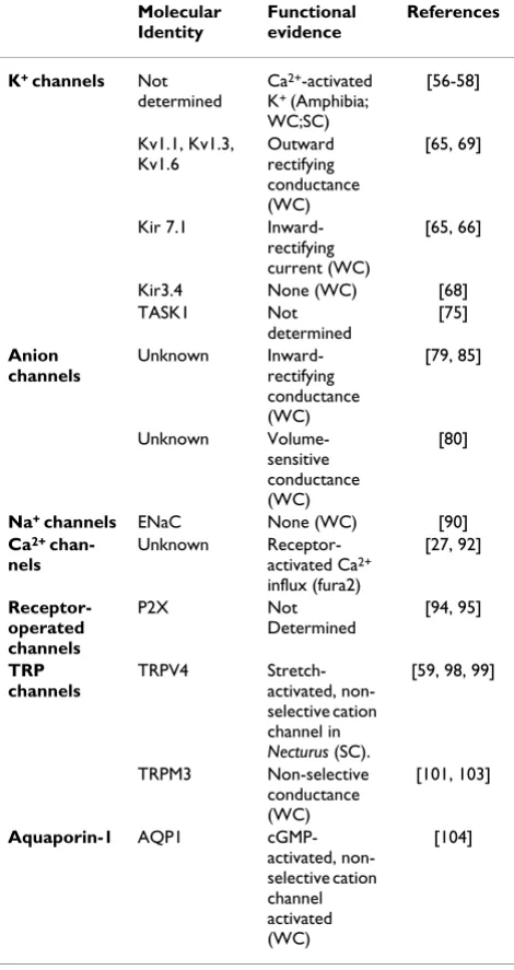
Two K+ conductances have been identified in the choroid plexus: Kv1 channel subunits mediate outward currents at depolarising potentials; Kir 7.1 carries an inward-rectifying conductance at hyperpolarising potentials. Both K+ channels are localised at the apical membrane where they may contribute to maintenance of the membrane potential while allowing the recycling of K+ pumped in by Na+-K+ ATPase. Two anion conductances have been identified in choroid plexus. Both have significant HCO3- permeability, and may play a role in CSF secretion. One conductance exhibits inward-rectification and is regulated by cyclic AMP. The other is carried by an outward-rectifying channel, which is activated by increases in cell volume. The molecular identity of the anion channels is not known, nor is it clear whether they are expressed in the apical or basolateral membrane. Recent molecular evidence indicates that choroid plexus also expresses the non-selective cation channels such as transient receptor potential channels (TRPV4 and TRPM3) and purinoceptor type 2 (P2X) receptor operated channels. In conclusion, good progress has been made in identifying the channels expressed in the choroid plexus, but determining the precise roles of these channels in CSF secretion remains a challenge for the future.
1. Introduction: 25 years of the patch clamp
method
It is now more than 25 years since the publication of the seminal paper which first described the patch clamp method for studying ion channels [1]. In recognition for their work in developing the patch clamp method Bert Sackmann and Erwin Neher, two of the authors on this
original paper, were awarded the 1991 Nobel Prize for Medicine. The impact of the method is perhaps most obvious in studies of the activity of individual ion chan-nels (single channel recording). This configuration of the method gives scientists the unique opportunity to study the activity of a single protein. Used in conjunction with recombinant DNA techniques, this method has vastly Published: 20 September 2007
Cerebrospinal Fluid Research 2007, 4:8 doi:10.1186/1743-8454-4-8
Received: 10 July 2007 Accepted: 20 September 2007
This article is available from: http://www.cerebrospinalfluidresearch.com/content/4/1/8
© 2007 Millar et al; licensee BioMed Central Ltd.
increased our understanding of how protein structure relates to channel function. In recognition of such studies the 2003 Nobel Prize for Chemistry was awarded to Rod McKinnon (jointly with Peter Agre).
Patch clamp methods, particularly whole cell methods, have also been important in determining the physiologi-cal roles of channels in mammalian cells. This is particu-larly true in secretory epithelia, where scientists such as Ole Petersen and Alain Marty pioneered the use of whole cell methods to study the mechanisms of secretion in exo-crine acinar cells [2,3]. In fact the one major refinement of the technique, the "perforated patch", was developed by Marty and Horn while working on lacrimal gland acinar cells [4]. The immense impact of patch clamp methods to disciplines such as physiology and neuroscience is per-haps best illustrated by the fact that a total of 100 000 papers which used the patch clamp method were pub-lished between 1981 and 2001 [5].
Patch clamp methods were originally applied to choroid plexus over 20 years ago; first in amphibian tissue and subsequently in mammalian tissue. The impact of the technique to understanding choroid plexus physiology, however, is less dramatic than that in studies of secretory, exocrine acinar cells. There are probably for two main rea-sons for this: 1) choroid plexus cells are less robust than exocrine acinar cells, thus it has been more difficult to per-form complex experiments, 2) exocrine acinar cells express relatively few channels, while in comparison the choroid plexus cells express a wide range of ion channels making the separation of distinct transport pathways more problematic. In recent years, however, the molecular structures of most ion channels have been determined. As a result molecular localisation techniques such as in situ
hybridisation, reverse transcriptase polymerase chain reaction (RT-PCR), Western blotting and immunocyto-chemistry have been employed to resolve many of the complexities of channel expression in the choroid plexus. This article will discuss these data. It will also speculate on future areas of development. To provide a frame work for this discussion we first give a brief general overview of ion channel structure and function.
The reader is reminded that while there is little doubt that ion channels have many important roles in the choroid plexus, many other transport proteins (pumps and carri-ers) are expressed in the choroid plexus. These are not dis-cussed in this article but are the subject of two recent reviews [6,7].
2 Ion channel diversity
Ion channels are expressed in all cells. They are integral membrane proteins that form selective pores in cell mem-branes (often as multimers), which facilitate the
move-ment of ions across the membrane down their electrochemical gradient. They are characterised by high rates of transport (millions of ions.s-1) compared to other transport proteins e.g. facilitated glucose (GLUT) trans-porters which transport about 100 molecules.s-1. This high rate of transport is important because it means that ion movements can create significant changes in the elec-trical properties of a cell, it also means that ion channels are exploited as a point of regulation in most cells. Thus many different mechanisms have evolved by which chan-nel activity can be modulated, e.g. voltage, ligand binding, phosphorylation and mechanical stress.
There is no simple, systematic nomenclature for ion chan-nels. However, in general they are classified primarily by reference to the ion to which they are selective, i.e. K+, Na+, Ca2+, anions. They are then sub-divided on the basis of functional properties such as a mechanism of regulation (e.g. Ca2+-activated) or a biophysical characteristic (e.g. inward rectifier). Even with increased knowledge of chan-nel molecular structure this simple classification based on selectivity still works well. However, there are two classes of channel that do not adhere to these simple rules: the receptor-operated channels (selective for either cations or anions) and novel transient receptor potential (TRP) channels (which discriminate poorly between monova-lent and divamonova-lent cations). Each broad group of channels has many members, and can be further sub-divided as will be seen below.
2.1 Potassium channels
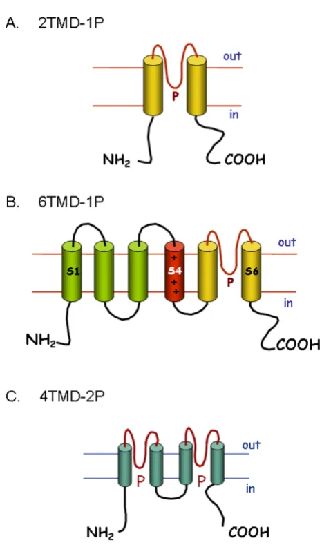
The potassium channels are the largest and most complex family of ion channels, represented by at least 70 loci in the human genome [8]. All K+ pore-forming channel pro-teins (α-subunits) share a conserved region of two trans-membrane domains (TMD) which are linked by an extracellular or pore (P) domain (Figure 1). This P domain is thought to form the pore region of the channel, in what is generally a tetrameric channel complex. K+ channels can be divided into three major families based on protein structure. The inward-rectifying channels (Kir) are structurally the simplest, with each subunit composed of the basic 2 TMD and a single P domain (2TMD-1P; see Figure 1A). A second large group of channels have an additional four TMDs adjacent to the 2TMD-1P structure these are the voltage-gated K+ or 6TMD-1P channels (Fig-ure 1B). The most recently identified family are twin-pore domain K+ channels (4TMD-2P), which show a repeat of the 2TMD-1P structure (Figure 1C).
2.1.1 Inward-rectifying K+ channels (2TMD-1P)
body. The molecular identities of Kir1.1 (ROMK; from kidney distal convoluted tubule) and Kir2.1 (from a mac-rophage cell line) were first determined in 1993 by expres-sion cloning [9,10]. Related channels in a total of seven sub-families (Kir1 to Kir7) have been subsequently cloned by homology methods [11]. Functional channels are formed by four α subunits. Each sub-family exhibits slight functional differences such as the degree of rectification and the mechanism of channel regulation. Two
particu-larly important subfamilies are the Kir 3 proteins which include the G-protein regulated channels (GIRKs) found in cardiac muscle, and Kir6.1 and Kir6.2 which combine with the sulphonylurea receptor proteins to produce func-tional ATP-sensitive K+ channels [11].
2.1.2 Voltage-gated K+ channels (6TMD-1P)
This is a large and diverse sub-family of K+ channels with some 40 members [8]. The first member of the family to be identified was the Shaker channel in Drosophila [12]. Homology methods subsequently identified other K+ channels in Drosophila and mammalian species. Most of the 6TMD-1P channels exhibit voltage-dependent gating, by virtue of the charged amino acids in the fourth TMD or S4 (Figure 1B). Functional channels are composed of a tetramer of α-subunits. Some β-subunits have also been identified which modify channel gating. The majority of the 6TMD-1P channels are classed as delayed-rectifier (Kv) channels, of which there are twelve families, i.e. Kv1 to Kv12 [8].
Kv1 to Kv4 represent the classical delayed-rectifier chan-nels which are widely distributed in many cell types around the body. These channels exhibit voltage-depend-ent kinetics, opening when the membrane potvoltage-depend-ential (Vm) is depolarised. The rate at which the channel opens on depolarisation in neurons, however, is slower than that for the activation of voltage-gated Na+ channels, hence the name delayed-rectifying channels. The pronounced out-ward-rectification of the current-voltage relationships for these channels is largely a product of the voltage-depend-ent opening of the channel. Kv5, 6, 8 and 9 are structurally related to the delayed-rectifiers, however, they do not act as channels but are classed as modifiers [8]. Kv7 includes five proteins, previously known as the KCNQ channels, which have roles such as damping neuronal activity. Kv10, Kv11 and Kv12 show less structural homology to the classic Kv channels and originally were classified as the
eag, erg and elk channels respectively [8].
A second major group in the 6TMD-1P family are the Ca2+-activated K+ channels: "maxi" Ca2+-activated K+ channels (BKCa), intermediate conductance Ca2+ -acti-vated K+ channels (IK
Ca) and small conductance Ca2+ -acti-vated K+ channels (SK
Ca) [13]. These channels are all activated by increases in intracellular Ca2+. In the case of BKCa this involves Ca2+ binding to the channel protein, whereas Ca2+ acts via calmodulin to open SK
Ca and IKCa [13]. BKCa is also activated by depolarising potentials because of the voltage-dependent binding of Ca2+ to the channel protein, but neither SKCa nor IKCa exhibit any voltage-dependence. The Ca2+-activated channels can be distinguished from one another by the size of the single channel conductance and by different sensitivities to a range of peptide toxins [13].
K+ channel structures Figure 1
K+ channel structures. Secondary structures of pore
2.1.3 Twin pore domain K+ channels (4TMD-2P)
This family of 15 channels was identified just over a dec-ade ago [14,15]. Each channel subunit has four TMDs and two P domains, i.e. they resemble two 2TMD-1P proteins linked together (Figure 1C). Dimers of these subunits form functional channels that are K+ selective. They were first described as "background" or "leak" K+ channels important in setting the resting Vm [16]. More recent studies suggest that they are regulated by a wide range of factors, e.g. pH, volatile anaesthetics and mechanical stress. They may therefore have specific roles in control-ling Vm and cellular activity in many different types of cell [15,16].
2.2 Sodium channels
2.2.1 Voltage-gated Na+ channels
Voltage-gated Na+ (Na
v) channels are activated and then display rapid inactivation at depolarising Vm. The activity of Nav channels is vital in the generation of action poten-tials in nerve cells. They are composed of a pore-forming
α-subunit which is usually associated with two modula-tory β-subunits, except in skeletal muscle where only one
β-subunit is required [17]. The α-subunit is composed of more than 1800 amino acids, which are divided into 4 homologous domains, each comprised of six transmem-brane spanning segments (S1–S6; Figure 2A). A pore loop links S5 and S6, whilst positive charged amino acids are abundant in S4 which acts the voltage sensor. The domains are arranged so that the pore loops from each domain form the pore of the channel. A short intracellular loop that links domains III and IV acts as the inactivation gate (red in Figure 2A). Nine different α-subunits have been identified and together they form the Nav gene fam-ily [17]. The Nav proteins show tissue specific distribution, but they are expressed only in excitable cells (e.g. nerve and muscle) and cells which exhibit action-like potentials (e.g. pancreatic β-cells) [17].
2.2.2 Epithelial Na+ channels
Epithelial Na+ channels(ENaC) were first described in frog skin, but are now known to be expressed in many mam-malian epithelia involved in Na+ absorption, e.g. distal convoluted tubule of the kidney, colon, lung and salivary gland duct [18]. By contrast to the Nav channels, ENaC are not gated by voltage and are characteristically blocked by low concentrations (<10 µM) of the diuretic drug amilo-ride. Three homologous subunits have been identified (α,
β and γ). Each subunit is comprised of two TMD and a complex extracellular loop [18]. Functional channels are thought to be heterotetramers of two α, one β and one γ subunits. This protein structure reveals that ENaC is a member of the degenerin (DEG) family of channels. This name originates from studies of nematodes where the expression of mutant DEG channels leads to cellular degeneration [18]. Other members of the DEG family in
mammals include acid-sensitive channels (ASIC) which may be involved in pain transduction [18].
2.3 Calcium channels
2.3.1 Voltage-gated Ca+ channels
Voltage-gated Ca2+ channels (Ca
v) are activated by Vm depolarisation and mediate Ca2+ influx into so called "excitable" cells. Functional channels are composed of four or five subunits: α1, α2, β, δ and in some cells γ (Fig-ure 2B). The α1 subunit is the largest subunit and deter-mines most of the functional properties of the channel: pore structure, gating and pharmacology. The α1-subunits share a similar structure to those of the α-subunits of
volt-Structures of A) voltage-gated sodium and B) voltage-gated Ca2+ channels
Figure 2
Structures of A) voltage-gated sodium and B) volt-age-gated Ca2+ channels. A) Secondary structure of the
age gated Na+ channels, i.e. they have four domains each composed of six transmembrane segments. Three families of α1 subunit have been identified [19]. The Cav1 family contains the four L-type Ca2+ channels which are expressed in muscle, neurons and endocrine cells. The neuronal specific P/Q, N and R type channels form the Cav2 family. While the three T-type channels expressed in neurons and muscle comprise the Cav3 family.
2.3.2 Capacitative or Store-Operated Ca2+ Entry
It was recognised in the early 1970's that agonist-evoked cytosolic Ca2+ mobilisation in non-excitable cells involves a transient Ca2+ release from intracellular stores, followed by a sustained Ca2+ entry [20,21]. This led to the concept of "capacitative Ca2+ entry" or store-operated Ca2+ entry (SOCE), by which the depletion of intracellular stores leads to sustained Ca2+ entry [22]. Electrophysiological studies have subsequently identified a Ca2+ release acti-vated current (ICRAC), which is characterised by inward rectification, very positive reversal potential (>30 mV), a high Ca2+ selectivity (P
Ca/PNa~1000), inhibition by La3+ and low single channel conductance (<100 femto sie-mens, fS) [23]. The molecular identity of this channel and the mechanism that couples store depletion to Ca2+ entry, however, have remained largely unknown until very recently.
The last two years have seen major advances in our under-standing of SOCE, with the discovery of two important proteins, stromal interation molecule (STIM1) and CRAC modulator (CRACM or Orai1) [24-28]. STIM1 contains a Ca2+-binding domain (EF hand) that has been suggested to sense endoplasmic reticulum Ca2+ store depletion [24,25]. Whereas Orai1, a four transmembrane domain protein, is suggested to be the pore forming subunit of the CRAC channel [27]. Interference RNA knockdown (siRNA) of either STIM1 or Orai1 significantly reduces SOCE and ICRAC [24,26], whereas co-expression of both STIM1 and Orai1 massively increases SOCE and ICRAC [29,30]. Mutagenesis of residues within Orai1, predicted to be important for Ca2+ binding within the pore of the channel, also markedly attenuated SOCE and ICRAC [27]. Collectively these data suggest that Orai1 acts as a Ca2+ entry channel [31,32].
2.4 Anion channels
Research performed over the last two decades has illumi-nated the importance of anion channels in many physio-logical and patho-physiolgical processes [33]. Knowledge of the molecular physiology of anion channels, however, is very limited compared to that of cation channels. Defin-itive information is available on the structure and func-tion of only two classes of channel (e.g. the cystic fibrosis transmembrane conductance regulator and the voltage dependent Cl- channels of the ClC family). The molecular
identity of many other channels is either controversial (e.g. Ca2+-activated Cl- channels) or unknown (e.g. vol-ume-sensitive anion channels).
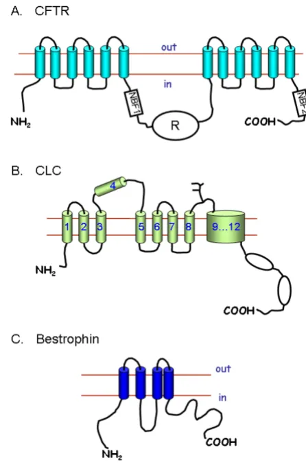
2.4.1 Cystic fibrosis transmembrane conductance regulator The cystic fibrosis transmembrane conductance regulator (CFTR) is the channel that is defective in the disease cystic fibrosis. The mRNA for the channel was identified by posi-tional cloning, and encodes a protein of 1280 amino acids (Figure 3A). The structure of the protein is unique for an ion channel, and CFTR is in fact an ATP binding cassette (ABC) protein [34]. This unusual structure meant that
Anion channel structures Figure 3
when first cloned the protein was described as conduct-ance regulator, and it was only after extensive series of experiments that channel function was established [35]. The CFTR channel has a single channel conductance of about 10 pico siemens (pS), it is primarily selective to Cl -, but also has a finite permeability to HCO3- [36]. Channel activity is increased by phosphorylation with protein kinase A of serine residues in the regulatory domain (R; Figure 3A) of the protein. Channel activity is also depend-ent on ATP binding to the nucleotide binding folds (NBF1 and NBF2; Figure 3A). Over a 1000 mutations have been identified in patients suffering from cystic fibrosis. These mutations can affect trafficking of CFTR to the membrane, channel regulation or single channel conductance [37].
CFTR is expressed in organ systems affected by cystic fibro-sis, e.g. airways epithelia, the exocrine pancreas, the small intestine, the biliary tract and the male reproductive tract [37]. However, it is also expressed in other tissues which are not thought to be affected by cystic fibrosis and where the function of CFTR remains unclear, e.g. cardiac muscle [38] and the kidney [39].
2.4.2 The CLC family of channels and transporters
The only family of anion channels which has been well characterised by both molecular and functional methods is the ClC family. The voltage-dependent channel ClC-0 was originally cloned by expression methods from the electric organ of the torpedo ray [33]. ClC-1 the mamma-lian skeletal muscle Cl- channel was subsequently cloned by homology methods [33]. Eight other members of the ClC family have now been identified, these include three Cl- channels: ClC-2 which is widely expressed, and ClC-Ka and ClC-Kb from kidney. The other ClC proteins (ClC-3 to ClC-7) were initially classed as channels, but they are now thought to mediate the exchange of Cl- and H+ [33,40,41].
The ClC channels are thought to have at least 11 trans-membrane spanning domains (Figure 3B), although pre-cise structure remains uncertain [33]. The ClC channels are thought to function as dimers with a functional pore structure in each subunit. Although all the ClC channels appear to have a similar structures, they are functionally quite different in terms of voltage-dependent gating e.g. 2 activates at hyperpolarising potentials whereas ClC-Kb is activated at depolarising potentials [33].
2.4.3 Ca2+-activated Cl- channels
The Ca2+-activated Cl- channels are a group of channels which have important functions in fluid secretion by some epithelial cells [2,3], stimulus contraction coupling in smooth muscle cells [42] and in olfaction [43]. The molecular identity of these channels has not been estab-lished. One group of candidate proteins is the ClCA
fam-ily of channels [42]. These proteins act as anion channels when expressed in mammalian cells, but their properties are significantly different from those of Ca2+-activated Cl -channels in native tissues [42]. A second family of pro-teins the bestrophins, may also act as Ca2+-activated Cl -channels. The structure (Figure 3C) and function of the bestrophin channels, however, has not been fully estab-lished [44]. The first bestrophin to be identified was best1 (in total there are four proteins encoded by the human genome: best1 to best 4), and mutations to this channel is associated with macular degeneration in the retina [44]. A very recent paper has shown that best 1 is expressed in a number of secretory epithelial cells, and that the use of siRNA against best 1 reduces the Ca2+-activated Cl- cur-rents in these cells [45].
2.4.4 Volume-sensitive anion channels (VSAC)
These channels appear to be ubiquitously expressed [33]. They make a major contribution to cell volume regula-tion, and may also play a critical role in the events of the cell cycle [46]. They display outward rectification and often time-dependent inactivation at extreme depolaris-ing Vm. A number of molecular candidates have been sug-gested including: p-glycoprotein, CLC3 and putative Cl -channel protein (pICln), but none has been proved to be VSAC [33]. Indeed pICln and p-glycoprotein are now described as regulators of VSAC [33], while ClC-3 is prob-ably an ion exchanger in the membranes intracellular organelles [41].
2.5 Receptor operated channels
The receptor operated channels (ROCs) are a diverse group of channels which are activated by the binding of an agonist to a receptor site that is part of the channel pro-tein. They have many important roles, principally at syn-apses of the central nervous system (CNS). By contrast to most other channels they have not been classified in terms of the permeating ion, but rather by the name of the acti-vating agonist. They can be divided into three major groupings: the classical ROCs, the glutamate receptors and the P2X receptors.
2.5.1 Classical ROCs
chan-nels [48]. These chanchan-nels are normally associated with synapses in the CNS, but there is evidence that ROCs are expressed in other cells. For instance nACh plays a key role in transmission at the neuromuscular junction, while GABAA is expressed in glial cells in the CNS, peripheral nerves [49] and other cells, e.g. glucagon secreting alpha-cells of the endocrine pancreas [50].
2.5.2 Glutmate receptors
The ionotropic glutamate receptors are functionally simi-lar to the classic ROCs. They have important roles at syn-apses of the CNS, and are activated by glutamate or aspartate. However, they have a different structure to the classic ROCs. Each subunit has only three TMD, and func-tional channels are heteromers in which four subunits assemble as a "dimer of dimers" [51]. The glutamate receptors can be divided into three sub-families on the basis of their activation by different selective agonists: kai-nate, N-methyl-D-aspartate (NMDA) and α -amino-3-hydroxy-5-methyl-4-isoxazole propionic acid (AMPA). The different sub-families also show slightly different kinetic properties and ion selectivity, e.g. the AMPA and kainate channels are selective for monovalent cations and display rapid activation, whereas the NMDA channels exhibit slower activation and are permeable to both Na+ and Ca2+.
2.5.3. Type 2 purinoceptors
Type 2 purinoceptors (P2X receptors) are receptors for the purine nucleotides. They can be functionally and structur-ally divided into two families: the P2Y family are G-pro-tein coupled receptors which are activated by ATP, UTP, ADP and UDP; and the P2X receptors which are receptor-operated channels which are activated primarily by ATP [52]. A total of seven P2X receptor proteins have been identified [53]. Each P2X protein has 2 TMD and func-tional channels are composed of trimers of three identical subunits, or a combination two different subunits. All P2X receptor channels are permeable to small monova-lent cations, and some are also permeable to Ca2+ and ani-ons. They are widely distributed throughout the body and have diverse roles, e.g. transmission in the autonomic nervous system and sensing tissue damage [52,53].
2.6 Transient receptor potential channels
Transient receptor potential (TRP) channels were first identified in Drosophila, where they have a role in photore-ception in the visual system. Six TRP protein families have been identified in mammals: canonical TRP channels (TRPC) which are similar to the Drosophila channel, the vallanoid receptors (TRPV), melastatin TRPs (TRPM), the mucolilpins (TRPML), the polycystins (TRPP) and the ankyrin transmembrane proteins (ANKTM1 and TRPA1). All of these channels are predicted to have six TMD and all are thought to assemble as tetramers to form functional
channels. All are cation selective, but most discriminate poorly between cations [54]. TRPV5 and TRPV6, however, are selective for Ca2+ against monovalent cations, whereas TRPM4 and TRPM5 are selective for monovalent cations. The best characterised are the TRPV family: TRPV1 is the capsaicin receptor which is also activated by an increase in temperature, TRPV4 is thought to play a role in osmosens-ing, TRPV5 and TRPV6 are Ca2+-selective channels which have a central role in transepithelial Ca2+ transport in the kidney and intestine [55]. Other well characterised chan-nels are TRPM8 which is sensitive to menthol and cold, and TRPM6 which has a role in magnesium transport in the kidney [54].
3 Ion channel expression and function in the
choroid plexus
3.1 Single channel studies
The earliest patch clamp experiments on choroid plexus examined single channel activity in tissue from the amphibian "mudpuppy" Necturus maculosa (see Table 1). These studies identified the expression of BKCa channels with a conductance of 180 pS in the apical membrane of the epithelial cells [56,57]. Further whole-cell experi-ments indicated that these BKCa channels carried the bulk of the whole cell conductance in amphibian tissue [58]. Christensen et al [59] also identified at least three anion channels with different conductances in Necturus choroid plexus. The importance of these channels to whole cell conductance, however, was not determined. One final important observation to emerge from these single chan-nel experiments was the activation of non-selective cation channels by cell swelling [60]. Christensen [60] went on to show that Ca2+ influx via these non-selective cation channels caused the activation of BKCa. This sequence of events has since become something of a paradigm in the initiation of cell volume regulatory mechanisms in many cells.
3.2 Whole cell patch clamp experiments on mammalian choroid plexus
Single channel experiments provided our first insight into the diverse range of ion channels expressed in the choroid plexus. However, the lack of consistency between data from mammalian and amphibian choroid plexus, cou-pled to the difficulty in observing any single channel activ-ity at all in mammalian choroid plexus, prompted my laboratory to switch to the whole cell method to study the mammalian choroid plexus. These studies have yielded more consistent results, and we have identified the expres-sion of K+channels, anion channels and non-selective cat-ion channels in choroid plexus tissue from rats and mice (Table 1). Channel expression has also been studied using molecular localisation techniques, i.e. RT-PCR, northern blotting, Western blotting and immunocytochemistry (see Table 1). The remainder of this review discusses these data, and comments on the potential roles of the channels identified in the choroid plexus epithelium.
3.2.1 K+ channels in mammalian choroid plexus
K+ channels are thought to have a number of important roles in CSF secretion. First, they help regulate the nega-tive Vm, and hence contribute to the electrochemical gra-dient favouring anion efflux at the apical membrane. Second, they act as leak pathway in the apical membrane for K+ accumulated in the cell through the actions of the Na+-K+ ATPase (which is also located in the apical mem-brane of the choroid plexus) and thus prevent cell swell-ing as a result of K+ accumulation. Finally, they may participate in the transcellular transport (CSF to blood) of K+[64]. This is an important process which is potentially vital in maintaining the low [K+] of the CSF, as it is thought to counteract the paracellular leak of K+ from blood to CSF (i.e. ion movement through the junctional complexes between the cells). In a model for transcellular K+ transport, Zeuthen and Wright [64] proposed that K+ is actively pumped into cells from the CSF across the apical membrane by the Na+-K+ ATPase. Much of this K+ (about 90%) is recycled across the apical membrane through the K+ channels in this membrane. However, some of the K+ (the remaining 10%) will leave the cell across the basola-teral membrane. Thus there is a small net absorptive (CSF to blood) flux of K+ across the epithelium.
Kir channels
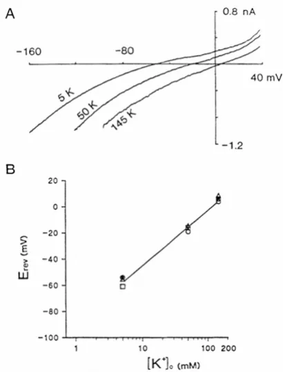
Kotera and Brown [65] characterised an inward-rectifying conductance observed in choroid plexus (Figure 4). This conductance is highly selective to K+ (Figure 4B), displays no time-dependent activation or inactivation, and is blocked by sub-millimolar concentrations of Cs+ or Ba2+ [65]. It therefore shares many properties with conduct-ances carried by many Kir channels. One distinguishing characteristic of the channel in the choroid plexus is that the chord conductance (i.e. the slope of the
current-volt-age relationship for the negative currents), is independent of the external K+ concentration (see Figure 4A), whereas for most Kir channels the conductance increases with K+. It is now thought that the choroid plexus conductance is carried mainly by Kir7.1 channels (Table 1).
The first indication for Kir7.1 involvement came from in situ hybridisation studies which showed that mRNA for Kir7.1 is highly expressed in the choroid plexus epithe-lium [66]. Döring et al [66] also showed that Kir7.1, when expressed in Xenopus oocytes, gives rise to an inward-recti-fying conductance with functional properties that are almost identical to those of the Kir in the choroid plexus, i.e. the conductance is independent of extracellular K+ activity. Nakamura et al [67] demonstrated that the Kir7.1 channel protein is expressed in the apical membrane of
Properties of the inward-rectifying K+ channel Kir7 Figure 4
Properties of the inward-rectifying K+ channel Kir7.1 expressed in rat choroid plexus epithelial cells. A) Current -voltage relationships with extracellular K+ of 5, 50 or 145 mM. The current-voltage relationships were produced by applying 0.8 s ramp potentials from Vm = -160 to 40 mV. B)
Kir reversal potentials (Erev) as a function of extracellular K+ activity ([K+]
the choroid plexus using immunocytochemical methods. In this membrane the Kir7.1 channel can contribute to the leak of K+ from the cells and help maintain a negative Vm.
Iizuka et al [68] reported Kir3.4 expression in the rat choroid plexus using both immunocytochemical and in situ hybridisation methods. The Kir3.4 channel forms heteromeric, G protein activated K+ channel with other Kir proteins (usually another member of the Kir 3 family). To date, however, electrophysiological studies have failed to identify a contribution from such channels to the whole cell conductance of choroid plexus cells. Furthermore, RT-PCR failed to identify expression of mRNA for Kir3.1 or Kir3.4 in rat choroid plexus (Speake and Brown, unpub-lished data).
Kv1 channels
Whole cell experiments also identified outward currents in rat choroid plexus, which exhibited time-dependent activation at depolarising potentials and inactivation at extreme depolarising potentials [65]. The channels carry-ing this conductance were blocked by TEA+, dendrotoxin-K and margatoxin, the latter two toxins being highly spe-cific blockers of Kv1.1 and Kv1.3 channels respectively [69]. Kv1 protein expression was therefore investigated by Western analysis and immunocytochemistry. Speake et al
[69] reported that Kv1.1, Kv1.3 and Kv1.6 (but not Kv1.4 and Kv1.5) were all expressed in rat choroid plexus (Kv1.2 expression was not determined). Furthermore the immu-nocytochemical studies (Figure 5) demonstrated that the expression of Kv1.1 and Kv1.3 is confined to the apical membrane [69]. Kv1 channels are only expressed in a few other types of epithelial cells [70-73] where their role is not understood. A recent study, however, has demon-strated that Kv7.1 channels are important in the regula-tion of the Vm which drives anion secreregula-tion in intestinal epithelial cells [72]. A similar role is envisaged for the Kv1.1, Kv1.3 and possibly Kv1.6 in the choroid plexus during CSF secretion [65,69].
Ca2+-activated K+ channels
BKCa are expressed in amphibian choroid plexus (see Sec-tion 3.1), however, there is no evidence for the expression of these channels in mammalian tissue. Thompson-Vest et al [74] have demonstrated the expression of IKCa in choroid plexus epithelium, expression however, appears to be confined to the cytoplasm of the cells. Indeed elec-trophysiological experiments have failed to identify any Ca2+-activated K+ channels in mammalian choroid plexus cells [65].
Tandem pore domain K+ channels
Soon after the discovery of the tandem pore domain chan-nels, a study of the rat CNS identified expression of the acid-sensitive K+ channel TASK-1 in the choroid plexus of
the third ventricle using immunocytochemical methods [75]. However, it is not clear from these studies whether TASK1 is expressed in the epithelial cells of the choroid plexus or in the underlying connective and vascular tissue. Furthermore a TASK1 component to the whole-cell K+ conductance has not yet been observed in patch clamp experiments (Millar & Brown, unpublished observation).
K+ channels in the basolateral membrane
Zeuthen & Wright [64] predicted that the basolateral membrane of the choroid plexus must also express K+ channels which are required to explain the net absorptive flux of K+ across the epithelium (CSF to Blood). To date K+ channel expression has not been observed in this mem-brane. A possible explanation is that in the mammalian choroid plexus K+ efflux at the basolateral membrane is mediated via the K+-Cl- cotransporter (KCC3) which is expressed at this membrane in rat choroid plexus [76]. It is possible, however, that K+ channels identified by molec-ular methods but not yet by electrophysiology may also contribute to K+ efflux at the basolateral membrane (e.g. TASK-1).
K+ channel conclusions
Mammalian choroid plexus epithelial cells exhibit two K+ conductances. One is carried by Kir7.1 channels, and one by Kv1.1 and Kv1.3 channel proteins. These channels are all expressed in the apical membrane of the choroid plexus (Figure 5) where they can mediate the reflux of K+
Immunoreactivity for: i) delayed-rectifier K+ channel Kv1 Figure 5
Immunoreactivity for: i) delayed-rectifier K+ channel Kv1.3 and ii) Kv1.1 channel subunits in sections of rat choroid plexus tissue. The expression of iii) Na+-K+ ATPase and iv) Cl--HCO
3- exchanger AE2 are shown as markers of the apical and basolateral membranes of the epithelial cells respectively. The arrows also indicate apical staining and the scale bars = 25 µm. Figure reproduced with permission from Speake et al
pumped into the cells by Na+, K+ ATPase, and help main-tain the intracellular negative Vm of the epithelial cell.
3.2.2 Anion channels
Anion channels are vital components of the secretory process in most epithelia, because they are normally the main route for anion efflux across the apical membrane [33]. Saito and Wright [77,78] proposed that HCO3-
per-meable, apical anion channels have a major role in CSF secretion by bull frog choroid plexus. Initial patch clamp experiments on mammalian choroid plexus showed that there was very little anion conductance in unstimulated cells (Figure 6A). Subsequent experiments, however, found that two types of anion channel could be activated: i) a cAMP-activated, inward-rectifying anion channel (Fig-ure 6B), and ii) a volume-sensitive anion channel (Fig(Fig-ure 6C).
Inward-rectifying anion conductance
Anion channels with inward-rectifying current-voltage relationships have been observed in whole cell recordings from the choroid plexus of rat [79], mouse [80] and pig [81] (Table 1). These channels: i) exhibit time-dependent activation at hyperpolarising Vm (Figure 6B), ii) have a uniquely high permeability to HCO3- (P
HCO3:PCl = 1.5),
Whole cell Cl- conductances in rat choroid plexus Figure 6
Whole cell Cl- conductances in rat choroid plexus. Current profiles and current-voltage relationships for: A) an unstimu-lated cell, B) a cell stimulated with protein kinase A and C) a cell swollen by reducing the osmolality of the extracellular solution. In each case voltage clamp experiments were per-formed in which Vm was held at -80 mV and then stepped for 1 s to Vm from -120 to +60 mV (at 20 mV increments). Figure reproduced with permission from Kibble et al [79].
Table 1: Ion channel expression in choroid plexus epithelial cells.
Molecular Identity
Functional evidence
References
K+ channels Not
determined
Ca2+-activated
K+ (Amphibia;
WC;SC)
[56-58]
Kv1.1, Kv1.3, Kv1.6
Outward rectifying conductance (WC)
[65, 69]
Kir 7.1 Inward-rectifying current (WC)
[65, 66]
Kir3.4 None (WC) [68] TASK1 Not
determined
[75]
Anion channels
Unknown Inward-rectifying conductance (WC)
[79, 85]
Unknown Volume-sensitive conductance (WC)
[80]
Na+ channels ENaC None (WC) [90]
Ca2+ chan-nels
Unknown Receptor-activated Ca2+
influx (fura2)
[27, 92]
Receptor-operated channels
P2X Not Determined
[94, 95]
TRP channels
TRPV4 Stretch-activated, non-selective cation channel in
Necturus (SC).
[59, 98, 99]
TRPM3 Non-selective conductance (WC)
[101, 103]
Aquaporin-1 AQP1
cGMP-activated, non-selective cation channel activated (WC)
[104]
WC = recorded by whole cell methods; SC = single channel recording; fura2 measurement of intracellular Ca2+ activity; None =
iii) are more permeant to I- than Cl- or Br-; iv) are blocked by the Cl- channel blockers DIDS and NPPB, v) are acti-vated by cAMP and protein kinase A, but inhibited by pro-tein kinase C [79,82,83]. Many of these properties are similar to those of the ClC-2 channel [33], the mRNA for which is expressed the choroid plexus [84,85]. However, the inward-rectifying conductance was unchanged in whole cell recordings in choroid plexus cells from ClC-2 knock-out mice, indicating that ClC2 channels do not contribute to the conductance [85]. Thus, the molecular identity of the inward-rectifying channel remains unknown. The potential role of ClC-2 in the choroid plexus is also unknown, however, the data from the knock-out mice suggest that this channel is not expressed in the plasma membrane of choroid plexus cells.
The regulation of the inward-rectifying channels by cAMP and their high permeability to HCO3-, indicates that they may be similar to the HCO3- channel thought to have a major role in CSF secretion by bullfrog choroid plexus [78]. The properties are also consistent with the observa-tion that Cl- efflux from the rat choroid plexus is stimu-lated by cAMP [86], and inhibited by agonists such as vasopressin which activate protein kinase C [87]. To par-ticipate in CSF secretion the inward-rectifying channels must be located in the apical membrane, but the site of their expression has yet to be determined. In this regard, recent immunocytochemical experiments have identified the expression of the electrogenic Na+-HCO
3- coporter (NBCe2) in rat choroid plexus [88]. This trans-porter may also have a role in HCO3- secretion [88]. It also generates small electrical currents which will contribute to the whole cell current, thus it is likely that the PHCO3:PCl for the inward-rectifying channel may be an overestimate of the true value.
Volume-sensitive anion conductance
Volume-sensitive anion channels are also expressed in choroid plexus cells from rats and mice [79,80] (Table 1). These channels are activated by cell swelling, and are dependent on intracellular ATP [80]. The channels exhibit slight outward-rectification (see Figure 6C), are blocked by DIDS and NPPB [80], and have significant HCO3- per-meability (Millar and Brown, unpublished observation). These properties are therefore similar to those of volume-sensitive anion channels found in many cells [33,46] and of the channels described by Saito & Wright [78]. Volume-sensitive channels have an important role in the regula-tory volume decrease observed in most cells in response to cell swelling [33,46]. The regulation of cell volume has not been studied in the mammalian choroid plexus, but it is conceivable that these channels could be involved in both volume regulation and CSF secretion.
CFTR and Ca2+-activated Cl- channels
In 1993 it was reported that CFTR was expressed in the choroid plexus on the basis of Western blotting and immunocytochemical experiments [89]. By contrast we were unable to detect mRNA for CFTR in rat choroid plexus. Furthermore, there were no differences between the anion currents in wild-type and CFTR knock-out mice [80]. Thus it is now thought that CFTR is not expressed in the choroid plexus epithelium, and that the antibody used in the immunocytochemical studies [89] may have lacked specificity for CFTR. There are no functional data to sup-port the expression of Ca2+-activated Cl- channels in the choroid plexus. The expression of the CLCAs and the bestrophins has not therefore been investigated.
Anion channel conclusions
An inward-rectifying anion conductance and a volume-sensitive anion conductance are expressed in the choroid plexus epithelium. The molecular identity of neither con-ductance has been determined, and the membrane in which they are expressed is also not known. Both conduct-ances, however, could contribute to the secretion of Cl -and HCO3- into the CSF, if they are expressed in the apical membrane of the epithelium.
3.2.3 Na+ channels
Choroid plexus epithelial cells do not display any electri-cal excitability. It is therefore highly unlikely that Nav channels will be expressed in these cells. This conclusion is supported by data from whole cell patch clamp studies, which have failed to reveal any transient currents at depo-larising potentials, which could be carried by Nav (or Cav channels).
A recent RT-PCR and immunolocalisation study (Table 1) suggested that ENaC is expressed in the choroid plexus epithelium [90]. Amiloride which blocks ENaC certainly inhibits Na+ transport into the choroid plexus [91]. This observation has always, however, been interpreted as being due to an effect of amiloride on Na+-H+ exchangers (NHE). We have therefore performed patch clamp experi-ments to investigate any contribution of ENaC to the whole-cell conductance of the choroid plexus cells. These studies showed that amiloride is without effect on the whole-cell conductance of mouse cells (Millar and Brown, unpublished observations). Thus, if ENaC is expressed in choroid plexus epithelial cells, it makes only a very minor contribution to the whole cell conductance.
3.2.4 Ca2+ channels
al [93] have reported agonist-induced increases in intrac-ellular Ca2+ activity in rat and sheep choroid plexus cells respectively (Table 1). In both studies the majority of the increase was thought to be due to Ca2+ release from intra-cellular stores, but a component may also be due to Ca2+ involved entry via store-operated channels. In neither study however, was this directly tested [92,93].
3.2.5 Receptor operated channels
RT-PCR experiments have determined the expression of mRNA for P2X1, P2X2, P2X4, P2X5 P2X6 and P2X7 in choroid plexus [94,95] (Table 1). Immunocytochemistry has also shown that the same P2X proteins are expressed in epithelial cells of the choroid plexus, and not in capil-lary endothelial cells [94,95]. The subcellular localisation of protein expression in the epithelium is not clear from data provided, and there appears to be expression on both apical and basolateral membranes [94]. There is however, some indication that expression may be greater on the apical membrane, particularly for P2X1 and P2X6 recep-tors[94,95]. If these data are correct then they suggest that the P2X receptors on the epithelial cells may respond to ATP in CSF, possibly as some sort of feedback loop in con-trolling the process of CSF secretion. Functional studies to investigate this possibility are therefore eagerly awaited.
Evidence for GABAA receptor expression in the choroid plexus comes from studies of benzodiazepine binding [96,97] and one of muscimol binding (a GABAA agonist) [98]. Furthermore, Williams et al [97] reported that ben-zodiazepines may inhibit the rate of CSF secretion. The possible expression of GABAA receptors in the choroid plexus, however, has not been substantiated by molecular or electrophysiological methods.
3.2.6 TRP channels
At least two TRP channels may be expressed in choroid plexus tissue (Table 1). TRPV4 protein has been identified in the third [99] and fourth ventricle [99,100] choroid plexus of mouse brain by immunochemistry. By contrast, in situ hybridisation methods were used to demonstrate the expression mRNA encoding TRPM3 in the lateral and third ventricle choroid plexus of mouse brain (see Figure 7) [101]. The TRPV4 and TRPM3 channels are both non-selective cation channels which exhibit a finite permeabil-ity to Ca2+ [102]. Both channels are activated by changes cell volume when the extracellular osmolality is per-turbed, and both may have a role in cell volume regula-tion and/or modulating ion and water transport across epithelial barriers [102].
Patch clamp experiments in our laboratory have identified a non-selective cation conductance in mouse choroid plexus cells. The kinetic properties of the conductance mean that it is difficult to differentiate from the
inward-rectifying anion conductance. Preliminary experiments however, indicate that the channel is permeable to Na+ or Cs+ but impermeable to the organic cation n-methyl-D-glucamine [103]. The conductance is also inhibited by 100 µM gadolinium (Gd3+) [103]. These data suggest that the conductance may be carried by TRPM3 or TRPV4 channels.
4 Unconventional ion channels
A surprising recent finding is that aquaproin 1 (AQP1) may act as both a water channel and a ion channel in the choroid plexus [104]. AQP1 was already known to be expressed at high concentrations in the apical membrane of the mammalian choroid plexus, and is thought to play an important role in water transport by the choroid plexus [105,106]. In 2000, however, Andrea Yool and colleagues reported that AQP1 can function as a cGMP-gated, non-selective cation channel when expressed in Xenopus
oocytes [107]. These studies were then extended to AQP1 In situ hybridisation for the melastatin transient receptor potential channel, TRPM3 in mouse brain
Figure 7
expressed in rat choroid plexus epithelial cells [104]. It was found that a non-selective cation conductance was activated by atrial naturetic peptide (ANP), which acts to increase intracellular concentrations of cGMP [104]). Fig-ure 8 shows the activation of the non-selective channel in choroid plexus cells by sodium nitroprusside (SNP, a nitric oxide donor which stimulates cGMP synthesis). The activated currents show no voltage-dependence (Figure 8B) and discriminate poorly between monovalent cations [104]. The AQP1 currents were inhibited by 200µM Cd2+ (Figure 8C) and 600 µM Cd2+ (Figure 8D). The conduct-ance observed was thought not to be due to the activation of endogenous channels by AQP1 because: 1) the channel properties are distinct from all known endogenous chan-nels, and 2) because the conductance could be reduced by siRNA targeted at AQP1 [104].
The biophysical relationship between the water channel and ion channel phenotype has not been established. The prevailing hypothesis is that the two different phenotypes represent differences in protein folding, with vast majority of proteins exhibiting the water channel phenotype [108]. The potential role of the AQP1 mediated conductance in the choroid plexus is also unknown. Boassa [104] did, however, observe that ANP reduced fluid secretion by choroid plexus cells, an effect which was partially reversed by 500 µM Cd2+. These data suggest that the activation of the non-selective conductance reduces secretion, possibly be dissipating the ion gradients across the cell membrane which are required for secretion.
5 Conclusion
The epithelial cells of the choroid plexus, which secrete CSF, express two K+ and two anion conductances. The properties of each conductance are such that they could all play a significant role in CSF secretion. The precise role of each, however, remains to be determined. In addition there is now evidence for the expression of a number of other channel proteins, e.g. P2X receptors and TRP chan-nels. Patch clamp experiments are required to determine the functional roles of these channels in the choroid plexus.
Abbreviations
ABC adenosine triphosphate binding cassette
ADP adenosine diphosphate
ATP adenosine triphosphate
BKCa "maxi" Ca2+-activated K+ channels
Cav voltage-gated Ca2+ channels
CFTR cystic fibrosis transmembrane conductance regula-tor
ClC a family of voltage-dependent channels and trans-porters
CNS central nervous system
DIDS 4-4' diisothiocyanatostilbene-2,2'-dislphonic acid (an anion channel blocker)
ENaC epithelial Na+ channels
AQP1 acts a non-selective cation ion conductance which can be activated by sodium nitroprusside (SNP) activates Figure 8
AQP1 acts a non-selective cation ion conductance which can be activated by sodium nitroprusside (SNP) activates. Cur-rents were recorded in: A (initial) unstimulated cells; B
(SNP 1 mM) in the presence of 1 mM SNP (a nitric oxide
donor which activates cGMP synthesis;); C (Cd2+ 200 µM)
in 1 mM SNP plus 200 µM Cd2+ and D (Cd2+ 600 µM) in 1
mM SNP plus 600 µM Cd2+). In each experiment Vm was stepped for 200 ms from -110 to +40 mV at 10 mV incre-ments. Figure reproduced with permission from Boassa et al
fS femto (10-15) siemen (a measure of electrical conduct-ance)
IKCa intermediate conductance Ca2+-activated K+ channels
Kir inward-rectifying K+ channel
Kv delayed-rectifier K+ channel
Nav voltage-gated Na+ channel
NPPB 5-nitro-2-(3-phenylpropylamino)benzoic acid (an anion channel blocker)
PHCO3:PCl channel permeability to HCO3- relative to Cl -permeability
P domain pore forming domain of an ion channel
pS pico (10-12) siemen
ROC receptor operated channel
RT-PCR reverse transcriptase polymerase chain reaction
S transmembrane segment
siRNA interference RNA knock down
SKCa small conductance Ca2+-activated K+ channels
SOCE store-operated Ca2+ entry
TMD transmembrane domain
TRP transient receptor potential
UDP uridine diphosphate
UTP uridine triphosphate
Vm membrane potential
This list only includes frequently used standard abbrevia-tions. Many channel proteins have abbreviated which are used as the accepted name, these are defined where they arise in the text.
Competing interests
The author(s) declare that they have no competing inter-ests.
Authors' contributions
IDM – Joint principal author
JIEB – Author of Section 2.3.2 and reading the manuscript
PDB – Joint principal author
All authors have read and approved the final version of the manuscript.
Acknowledgements
This work was supported by grant 070139/Z/02 from the Wellcome Trust. The work of J.I.E.B is funded by a grant from the BBSRC.
References
1. Hamill OP, Marty A, Neher E, Sakmann B: Improved patch-clamp techniques for high-resolution current recording from cells
and cell-free membrane patches. Pflugers Arch 1981,
391:85-100.
2. Petersen OH: Stimulus-secretion coupling: cytoplasmic cal-cium signals and the control of ion channels in exocrine aci-nar cells. J Physiol 1992, 448:1-51.
3. Marty A: Calcium release and internal calcium regulation in acinar cells of exocrine glands. J Membr Biol 1991, 124:189-197. 4. Marty A, Horn R: Muscarinic activation of ionic currents meas-ured by a new whole-cell recording. J Gen Physiol 1988,
92:145-159.
5. Hille B: Ion channels 3rd edition. Sunderland, MA: Sinauer Associates; 2001.
6. Brown PD, Davies SL, Speake T, Millar ID: Molecular mechanisms of cerebrospinal fluid production. Neuroscience 2004,
129:957-970.
7. Praetorius J: Water and solute secretion by the choroid plexus. Pflugers Arch 2007, 454:1-18.
8. Gutman GA, Chandy KG, Grissmer S, Lazdunski M, Mckinnon D, Pardo LA, Robertson GA, Rudy B, Sanguinetti MC, Stühmer W, Wang X: International Union of Pharmacology. LIII. Nomenclature and Molecular Relationships of Voltage-Gated Potassium Channels. Pharmacol Rev 2005, 57:473-508.
9. Ho K, Nichols CG, Lederer WJ, Lytton J, Vassilev PM, Kanazirska MV, Hebert SC: Cloning and expression of an inwardly rectifying ATP-regulated potassium channel. Nature 1993, 362:31-38. 10. Kubo Y, Baldwin TJ, Jan YN, Jan LY: Primary structure and
func-tional expression of a mouse inward rectifier. Nature 1993,
362:127-133.
11. Kubo Y, Adelman JP, Clapham DE, Jan LY, Karschin A, Kurachi Y, Laz-dunski M, Nichols CG, Seino S, Vandenberg CA: International Union of Pharmacology. LIV. Nomenclature and Molecular Relationships of Inwardly Rectifying Potassium Channels.
Pharmacol Rev 2005, 57:509-526.
12. Papazian DM, Schwarz TL, Tempel BL, Jan YN, Jan LY: Cloning of genomic and complementary DNA from Shaker, a putative potassium channel gene from Drosophila. Science 1987,
237:749-753.
13. Wei AG, Gutman GA, Aldrich R, Chandy KG, Grissmer S, Wulff H:
International Union of Pharmacology. LII. Nomenclature and molecular relationships of calcium-activated potassium channels. Pharmacol Rev 2005, 57:463-472.
14. Ketchum KA, Joiner WJ, Sellers AJ, Kaczmarek LK, Goldstein S: A new family of outwardly rectifying potassium channel
pro-teins with two pore domains in tandem. Nature 1995,
376:690-695.
15. Goldstein SA, Bayliss DA, Kim D, Lesage F, Plant LD, Rajan S: Inter-national Union of Pharmacology. LV. Nomenclature and molecular relationships of two-P potassium channels. Phar-macol Rev 2005, 57:527-540.
16. Lesage F, Lazdunski M: Molecular and functional properties of two-pore-domain potassium channels. Am J Physiol Physiol 2000,
279:F793-F801.
18. Kellenberger S, Schild L: Epithelial sodium channel/degenerin family of ion channels: a variety of functions for a shared structure. Physiol Rev 2002, 82:735-767.
19. Catterall WA, Perez-Reyes E, Snutch TP, Striessnig J: International Union of Pharmacology. XLVIII. Nomenclature and struc-ture-function relationships of voltage-gated calcium chan-nels. Pharmacol Rev 2005, 57:411-425.
20. Nielsen SP, Petersen OH: Transport of calcium in the perfused submandibular gland of the cat. J Physiol 1972, 223:685-697. 21. Case RM, Clausen T: The relationship between calcium
exchange and enzyme secretion in the isolated rat pancreas.
J Physiol 1973, 235:75-102.
22. Putney JW Jr: A model for receptor-regulated calcium entry.
Cell Calcium 1986, 7:1-12.
23. Prakriya M, Lewis RS: CRAC channels: activation, permeation, and the search for a molecular identity. Cell Calcium 2003,
33:311-321.
24. Roos J, DiGregorio PJ, Yeromin AV, Ohlsen K, Lioudyno M, Zhang S, Safrina O, Kozak JA, Wagner SL, Cahalan MD, et al.: STIM1 an
essential and conserved component of store-operated Ca2+
channel function. J Cell Biol 2005, 169:435-445.
25. Zhang SL, Yu Y, Roos J, Kozak JA, Deerinck TJ, Ellisman MH, Stauder-man KA, Cahalan MD: STIM1 is a Ca2+ sensor that activates
CRAC channels and migrates from the Ca2+ store to the
plasma membrane. Nature 2005, 437:902-905.
26. Vig M, Peinelt C, Beck A, Koomoa DL, Rabah D, Koblan-Huberson M, Kraft S, Turner H, Fleig A, Penner R, Kinet JP: CRACM1 is a plasma membrane protein essential for store-operated Ca2+ entry. Science 2006, 312:1220-1223.
27. Yeromin AV, Zhang SL, Jiang W, Yu Y, Safrina O, Cahalan MD:
Molecular identification of the CRAC channel by altered ion selectivity in a mutant of Orai. Nature 2006, 443:226-229. 28. Zhang SL, Yeromin AV, Zhang XH, Yu Y, Safrina O, Penna A, Roos J,
Stauderman KA, Cahalan MD: Genome-wide RNAi screen of Ca2+ influx identifies genes that regulate Ca2+
release-acti-vated Ca2+ channel activity. Proc Natl Acad Sci USA 2006,
103:9357-9362.
29. Soboloff J, Spassova MA, Tang XD, Hewavitharana T, Xu W, Gill DL:
Orai1 and STIM reconstitute store-operated calcium chan-nel function. J Biol Chem 2006, 281:20661-20665.
30. Mercer JC, Dehaven WI, Smyth JT, Wedel B, Boyles RR, Bird GS, Putney JW: Large store-operated calcium selective currents due to co-expression of Orai1 or Orai2 with the intracellular calcium sensor, Stim1. J Biol Chem 2006, 281:24979-24990. 31. Soboloff J, Spassova MA, Dziadek MA, Gill DL: Calcium signals
mediated by STIM and Orai proteins-A new paradigm in inter-organelle communication. Biochim Biophys Acta 2006,
1763:1161-1168.
32. Smyth JT, Dehaven WI, Jones BF, Mercer JC, Trebak M, Vazquez G, Putney JW: Emerging perspectives in store-operated Ca2+
entry: Roles of Orai, Stim and TRP. Biochim Biophys Acta 2006,
1763:1147-1160.
33. Jentsch TJ, Stein V, Weinreich F, Zdebik AA: Molecular structure and physiological function of chloride channels. Physiol Rev 2002, 82:503-568.
34. Sheppard DN, Welsh MJ: Structure and function of the CFTR chloride channel. Physiol Rev 1999, 79(1 Suppl):S23-S45. 35. Fuller CM, Benos DJ: CFTR! Am J Physiol 1992, 263:C267-C286. 36. Linsdell P, Tabcharani JA, Rommens JM, Hou YX, Chang XB, Tsui LC,
Riordan JR, Hanrahan JW: Permeability of wild-type and mutant cystic fibrosis transmembrane conductance regulator chlo-ride channels to polyatomic anions. J Gen Physiol 1997,
110:355-364.
37. Rowe SM, Miller S, Sorscher EJ: Cystic fibrosis. N Engl J Med 2005,
352:1992-2001.
38. Gadsby DC, Nagel G, Hwang TC: The CFTR chloride channel of mammalian heart. Annu Rev Physiol 1995, 57:387-416.
39. Devuyst O, Guggino WB: Chloride channels in the kidney: les-sons learned from knockout animals. Am J Physiol 2002,
283:F1176-F1191.
40. Picollo A, Pusch M: Chloride/proton antiporter activity of
mammalian CLC proteins ClC-4 and ClC-5. Nature 2005,
436:420-423.
41. Jentsch TJ: Chloride and the endosomal-lysosomal pathway: emerging roles of CLC chloride transporters. J Physiol 2007,
578:633-640.
42. Leblanc N, Ledoux J, Saleh S, Sanguinetti A, Angermann J, O'Driscoll K, Britton F, Perrino BA, Greenwood IA: Regulation of calcium-activated chloride channels in smooth muscle cells: a com-plex picture is emerging. Can J Physiol Pharmacol 2005,
83:541-556.
43. Reisert J, Bauer PJ, Yau KW, Frings S: The Ca-activated Cl chan-nel and its control in rat olfactory receptor neurons. J Gen Physiol 2003, 122:349-363.
44. Hartzell C, Qu Z, Putzier I, Artinian L, Chien LT, Cui Y: Looking chloride channels straight in the eye: bestrophins, lipofusci-nosis, and retinal degeneration. Physiology 2005, 20:292-302. 45. Soria RB, Spitzner M, Schreiber R, Kunzelmann K: Bestrophin 1
enables Ca2+ activated Cl- conductance in epithelia. J Biol Chem 2006. Published on line 09/06.
46. Okada Y: Volume expansion-sensing outward-rectifier Cl-channel: fresh start to the molecular identity and volume sensor. Am J Physiol 1997, 273:C755-C789.
47. Corringer PJ, Le Novere N, Changeux JP: Nicotinic receptors at the amino acid level. Annu Rev Pharmacol Toxicol 2000, 40:431-458. 48. Lynch JW: Molecular Structure and Function of the Glycine
Receptor Chloride Channel. Physiol Rev 2004, 84:1051-1095. 49. Magnaghi V, Ballabio M, Consoli A, Lambert JJ, Roglio I, Melcangi RC:
GABA receptor-mediated effects in the peripheral nervous system: A cross-interaction with neuroactive steroids. J Mol Neurosci 2006, 28:89-102.
50. Wendt A, Birnir B, Buschard K, Gromada J, Salehi A, Sewing S, Rors-man P, Braun M: Glucose inhibition of glucagon secretion from rat alpha-cells is mediated by GABA released from neigh-boring beta-cells. Diabetes 2004, 53:1038-1045.
51. Furukawa H, Singh SK, Mancusso R, Gouaux E: Subunit
arrange-ment and function in NMDA receptors. Nature 2005,
438:185-192.
52. Khakh BS, North RA: P2X receptors as cell-surface ATP sen-sors in health and disease. Nature 2006, 442:527-532.
53. North RA: Molecular physiology of P2X receptors. Physiol Rev 2002, 82:1013-1067.
54. Clapham DE, Julius D, Montell G, Schultz G: International Union of Pharmacology. XLIX. Nomenclature and Structure-Func-tion RelaStructure-Func-tionships of Transient Receptor Potential Channels.
Pharmacol Rev 2005, 57:427-450.
55. Hoenderop JG, Nilius B, Bindels RJ: Calcium absorption across epithelia. Physiol Rev 2005, 85:373-422.
56. Christensen O, Zeuthen T: Maxi K+ channels in leaky epithelia
are regulated by intracellular Ca2+, pH and membrane
potential. Pflugers Arch 1987, 408:.
57. Brown PD, Loo DD, Wright EM: Ca2+-activated K+ channels in
the apical membrane of Necturus choroid plexus. J Membr Biol 1988, 105:207-219.
58. Loo DD, Brown PD, Wright EM: Ca2+-activated K+ currents in Necturus choroid plexus. J Membr Biol 1988, 105:221-231. 59. Christensen O, Simon M, Randlev T: Anion channels in a leaky
epithelium. A patch-clamp study of choroid plexus. Pflugers Arch 1989, 415:37-46.
60. Christensen O: Mediation of cell volume regulation by Ca2+
influx through stretch-activated channels. Nature 1987,
330:66-68.
61. Garner C, Brown PD: Two types of chloride channel in the api-cal membrane of rat choroid plexus epithelial cells. Brain Res 1992, 591:137-145.
62. Garner C, Feniuk W, Brown PD: Serotonin activates Cl- channels
in the apical membrane of rat choroid plexus epithelial cells.
Eur J Phamacol 1993, 239:31-37.
63. Hung BC, Loo DD, Wright EM: Regulation of mouse choroid plexus apical Cl- and K+ channels by serotonin. Brain Res 1993,
617:285-295.
64. Zeuthen T, Wright EM: Epithelial Potassium transport: tracer and electrophysiological studies in choroid plexus. J Mem-brane Biol 1981, 60:105-128.
65. Kotera T, Brown PD: Two types of potassium current in rat choroid plexus epithelial cells. Pflügers Archiv 1994, 237:317-324. 66. Döring F, Derst C, Wischmeyer E, Karschin C, Schneggenburger R, Daut J, Karschin A: The epithelial inward rectifier channel Kir 7.1 displays unusual K+ permeation properties. J Neurosci 1998,
18:8625-8636.
67. Nakamura N, Suzuki Y, Sakuta H, Ookata K, Kawahara K, Hirose S: