Open Access
Research
Iterleukin 1 alpha is a marker of endothelial cellular senescent
Massimo Mariotti*, Sara Castiglioni, Daniela Bernardini and
Jeanette AM Maier
Address: Department of Preclinical Sciences, University of Milan Medical School, Via GB Grassi, 74 Milan, Italy
Email: Massimo Mariotti* - [email protected]; Sara Castiglioni - [email protected]; Daniela Bernardini - [email protected]; Jeanette AM Maier - [email protected] * Corresponding author
Abstract
Background: The functional changes associated with endothelial senescence may be involved in human aging and age-related vascular disorders. Since the inflammatory cytokine interleukin (IL-)1 inhibits endothelial growth, we evaluated the expression of IL-1α, IL-1β and their antagonist, the IL-1 receptor antagonist (IL-1ra), in endothelial in vitro senescence and quiescence. We also examined the expression of IL-1α in human senescent and progeric fibroblasts.
Results: We found that the overexpression of IL-1α specifically characterizes endothelial senescence. No modulation of this cytokine was observed in endothelial quiescence and in senescent or progeric human fibroblasts. The expression of IL-1β and IL-1ra was also assessed and found not to be affected by senescence.
Conclusion: Our results indicate that a dysfunction of the cytokine network associates with aging and point to a specific role of IL-1α in endothelial senescence.
Introduction
The endothelium is a dynamic, heterogeneous, dissemi-nated organ that possesses vital secretory, synthetic, meta-bolic and immunologic functions. In vivo endothelial cells (EC) represent a large population of quiescent cells lining the vessels. Macrovascular EC rarely divide with a turnover rate of approximately once every three years [1], although replication is increased under conditions that favor atherogenesis, such as hypertension, high cholesterol lev-els and anatomical branch points. In vitro, EC have a finite number of cell replication reaching replicative senescence [2]. This cessation of cell division is accompa-nied by a specific set of changes in cell function, morphol-ogy and gene expression [3,1] that may contribute to age-associated diseases, including atherosclerosis. Interest-ingly, vascular endothelial cells with
senescence-associ-ated phenotypes have been detected in the atherosclerotic regions of human aorta [4] and coronary arteries [5]. Accordingly, multiple baloon endothelial denudation in non-atheromatous rabbit carotid arteries promoted the accumulation of senescent cells in the arterial wall [6].
Endothelial senescence is modulated in part by the inflammatory cytokine interleukin (IL-)1α [2,7]. IL-1 and its family members are expressed in human atheroscle-rotic vessels, mainly in the endothelium [8]. It is notewor-thy that EC replicative senescence and IL-1 have been associated with atherosclerosis.
Although senescence and quiescence have a common denominator represented by the inhibition of cell growth, the two processes are different, because senescence is
char-Published: 06 April 2006
Immunity & Ageing 2006, 3:4 doi:10.1186/1742-4933-3-4
Received: 28 October 2005 Accepted: 06 April 2006
This article is available from: http://www.immunityageing.com/content/3/1/4
© 2006 Mariotti et al; licensee BioMed Central Ltd.
acterized by an irreversible growth arrest as well as by a specific gene expression profile [3].
This paper addresses the relation between IL-1α, IL-1β
and IL-1ra expression and macrovascular endothelial cell quiescence and senescence. We also examined the expres-sion of IL-1α in human senescent and progeric fibroblasts. We conclude that the overexpression of IL-1α is a specific marker of in vitro endothelial senescence.
Methods
Cell culture
Human umbilical vein EC (HUVEC) were cultured in M199 containing 10% fetal calf serum (FCS), Endothelial
Cell Growth Supplement (150 µg/ml) and heparin (5 U/
ml) on 2% gelatin coated dishes. All culture reagents were from Gibco. Human fibroblasts from progeric individuals (GM0498, 3 year-old male; GM2037, 13 year old male) and age-matched controls (AG6917, 3 year-old male; AG3513, 13 year old male) were from ATCC and cultured in D-MEM containing 20% FCS. Human dermal
fibrob-lasts were isolated and propagated in D-MEM with 10% FCS until they reached cellular senescence [9].
The population doublings (PD) were calculated as log2
(number of cells at time of subculture/number of cells plated). The senescent phenotype was assessed by evaluat-ing the senescence-associated (SA)-beta galactosidase activity as described [10].
Northern blot and RT-PCR analysis
HUVEC were rinsed with phosphate buffered saline and lyzed in RNAzol (Gibco). PolyA+ RNA was purified on
oli-godT columns, electrophoresed on a 1% agarose gel con-taining 2.2 M formaldehyde, capillary blotted onto nylon
membranes and UV crosslinked. IL-1α and GAPDH
cDNAs were labelled with a random primer labeling kit (Ambion). Filters were hybridized in 0.5 M sodium phos-phate (pH 7.2) containing 7% SDS, 1 mM EDTA and 20% formamide at 65°C for 20 h and extensively washed at high stringency before autoradiography. The results were quantitated by densitometry. To establish whether com-parable amounts of RNA had been loaded, the ratio
GAPDH/IL-1α was evaluated. For RT-PCR, 1 µg of total
RNA was reverse transcribed and PCR amplification was carried out using 1/50 of the final RT reaction. Each amplification cycle consisted of 30 sec at 95°C, 30 sec at 52°C and 1 min at 72°C using 30 pmol of each primer. The reaction was stopped after 15 or 30 cycles. One fifth of the reaction mix was separated on a 1% agarose gel. The
primers used to amplify IL-1β are the following:
GACTTGTTCTTTGAAGTCGAT-3' (sense) and 5'-TAGAGTGGGCTTATCATCTTT-3' (reverse). The primers for IL-1ra are: 5'-ATGGAAATCTGCAGAGGCCTCCG-CAGT-3' (sense) and 5'-CTGGTCAGCTTCCATCGCTGT-GCAGAGGAA-3' (antisense). The sequence of the GAPDH primers has been published [2].
Western blot
HUVEC were lysed in 10 mM Tris-HCl (pH 7.4)
contain-ing 3 mM MgCl2, 10 mM NaCl, 0,1% SDS, 0,1% Triton
X-100, 0,5 mM EDTA and protein inhibitors, separated on 15% SDS-PAGE and transferred to nitrocellulose sheets. Western blot analysis was performed using polyclonal goat antibodies against IL-1α (Santa Cruz-TebuBio). Sec-ondary antibodies were labeled with horseradish peroxi-dase (Amersham Pharmacia Biotech). The SuperSignal chemiluminescence kit (Pierce) was used to detect immu-noreactive proteins following the manufacturer's instruc-tions. The blots were stripped and incubated with an anti-actin antibody (Santa Cruz – Tebu-bio) to show that com-parable amounts of protein were loaded per lane. Densit-ometric analysis was performed to better quantitate the results. All the western blots have been repeated at least three times on cell extracts from different experiments.
IL-1α expression in HUVEC Figure 1
IL-1α expression in HUVEC. (A) 3 µg of polyA+ RNA
Results
IL-1α levels in proliferating vs quiescent HUVEC
On culture plates, HUVEC grow until they form a perfect monolayer. At this stage, cells stop growing and become quiescent. This pattern correlates with the arrest of
thymi-dine incorporation (not shown). Since IL-1α inhibits
endothelial proliferation, we evaluated whether IL-1α was modulated in actively proliferating versus quiescent
HUVEC, both at PD 20. PolyA+ RNA was separated by
electrophoresis and Northern blot was performed. We detected no modulation of IL-1α RNA in quiescent vs pro-liferating cells (Fig. 1A). Accordingly, the protein levels of IL-1 were comparable in quiescent and proliferating cells (Fig. 1B). Similar results were obtained in HUVEC cells when quiescence was reached by growth factor with-drawal (not shown).
IL-1s expression in HUVEC cells at various PD
Since cellular senescence correlates with the decline of the proliferating rate and an antisense against IL-1α extends
endothelial lifespan [2], we evaluated IL-1α levels in
HUVEC at different PD. As shown in figure 1A, a gradual increase of IL-1α mRNA paralleled the increase of PD, as
detected by Northern blot performed on polyA+ RNA.
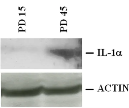
Accordingly, we detected higher amounts of IL-1α in
senescent (PD 45) than in young (PD 15) cells by western analysis (Fig. 2).
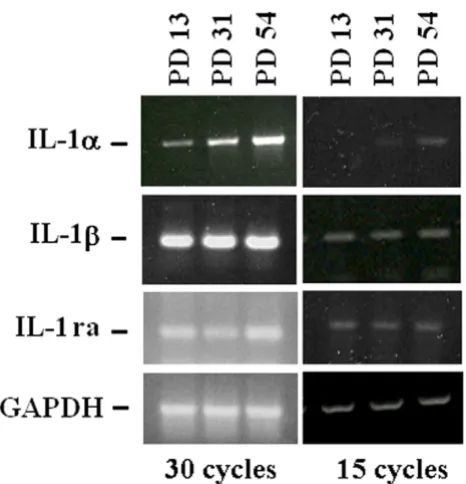
Because i) IL-1α shares many activities with IL-1β and ii) their activities are antagonized by the IL-1 receptor antag-onist (IL-1ra), we determined the expression of the mem-bers of the IL-1 family, i.e. IL-1α, IL-1β and IL-1ra by RT-PCR in HUVEC at different PDs. To provide a better quan-tification, the PCR reaction was stopped after 15 or 30 cycles. In both conditions, we found no significant mod-ulation of the total amounts of IL-1β and IL-1ra in young and senescent endothelial cells, while we confirmed the induction of IL-1α expression (Fig. 3).
IL-1s in young, senescent and progeric human fibroblasts
To determine whether the relationship between IL-1s and senescence occurred also in fibroblasts, we examined young and senescent human dermal fibroblasts for their expression of IL-1α by Northern on polyA+ RNA and
west-ern blot and found no differences (Fig. 4A and 4B). Prog-eria is a rare premature aging syndrome which is characterized by external stigmata of aging and death within the fierst two decades of life [11]. We therefore evaluated IL-1α expression in dermal fibroblasts derived from progeric individuals. Figure 4A and 4B show that
IL-1α mRNA and protein levels are not significantly
modu-lated in progeric vs age-matched dermal fibroblasts.
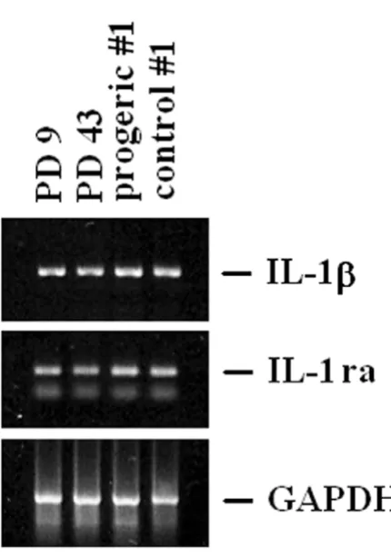
By RT-PCR, we show that no modulation occurs in the steady state levels of IL-1β and IL-1ra (Fig. 5) in senescent and progeric fibroblasts.
Discussion
The results of the present study indicate that the overex-pression of IL-1α specifically characterizes endothelial senescence. No modulation of this cytokine was observed in endothelial quiescence and in senescent or progeric human fibroblasts.
IL-1α shares many activity with IL-1β, since they act by binding to a common receptor, the type I IL-1 receptor [12]. A third member of the family, the IL-1 receptor antagonist (IL-1ra), also binds to the type I IL-1 receptor and blocks the receptor, preventing the action of the ago-nist IL-1s [12]. In senescent endothelial cells we did not
detect any modulation of the mRNA levels for IL-1β and
IL-1ra. We therefore propose that IL-1α could be used as a
marker of endothelial senescence. IL-1α causes multiple
responses in vascular endothelial cells including inhibi-tion of cell proliferainhibi-tion [13], inducinhibi-tion of adhesion mol-ecules which bind leukocytes [14] and promotion of thrombus formation [15]. Indeed, in vitro, IL-1α overex-pression has been linked to endothelial lifespan and to several dysfunctions [2]. It is noteworthy that senescent endothelial cells are found on the surface of atheroscle-rotic plaques [5] and that IL-1 is produced by endothe-lium on aged coronary arteries [8].
IL-1α total amounts in young and senescent HUVEC Figure 2
Increased vascular production of pro-inflammatory cytokines may contribute to the increased plasma levels of these mediators in aging [16,17]. We propose that the upregulation of IL-1 can be linked to the activation of var-ious pathophysiological programs that underlie the com-plex biological phenomenon described as "vascular aging". Proinflammatory status of aged arteries may shift the intravascular environment from a hemodynamically stable state to a pro-coagulant, pro-oxidant state which may favour an exaggerated response to vessel injury, pro-moting the development of ischemic heart disease in the elderly.
IL-1ra allelic polymorphisms affect replicative lifespan of human EC [10]. The polymorphism IL-1RN*2*2, which decreases the levels of IL-1ra, was associated with increased numbers of senescent endothelial cells and an inhibition of proliferation, while the addition of IL-1ra restored the proliferative potential of the cells and extended their lifespan [10]. Because endothelial turnover is enhanced under conditions that favour atherogenesis thus leading to a senescent phenotype, it is noteworthy
that the IL-1RN*2 allele is associated with atherosclerotic coronary disease [18]. All together, these data suggest that 1ra may prevent the senescent-promoting effects of IL-1 in the endothelium.
We did not detect any modulation of the steady state lev-els of IL-1s in in vitro aged human dermal and in progeric fibroblasts. This is in agreement with a previous study per-formed on IMR-90 cells, demonstrating that the human diploid fibroblast senescence pathway is independent of IL-1α mRNA levels [19]. On the contrary, expression of
IL-1β and IL-1ra was induced in senescent mouse embryonic
fibroblasts [20]. Interestingly, the IL-1ra-/- mice presented
early mortality compared to wild-type mice and acceler-ated senescence was observed in IL-1ra deficient fibrob-lasts [21]. All together these data indicate that a dysfunction of the cytokine network associates with aging and point to a specific role of IL-1α in endothelial senes-cence.
IL-1α expression in young, senescent and progeric human dermal fibroblasts
Figure 4
IL-1α expression in young, senescent and progeric human dermal fibroblasts. (A) 3 µg of polyA+ RNA were
utilized for Northern analysis. GAPDH hybridization shows that comparable amounts of RNA were loaded. PD 9: young fibroblasts; PD 43: senescent fibroblasts. Two different prog-eric cell types and age-matched controls have been utilized as described. Progeric and control #1: GM0498 and AG6917, respectively; progeric and control #2: GM2037 and AG3513, respectively. (B) Cell lysates (250 µg) from young, senescent and progeric (#1: GM0498 and AG6917) dermal fibroblasts were separated by a 15% SDS-PAGE and western blot was performed. After stripping, the blot was incubated with an anti-actin antibody to show that comparable amounts of pro-tein were loaded per lane. PD 9: young fibroblasts; PD 43: senescent fibroblasts; progeric and control #1: GM0498 and AG6917.
IL-1α, IL-1β and IL-1ra expression in young and senescen-tHUVEC
Figure 3
Publish with BioMed Central and every scientist can read your work free of charge "BioMed Central will be the most significant development for disseminating the results of biomedical researc h in our lifetime."
Sir Paul Nurse, Cancer Research UK
Your research papers will be:
available free of charge to the entire biomedical community
peer reviewed and published immediately upon acceptance
cited in PubMed and archived on PubMed Central
yours — you keep the copyright
Submit your manuscript here:
http://www.biomedcentral.com/info/publishing_adv.asp
BioMedcentral
Abbreviations
interleukin-1 IL-1
IL-1 receptor antagonist IL-1ra
endothelial cells EC
human umbilical vein EC HUVEC
reverse transcription- polymerase chain reaction RT-PCR
References
1. Foreman KE, Tang J: Molecular mechanisms of replicative senescence in endothelial cells. Exp Gerontol 2003, 38:1251-1257.
2. Maier JAM, Voulalas P, Roeder D, Maciag T: Extension of the life-span of human endothelial cells by an interleukin-1 alpha antisense oligomer. Science 1990, 249:1570-1574.
3. Shelton DN, Chang E, Whittier PS, Choi D, Funk WD: Microarray analysis of replicative senescence. Curr Biol 1999, 9:939-945. 4. Vasile E, Tomita Y, Brown LF, Kocher O, Dvorak HF: Differential
expression of thymosin beta-10 by early passage and senes-cent vascular endothelium is modulated by VPF/VEGF: evi-dence for senescent endothelial cells in vivo at sites of atherosclerosis. FASEB J 2001, 15:458-466.
5. Minamino T, Miyauchi H, Yoshida T, Ishida Y, Yoshida H, Komuro I: Endothelial cell senescence in human atherosclerosis: role of telomere in endothelial dysfunction. Circulation 2002, 105:1541-1544.
6. Fenton M, Barker S, Kurz DJ, Erusalimsky JD: Cellular senescence after single and repeated balloon catheter denudations of rabbit carotid arteries. Arterioscler Thromb Vasc Biol 2001, 21:220-226.
7. Garfinkel S, Brown S, Wessendorf JH, Maciag T: Post-transcrip-tional regulation of interleukin 1 alpha in various strains of young and senescent human umbilical vein endothelial cells.
Proc Natl Acad Sci U S A 1994, 91:1559-1563.
8. Csiszar A, Ungvari Z, Koller A, Edwards JG, Kaley G: Aging-induced proinflammatory shift in cytokine expression profile in coro-nary arteries. FASEB J 2003, 17:1183-1185.
9. Comi P, Chiaramonte R, Maier JAM: Senescence-dependent reg-ulation of type 1 plasminogen activator inhibitor in human vascular endothelial cells. Exp Cell Res 1995, 219:304-308. 10. Dewberry RM, Crossman DC, Francis SE: Interleukin-1 receptor
antagonist (IL-1RN) genotype modulates the replicative capacity of human endothelial cells. Circ Res 2003, 92:1285-1287.
11. Beauregard S, Gilchrest BA: Syndromes of premature aging.
Dermatol Clin 1987, 5:109-121.
12. Fantuzzi G: Lessons from interleukin-deficient mice: the inter-leukin-1 system. Acta Physiol Scand 2001, 173(1):5-9.
13. Cozzolino F, Torcia M, Aldinucci D, Ziche M, Almerigogna F, Bani D, Stern DM: Interleukin 1 is an autocrine regulator of human endothelial cell growth. Proc Natl Acad Sci U S A 1990, 87:6487-6491.
14. Bevilacqua MP, Pober JS, Majeau GR, Cotran RS, Gimbrone MA Jr: Interleukin 1 (IL-1) induces biosynthesis and cell surface expression of procoagulant activity in human vascular endothelial cells. J Exp Med 1984, 160:618-623.
15. Schorer AE, White JG: Interleukin 1 enhances arterial throm-bogenicity in vitro. Thromb Res 1989, 56:515-22.
16. Gerli R, Monti D, Bistoni O, Mazzone AM, Peri G, Cossarizza A, Di Gioacchino M, Cesarotti ME, Doni A, Mantovani A, Franceschi C, Paganelli R: Chemokines, sTNF-Rs and sCD30 serum levels in healthy aged people and centenarians. Mech Ageing Dev 2000, 121:37-46.
17. Straub RH, Cutolo M, Zietz B, Scholmerich J: The process of aging changes the interplay of the immune, endocrine and nervous systems. Mech Ageing Dev 2001, 122:1591-1611.
18. Francis SE, Camp NJ, Dewberry RM, Gunn J, Syrris P, Carter ND, Jef-fery S, Kaski JC, Cumberland DC, Duff GW, Crossman DC: Inter-leukin-1 receptor antagonist gene polymorphism and coronary artery disease. Circulation 1999, 99:861-866.
19. Garfinkel S, Wessendorf JH, Hu X, Maciag T: The human diploid fibroblast senescence pathway is independent of interleukin-IL-1β and IL-1ra expression in young, senescent and progeric
human dermal fibroblasts Figure 5
Publish with BioMed Central and every scientist can read your work free of charge "BioMed Central will be the most significant development for disseminating the results of biomedical researc h in our lifetime."
Sir Paul Nurse, Cancer Research UK
Your research papers will be:
available free of charge to the entire biomedical community
peer reviewed and published immediately upon acceptance
cited in PubMed and archived on PubMed Central
yours — you keep the copyright
Submit your manuscript here:
http://www.biomedcentral.com/info/publishing_adv.asp
BioMedcentral 1 alpha mRNA levels and tyrosine phosphorylation of
FGFR-1 substrates. Biochim Biophys Acta 1996, 1314:109-119.
20. Uekawa N, Nishikimi A, Isobe K, Iwakura Y, Maruyama M: Involve-ment of IL-1 family proteins in p38 linked cellular senescence of mouse embryonic fibroblasts. FEBS Lett 2004, 575:30-34. 21. Irikura VM, Lagraoui M, Hirsh D: The epistatic interrelationships