R E V I E W
Open Access
Structural aspects of plasticity in the
nervous system of
Drosophila
Atsushi Sugie
1,2†, Giovanni Marchetti
3†and Gaia Tavosanis
3*Abstract
Neurons extend and retract dynamically their neurites during development to form complex morphologies and to reach out to their appropriate synaptic partners. Their capacity to undergo structural rearrangements is in part maintained during adult life when it supports the animal’s ability to adapt to a changing environment or to form lasting memories. Nonetheless, the signals triggering structural plasticity and the mechanisms that support it are not yet fully understood at the molecular level. Here, we focus on the nervous system of the fruit fly to ask to which extent activity modulates neuronal morphology and connectivity during development. Further, we summarize the evidence indicating that the adult nervous system of flies retains some capacity for structural plasticity at the synaptic or circuit level. For simplicity, we selected examples mostly derived from studies on the visual system and on the mushroom body, two regions of the fly brain with extensively studied neuroanatomy.
Keywords:Structural plasticity, Drosophila, Photoreceptors, Synapse, Active zone, Mushroom body, Mushroom body calyx, Learning
Background
The establishment of a functional neuronal circuit is a dynamic process, including an extensive structural re-modeling and refinement of neuronal connections. Intrinsic differentiation programs and stereotypic mo-lecular pathways contribute the groundwork of pattern-ing the nervous system durpattern-ing development, includpattern-ing the guidance of axons and dendrites over long distances or the recognition of appropriate connection partners. In addition, information derived from functional cues controls the refinement of the circuit. Even after the major task of assembling a fully functional network has been achieved, the nervous system retains the capacity of undergoing not only functional, but also structural modifications related, for instance, to adaptation or learning. The role of activity in the developmental re-finement of neuronal morphology and of the connec-tions within a circuit (and possibly also the setting up of circuits; [1]) as well as in the initiation of structural re-modeling during adult life is undisputed [2]. In intricate neuropils, dense with axons and dendrites of different
neuronal types, the feed-back derived from activity ap-pears to be an important element to define which connec-tions can be stabilized and which ones removed [3–5]. Nonetheless, the cellular mechanisms initiated by activity to drive structural remodeling during development and in the course of adult life are not fully elucidated. Here, we review the literature supporting structural plasticity in the fruit fly Drosophila, a system offering major advantages for genetic and molecular analysis. Where appropriate, we include comparisons with other invertebrate and verte-brate systems to highlight evolutionary conserved mecha-nisms. Thanks to the stereotypy of the “macroscopic” organization of the fly’s nervous system, work carried out usingDrosophilaled to major breakthroughs in the identi-fication of conserved molecular cascades and mechanisms that orchestrate genetically controlled developmental pro-grams. Possibly due to this emphasis on stereotypy, the role of signals providing feed-back information about functional connections during fly nervous system develop-ment has not been investigated as deeply. Nonetheless, multiple examples of activity controlling neuronal com-plexity during development have emerged [6]. For in-stance, dendrite elaboration of fly larval motorneurons as well as of the wide-field serotonergic neuron CSDn in the Drosophila central nervous system can be affected by the
* Correspondence:[email protected]
†Atsushi Sugie and Giovanni Marchetti contributed equally to this work.
3Center for Neurodegenerative Diseases (DZNE), 53127 Bonn, Germany
Full list of author information is available at the end of the article
level of input signals and actually by input activity during development [7,8]. Similarly, exposure of the larva to dif-ferent light regimes modifies the total dendrite length of ventral lateral neurons (LNv), postsynaptic to the photore-ceptors [9]. The accessibility of the neuromuscular junction (NMJ) of larvae allows for detailed molecular, morpho-logical and functional analysis [10]. The level of activity in the motorneuron can modulate the number of boutons formed and the density of synaptic release sites at the NMJ, providing a clear example of activity-related structural con-trol [11–13]. In this context, postsynaptically-derived sig-nals carried by the Wnt and BMP signaling pathways, modulate the presynaptic terminal at the NMJ [14–16].
Evidence for structural rearrangements in the nervous system of the adult fly after development is completed has been rather limited and it is mostly related to adap-tive phenomena. As an example, prolonged exposure to a given odor induces increased size and synaptic density in discrete glomeruli of the antennal lobe, the first olfac-tory processing center [17, 18]. Nonetheless, the behav-ior of adult flies (as well as of larvae) can be modified by experience in a non-adaptive fashion. In fact, flies can learn multiple types of cues and form lasting memories, a capacity that might require structural modifications in the neurons and the circuits involved [19–21].
Recent large-scale efforts are yielding complete maps at synaptic-resolution of circuits within the adult fly cen-tral nervous system, including areas involved in memory formation [22, 23]. This information can be combined with the availability of tools to visualize, manipulate and control the activity of restricted and defined populations of neurons in this system [24–27]. Thus, novel insights to the fundamental understanding of information pro-cessing and of learning are starting to be produced and much more is expected in the coming years [22,28–30]. Importantly, the high-resolution description of circuits obtained in electron microscopy images and with tools to highlight synaptic components is challenging the idea of circuit stereotypy in the fly nervous system. As an ex-ample, the detailed study of motorneuron network in the ventral nerve cord of the larva revealed a high de-gree of variability in terms of synaptic connections [31].
Taken together, it appears that it is the right time to approach the non-stereotypy and plasticity of neurons in the adult fly nervous system.
For the purpose of this review, we define structural plas-ticity as the changes that include physical remodeling of recognizable structures. In particular, we concentrate on large-scale changes that might involve neuronal processes, their connections and circuit subroutines and on molecular changes that affect particularly the structural organization of the presynapse. Certain types of functional plasticity involve structural changes, as the formation of new dendritic spines [32] or the reorganization of the
molecular components of the synapse [33,34]. In this re-view we will select the aspects that deal in particular with the structural components of functional and synaptic plas-ticity. We chose to focus on two centers of the fly nervous system to summarize the current evidence in support of an influence of activity during development and of plastic changes in the adult nervous system in adaptive or learning conditions.
The establishment of circuits
The ease of manipulating their input makes sensory systems particularly suitable for the study of activity-dependent processes involved in neuronal circuit assem-bly, refinement and plasticity. In this review we concen-trate therefore our attention on the fly adult visual system and on the pathways that deliver olfactory information to the mushroom body (MB), involved in memory processing. Sensory information is initially encoded in discrete stereotypic pathways. For instance, the presence of a bright signal in the visual field or the specific odorant present in the air flux activates defined subroutines within the visual or olfactory circuits, respectively. To maintain the initial specificity of information and to transmit it precisely towards higher processing centers, circuits are assembled with remarkable precision during development. Correct axon and dendrite targeting to the appropriate region, pairing of the suitable synaptic partners and synaptogenesis are all highly regulated developmental steps (Fig. 1). In principle, targeting and recognition of processes to form functional connections can be achieved through genetically defined pathways. For instance, specific tags and receptors allow the cor-rect partner neurons to recognize each other. Alterna-tively, guidance signals could support the formation of initially sloppy maps, which are subsequently refined. In this case, the evaluation of the functional performance of a given connection or of the circuit is likely to be a highly valuable factor for deciding whether the connec-tion should be maintained or removed [35].
Here, we address how much these two potential mecha-nisms contribute to the assembly of circuits in the visual system or in the MB. While the extant literature regarding the molecular mechanisms of genetically controlled pro-grams is abundant, particularly for the visual system, the information about activity-dependent circuit assembly control is rather scant in Drosophila. We put our em-phasis primarily on this second, less explored aspect.
The initial connectivity in the visual system is independent of activity
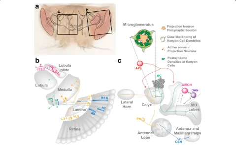
(R1 to R8). R1–6 project into the first optic ganglion, the lamina, while R7 and R8 project their axons to the M6 and M3 layers, respectively, of the medulla, the second optic gan-glion. The five subtypes of lamina neurons (L1 to L5) project into distinct layers in the distal medulla (Fig.2b). Within the
third optic ganglion, the lobula complex, the lobula plate tangential cells (LPTCs) integrate information from R1–6 to compute the direction of optic flow (Fig.2b) [37].
The pairing of appropriate synaptic partners is an es-sential aspect for the establishment of functional circuits
Fig. 1Activity dependent modulation of neuronal connectivity during development in theDrosophilavisual and MB circuits. Steps supporting the establishment of neuronal circuits in the adultDrosophilavisual and olfactory systems during development
a
b
c
(Fig.1). How much of this recognition is driven by genetic programs as opposed to functional cues in the visual sys-tem? As a striking example of circuit assembly controlled by recognition among identity tags, each different synaptic pair in the medulla expresses distinct Immunoglobulin superfamily cell adhesion molecules (21 Dprs and 9 DIPs) for precise synaptic partner matching [21, 38, 39]. Once appropriate partners have come to close proximity, cell adhesion molecules contribute to synaptic formation among them [40]. Interestingly, also functional com-ponents of the presynaptic active zone (AZ) such as DLiprin-αand DSyd-1 are required not only for synaptic vesicle (SV) clustering at R7 axon terminal synapses, but also for axon targeting [41,42]. These data are intriguing as they suggest a negative relationship between synapse assembly and axon extension. Taken together, cell surface molecule diversity contributes to axon targeting, pairing of synaptic partners and synaptogenesis, suggesting a robust genetically controlled program supporting these events.
Activity-dependent fine-tuning of neuronal circuits plays a role during the development of the visual system in ver-tebrates [43–45]. Several studies addressed whether neur-onal activity is relevant for precisely assembling neurneur-onal circuits in theDrosophilavisual system as well. They pro-vide epro-vidence that neuronal circuit formation is independ-ent of neuronal activity in the visual system, especially for the photoreceptors R1–6 [46]. The number of synapses in R1-R6 and the downstream circuit organization has been investigated in a series of neuronal activity mutants, in-cluding Phospholipase CnorpA[47] and Ca2+channelstrp and trpl[48, 49] mutants that suppress the generation of electropotentials, or histidine decarboxylase hdc [50, 51] and the Ca2+ sensor synaptotagmin sytAX4 [52] mutants that inhibit neurotransmitter release. All those mutants show no obvious defect in R1-R6 axon targeting or in the number of presynaptic AZs in the lamina [46]. Also deeper in the visual system, the complexity of LPTC dendrites is not affected by a constant darkness (DD) regime. In addition, LPTC dendritic spine structure and density remain unchanged after genetically-induced visual depri-vation elicited by the expression ofhead involution defective (hid) in the eye [53]. From these studies, axon projection or dendrite arborization in the fly visual system seems to be defined largely independently of activity (Fig.1).
Contribution of experience to larval visual system connectivity
Although activity seems dispensable for the establishment of connectivity in the adult visual system, recent work points to its involvement of activity within larval visual circuits to guarantee the establishment of correct morph-ologies. The larval optic nerve, called Bolwig’s nerve (BN), projects into the central brain along a simple invariant path. The BN is required for the appropriate arborization
of a serotonergic neuron and for the development of the dendritic tree of the circadian pacemakers, ventral lateral neurons (LN(v)s) [54,55]. Suppression of synaptic activity in the presynaptic BN disrupts the dendritic arborization of the postsynaptic neurons in the larval visual system [56]. In this study, tetanus toxin light chain (TeTxLC), which blocks synaptic release by cleaving neuronal-Synap-tobrevin, was expressed in photoreceptors leading to a re-duction of the dendritic arborization of the serotonergic neuron. In contrast, attenuation of evoked activity by the expression of a genetically modified Shaker K+ channel (EKO channel) in photoreceptors did not alter the den-drites of this serotonergic neuron. While the possibility of a broader effect of Synaptobrevin inhibition remains, these results suggest that spontaneous synaptic activity could promote dendrite arborization in the serotonergic neuron. Also the arborization of the dendrites of the ventral lateral neurons LN(v)s at the third instar larval stage depends on activity. In fact, prolonged light exposure reduced, while constant darkness increased the LN(v) dendritic length via the cyclic adenosine monophosphate (cAMP) pathway [9]. Larvae are continuously exposed to sensory stimuli. Thus, experience might contribute to the adjustment of neuronal connectivity to guarantee appropriate synaptic strength in a variety of environments (Fig.1).
Activity-dependent development and maturation of the olfactory and mushroom body circuits
calyx, peduncle, and lobes (Fig.2c) [67–69]. The output of approximately 2000 KCs per adult brain hemisphere converges onto a population of only 34 MB output neu-rons (MBONs) of 21 anatomically distinct types [27] (Fig.2c).
Neural activity appears to be largely dispensable dur-ing metamorphosis for the establishment of the adult fly olfactory circuit [70]. For instance, the glomerular map
in the Drosophila AL was not modified when all
odor-evoked activity was eliminated or when input or output neurons were removed [71–73]. However, com-plementary work in social insects suggests that the pres-ence and function of olfactory sensory neurons (OSNs) is fundamental for the development of the olfactory cir-cuit. In particular, ants carrying mutations in the highly conserved co-receptor of odorant receptors (ORs) Orco, showed a striking reduction in the AL glomeruli number associated with deficiencies in social behavior [74]. Simi-larly, surgical removal of the antenna of honeybees at dif-ferent time points during pupal development led to decreased synapse density in the AL in a stage-dependent manner [75]. Clearly, further studies are needed towards a comprehensive view of the role of neural activity in adult olfactory circuit wiring in insects.
After metamorphosis the adult fly emerges from the pupal case with a formed olfactory circuit. It appears none-theless that the first days of adult life represent a critical period in which the olfactory circuit can undergo activity-dependent refinement. For instance, prolonged ex-posure to CO2causes activity-dependent volume increase
of the CO2-responding AL glomerulus. Those changes are
reversible and occur in a critical time window correspond-ing to early adult life. In fact, exposure-induced plasticity in the CO2-responding glomerulus was not observed in
flies 11 days post eclosion [18]. Whole-cell recordings of cultured MB neurons derived from late stage Drosophila pupae reveal spontaneous Ca2+transients that might play a role in the maturation of the adult circuit [76]. At the mo-lecular level, the RNA-binding protein Fragile X Mental Retardation Protein (FMRP) regulates MB circuit refine-ment in an activity-dependent manner [77]. FMRP is re-quired at late pupal stages and during early adult life to control MB axonal pruning and presynaptic refinement in the MB calyx [77, 78]. Repressing PN activity during the first day after pupal eclosion results in enhancement of presynaptic axonal branching [78]. In addition, blocking PN synaptic vesicle release post-eclosion for 5 days yields increased bouton size [79]. Thus, the time following eclosion could represent a period in which theDrosophila olfactory circuit is evaluated and adapted to the local envir-onment. Similar critical periods have been documented for the development of the mammalian cortex and olfactory bulb [80,81]. In all these model systems, the critical period likely allows the animal to compare the developmentally
determined network template with external conditions and make activity-dependent adjustments that reflect the external environment.
Plasticity during adult life and ageing
Even after functional circuits have been established dur-ing development and refined durdur-ing a critical period, they can still undergo structural and functional changes to allow the animal to adapt to a modified sensory envir-onment or store relevant information to modify future behavior. While studies investigating functional plasticity in Drosophila have a long tradition, evidence for struc-tural plasticity in the adult nervous system has been rather fragmentary.
Structural plasticity in the adult visual system
Visual experience during early adult life can modulate behavior inDrosophila. In visually guided choice behav-ior tests, flies reared in darkness (DD) are attracted to wider vertical black lines against a white background compared to control flies reared in a regular light-dark cycle (LD), providing evidence for developmental visual plasticity in this system [82, 83]. DD reared flies also show lower preference for visible light in comparison to flies reared in an LD cycle in a Y-maze apparatus designed to test phototaxis preference behavior [84]. The plasticity of phototaxis preference is reversible in adult flies and can be modulated by the expression levels of N-methyl-D-aspartate receptor 1 (NMDAR1) [84]. Taken together, light exposure conditions during early adult life can modulate adult visual behavior, suggesting some plasticity in circuit function.
recruitment of AZ machinery such as the voltage-gated Ca2+ channels in hippocampal neurons, revealed with super-resolution imaging [33]. In turn, the probability of neurotransmitter release correlates with the amount of Bassoon or RIM in rat or mouse neuronal cultures [94–96]. In this system, also the localization of Liprin-α2 at AZs depends on activity and the expression level of Liprin-α2 regulates the probability of SV release [97]. Taken together, these studies performed with rodent neu-rons indicate that the level of activity in the presynaptic neuron controls the abundance of AZ proteins, which in turn affects the probability of SV release at the synapse.
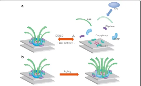
Notably, an activity-dependent remodeling of the AZ proteins has been described recently in the adult fly vis-ual system. The presynaptic AZ in insects is marked by the presence of a T-shaped structure formed by the ELKS family Brp protein [98, 99] (Fig.3). In spite of its complexity [10,100,101], the T-bar can dynamically dis-assemble and dis-assemble. Rapid shifts from a dark re-gime to light or vice versa can induce reversible changes in the size and number of presynaptic T-bars in the photoreceptor neurons of the house fly (Musca domes-tica) within minutes [102]. The structural changes are reflected by measurable changes in protein abundance.
Already a short light stimulation significantly increases the level of BRP, Synapsin and Dlg proteins in the Drosophilalamina even just for 15 min exposure to light [103]. In addition to these rapid changes, late-onset modifications alter synaptic composition by prolonged light exposure. A subset of AZ components as BRP, DLiprin-α, and DRBP are lost from the AZ in this condi-tion, while DSysd-1 or the voltage-gated Ca2+ channel Cacophony is not (Fig. 3a). This presynaptic remodeling is triggered by a postsynaptic signal that elicits microtubule destabilization in the presynaptic photoreceptors via the di-vergent canonical Wnt pathway (Fig.3a) [104,105]. Only a subset of AZ undergoes these reversible modifications and loses their T-bar. Since loss of BRP suppresses transmission from the AZ [98,99], it is expected that the final outcome of these changes is a reduction in transmission, potentially supporting homeostasis in the circuit. Taken together, light exposure can induce activity-regulated structural changes in the fly photoreceptor AZs.
Structural plasticity in the mushroom body calyx
The MB has been most extensively studied in the context of associative memory that utilizes olfactory or other sen-sory information to guide future behavior [106–110].
a
b
Coincidence detection of sensory information (odor) and value (reward or punishment) initiating memory forma-tion involves the MB output synapses and circuits at the lobes [111,112]. The recurrent circuits between MBONS, dopaminergic neurons (DANs) and KCs contribute to memory consolidation [29] and to its re-consolidation after re-evaluation [30]. The contribution of additional cir-cuits to multiple aspects of memory representations will require future investigations.
The MB calyx is involved in the extraction of a sparse code from the sensory information derived from the ol-factory PNs [113, 114]. Electrophysiological recordings in PNs and KC upon odor exposure reveal that the MB transforms the flood of odor-elicited activation of the PNs into a sparse representation of the odor in the KCs [113,115,116]. This sparse format of sensory coding is a widely observed feature in vertebrate cortical areas sug-gesting that minimizing the overlap between representa-tion patterns of different stimuli maximizes memory capacity [117]. Sparse KC activation is important for odor discrimination after associative olfactory learning [118]. Recently generated genetically-encoded functional reporters targeted to either pre- or postsynaptic compartments allow to monitor Ca2+ dynamics during olfactory processing in the adult fly brain. Imaging of odor-evoked activity of synapses in the MB calyx revealed experience-dependent changes in pre- and postsynaptic activity [79]. The reduced anatomical complexity and the ability to monitor physiological changes in identified neurons suggests that studies in the fly will likely deliver important contributions to the understanding of how activity-dependent functional plasticity is generated, rein-forced and maintained in a neuronal circuit.
An additional major feature of the insect MB calyx is that it displays no obvious stereotypy. While subsets of PNs, project to loosely defined calycal regions [119,120] the identity of their postsynaptic KCs cannot be foretold [66,113]. KC subtypes project their dendrites also to ap-proximate layers within the calyx [119–121]. Nonethe-less, a single PN bouton is predicted to contact different types of KCs [63]. Although, it remains conceivable that the genetic tools available do not allow yet recognizing existing stereotypy, anatomical and functional data sup-port the view that PN/ KC connections are not prede-fined [66,113]. Which are the rules that control setting up such a non-stereotypical circuit remains an open and fascinating question.
Experience dependent structural plasticity in the MB calyx has been explored extensively in social insects such as bees and ants. Those studies indicate that the volume of the MB calyx is modulated by experience. The initial exploration of the foraging area by these insects corre-lates with an increase in MB calyx volume [122–125]. In honeybees, the experience-dependent volume increase of
the MB calyx correlates with activity mediated by mus-carinic cholinergic pathways [126]. The core functional unit of the MB calyx is the synapse formed by individual PN boutons and multiple KCs dendrite endings of claw shape (Fig.2c) [62,63,127]. The resulting large synaptic complex, the microglomerulus (MG), also comprises in-put of additional extrinsic neurons that in Drosophila have not yet been unequivocally identified [63, 127]. MGs are readily detectable using antibodies recognizing presynaptic markers such as Synapsin in the PN bouton or by highlighting actin in the KC dendrites [128, 129]. Using such tools, it was possible to show that PN bou-ton size and postsynaptic densities increase during the behavioral transition of honeybees from nursing to for-aging [130, 131]. Such findings suggest that the MGs might be a major component of MB calyx plasticity. Im-portantly, in bees the establishment of long-term olfac-tory associative memories correlates with an increase in the density of MGs, specifically in the calycal region responding to olfactory stimuli [132]. Collectively these data, together with comparable results obtained in other insect species [122, 133, 134], point to the MG as sites of structural plasticity related to experience and learning. Whether MGs size and number might be directly af-fected by experience or in learning has not been directly tested in Drosophila. However, MG properties can be modulated by input activity also in this system. The use of genetic tools to specifically label subsets of PNs and their presynaptic structures, in combination with post-synaptic markers expressed in KCs, allows to image MGs in the adult fly calyx at high resolution [135]. Pro-longed deprivation of PN synaptic input in the adult MB calyx leads to increased MG number and enlarged pre and postsynaptic elements in the silenced MGs [79,135]. These effects could represent a homeostatic response to decreased neuronal activity. They suggest that olfactory experience encoded by PN neuron activity induces MG structural changes [79, 135]. However, how functional plasticity in response to odor stimulation correlates with structural modifications remains to be tested.
AZ size. Conversely, artificially increased expression of AZ components BRP or RIM-BP in young flies, mimicked the reduced learning performance of aged flies [138]. These data point to the fact that AZs undergo structural changes during ageing (Fig. 3b). They furthermore indi-cate a role of the presynaptic AZ scaffold in regulating synaptic plasticity during olfactory memory formation and reveal that calycal synapses can modulate memory cap-acity. Finally, they suggest that re-establishing appropriate presynaptic function might significantly contribute to re-storing cognitive impairment associated with ageing.
Conclusions
Thanks to the relatively small size of its nervous system and to coordinated efforts, the reconstruction of circuits within the brain of Drosophila is proceeding at an im-pressive pace [22, 23]. Large-scale approaches based on electron microscopy are providing maps of every single synapse in large parts of the nervous system. This level of resolution raises now even more clearly the question of stereotypy of neuronal processes and circuits among animals and thus of degrees of freedom in circuit estab-lishment during development- and of plasticity in face of changing experience during adult life.
How much freedom is allowed in setting up connections during development? Answering this question will require a systematic analysis of neuronal morphology and of known connections, ideally at the synaptic level, in a number of an-imals or at different developmental stages. Such studies are starting to appear [31, 140]. It is possible that certain cir-cuits allow little discrepancy from a basic scheme, while others afford larger degrees of freedom during develop-ment. The randomly set up MB calyx would be a good can-didate for the latter scenario. What are the mechanisms that control non-stereotyped circuits to attain a balanced level of activity and produce meaningful signals?
In addition to these potentially nervous system-intrinsic levels of control, environmental factors might well play a role in modulating neuron differentiation and circuit as-sembly. In honeybees, for instance, MG density and size in the adult MB calyx depend on temperature and light experienced by the animals during development [141]. A striking example of control exerted by the growth condi-tions on the development of the nervous system recently emerged from studies on fate decisions during neuroblast divisions [142–144]. The nutritional state of the animal determines the timing of peaks of production of the hor-mone Ecdysone [145, 146]. In turn, it is the response to Ecdysone that initiates the fate switch fromγto α’β’and then toαβneurons during neuroblast divisions [142].
As detailed in this review, a lot of the work to respond to the questions of to which extent and how experience modulates neuronal circuit development lies ahead of us.
Addressing systematically structural plasticity in the adult nervous system presents a number of even more complex challenges. Faced with circuits and connections that are not stereotyped to the synaptic level or in some cases, as in the calyx, that present little stereotypy, the potential of identifying eventual small modifications will be limited. Fortunately, many of the tools necessary are becoming rapidly available. Fly lines that allow manipu-lating specifically and independently pre- and postsynap-tic partners were recently generated [147, 148] and they will allow to concentrate on reproducible connections. So-phisticated tools for localizing AZ components and some postsynaptic markers have been produced over the past years [10]. Functional imaging in the fly brain can be car-ried out especially in more accessible brain regions [79, 116, 149]. Activity-dependent gene expression profiles were described in subsets of neurons in the adult fly brain [150]. Functional analysis of the identified genes might in the future shed light on activity-dependent structural re-finement processes. With the repertoire of genetic tools in Drosophila, a large-scale interrogation of the signals that trigger structural plasticity, its molecular and cell bio-logical mechanisms, as well as the cause-effect relation-ship between structural changes and their functional and behavioral consequences might be at hand.
Abbreviations
AL:Antennal lobe; APL: Anterior paired lateral; AZ: Active zone; BN: Bolwig’s nerve; cAMP: Cyclic adenosine monophosphate; DD: Constant darkness; FMRP: Fragile X Mental Retardation Protein; KCs: Kenyon cells; LD: 12 h light/ 12 h dark cycle; LL: Constant light; LNv: Ventral lateral neurons; LPTCs: Lobula plate tangential cells; MB: Mushroom body; MBONs: MB output neurons; MG: Microglomerulus; NMDAR1: N-methyl-D-aspartate receptor 1;
NMJ: Neuromuscular junction; ORs: Odorant receptors; OSNs: Olfactory sensory neurons; PNs: Projection neurons; PPB: Phototaxis preference behavior; SV: Synaptic vesicle; TeTxLC: Tetanus toxin light chain
Acknowledgements
We wish to thank the members of the Tavosanis lab for discussions. We are grateful to L. Baltruschat, F. Bradke, T. Suzuki and M. Pankratz for critically reading the manuscript.
Funding
Our work is funded by institutional core funding form the DZNE (G.M and G.T) and JSPS KAKENHI Grant Number 17H04983 (A.S).
Authors’contributions
All three authors contributed to the writing of the manuscript. All authors read and approved the final manuscript.
Ethics approval and consent to participate
Not applicable
Consent for publication
Not applicable
Competing interests
The authors declare that they have no competing interests.
Publisher’s Note
Author details
1Center for Transdisciplinary Research, Niigata University, Niigata 951-8585,
Japan.2Brain Research Institute, Niigata University, Niigata 951-8585, Japan. 3
Center for Neurodegenerative Diseases (DZNE), 53127 Bonn, Germany.
Received: 22 December 2017 Accepted: 12 June 2018
References
1. Andreae LC, Burrone J. The role of spontaneous neurotransmission in synapse and circuit development. J Neurosci Res. 2017;96:354–9. 2. Andersen N, Krauth N, Nabavi S. Hebbian plasticity in vivo: relevance and
induction. Curr Opin Neurobiol. 2017;45:188–92.
3. Katz LC, Shatz CJ. Synaptic activity and the construction of cortical circuits. Science. 1996;274(5290):1133–8.
4. Sanes JR, Lichtman JW. Development of the vertebrate neuromuscular junction. Annu Rev Neurosci. 1999;22:389–442.
5. Huberman AD, Feller MB, Chapman B. Mechanisms underlying development of visual maps and receptive fields. Annu Rev Neurosci. 2008;31:479–509. 6. Tavosanis G. Dendritic structural plasticity. Dev Neurobiol. 2012;72(1):73–86. 7. Tripodi M, et al. Structural homeostasis: compensatory adjustments of
dendritic arbor geometry in response to variations of synaptic input. PLoS Biol. 2008;6(10):e260.
8. Singh AP, VijayRaghavan K, Rodrigues V. Dendritic refinement of an identified neuron in the Drosophila CNS is regulated by neuronal activity and Wnt signaling. Development. 2010;137(8):1351–60.
9. Yuan Q, et al. Light-induced structural and functional plasticity in Drosophila larval visual system. Science. 2011;333(6048):1458–62.
10. Van Vactor D, Sigrist SJ. Presynaptic morphogenesis, active zone organization and structural plasticity in Drosophila. Curr Opin Neurobiol. 2017;43:119–29.
11. Budnik V, Zhong Y, Wu CF. Morphological plasticity of motor axons in Drosophila mutants with altered excitability. J Neurosci. 1990;10(11):3754–68. 12. Sigrist SJ, et al. Experience-dependent strengthening of Drosophila
neuromuscular junctions. J Neurosci. 2003;23(16):6546–56.
13. Weyhersmuller A, et al. Rapid active zone remodeling during synaptic plasticity. J Neurosci. 2011;31(16):6041–52.
14. Ataman B, et al. Rapid activity-dependent modifications in synaptic structure and function require bidirectional Wnt signaling. Neuron. 2008;57(5):705–18.
15. Aberle H, et al. Wishful thinking encodes a BMP type II receptor that regulates synaptic growth in Drosophila. Neuron. 2002;33(4):545–58. 16. Packard M, et al. The Drosophila Wnt, wingless, provides an essential signal
for pre- and postsynaptic differentiation. Cell. 2002;111(3):319–30. 17. Devaud JM, Acebes A, Ferrus A. Odor exposure causes central adaptation
and morphological changes in selected olfactory glomeruli in Drosophila. J Neurosci. 2001;21(16):6274–82.
18. Sachse S, et al. Activity-dependent plasticity in an olfactory circuit. Neuron. 2007;56(5):838–50.
19. Bailey CH, Chen M. Long-term sensitization in Aplysia increases the number of presynaptic contacts onto the identified gill motor neuron L7. Proc Natl Acad Sci U S A. 1988;85(23):9356–9.
20. Bailey CH, Chen M. Long-term memory in Aplysia modulates the total number of varicosities of single identified sensory neurons. Proc Natl Acad Sci U S A. 1988;85(7):2373–7.
21. Bailey CH, Kandel ER, Harris KM. Structural components of synaptic plasticity and memory consolidation. Cold Spring Harb Perspect Biol. 2015;7(7):a021758.
22. Takemura SY, et al. A connectome of a learning and memory center in the adult Drosophila brain. Elife. 2017;6:e26975.
23. Takemura SY, et al. The comprehensive connectome of a neural substrate for 'ON' motion detection in Drosophila. Elife. 2017;6:e24394.
24. Jenett A, et al. A GAL4-driver line resource for Drosophila neurobiology. Cell Rep. 2012;2(4):991–1001.
25. Manning L, et al. A resource for manipulating gene expression and analyzing cis-regulatory modules in the Drosophila CNS. Cell Rep. 2012;2(4):1002–13.
26. Aso Y, et al. The neuronal architecture of the mushroom body provides a logic for associative learning. Elife. 2014;3:e04577.
27. Aso Y, et al. Mushroom body output neurons encode valence and guide memory-based action selection in Drosophila. Elife. 2014;3:e04580.
28. Strother JA, et al. The emergence of directional selectivity in the visual motion pathway of Drosophila. Neuron. 2017;94(1):168–82. e10 29. Ichinose T, et al. Reward signal in a recurrent circuit drives appetitive
long-term memory formation. Elife. 2015;4:e10719.
30. Felsenberg J, et al. Re-evaluation of learned information in Drosophila. Nature. 2017;544(7649):240–4.
31. Couton L, et al. Development of connectivity in a motoneuronal network in Drosophila larvae. Curr Biol. 2015;25(5):568–76.
32. Poo MM, et al. What is memory? The present state of the engram. BMC Biol. 2016;14:40.
33. Glebov OO, et al. Nanoscale structural plasticity of the active zone matrix modulates presynaptic function. Cell Rep. 2017;18(11):2715–28. 34. Michel K, et al. The presynaptic active zone: a dynamic scaffold that
regulates synaptic efficacy. Exp Cell Res. 2015;335(2):157–64.
35. Kutsarova E, Munz M, Ruthazer ES. Rules for shaping neural connections in the developing brain. Front Neural Circuits. 2016;10:111.
36. Sanes JR, Zipursky SL. Design principles of insect and vertebrate visual systems. Neuron. 2010;66(1):15–36.
37. Borst A. Neural circuits for elementary motion detection. J Neurogenet. 2014;28(3–4):361–73.
38. Carrillo RA, et al. Control of synaptic connectivity by a network of Drosophila IgSF cell surface proteins. Cell. 2015;163(7):1770–82. 39. Morey M. Dpr-DIP matching expression in Drosophila synaptic pairs.
Fly (Austin). 2017;11(1):19–26.
40. Yogev S, Shen K. Cellular and molecular mechanisms of synaptic specificity. Annu Rev Cell Dev Biol. 2014;30:417–37.
41. Holbrook S, et al. Loss of syd-1 from R7 neurons disrupts two distinct phases of presynaptic development. J Neurosci. 2012;32(50):18101–11. 42. Kniss JS, Holbrook S, Herman TG. R7 photoreceptor axon growth is
temporally controlled by the transcription factor Ttk69, which inhibits growth in part by promoting transforming growth factor-beta/activin signaling. J Neurosci. 2013;33(4):1509–20.
43. Cang J, Feldheim DA. Developmental mechanisms of topographic map formation and alignment. Annu Rev Neurosci. 2013;36:51–77.
44. Okawa H, et al. Illuminating the multifaceted roles of neurotransmission in shaping neuronal circuitry. Neuron. 2014;83(6):1303–18.
45. Owens MT, et al. Stochastic interaction between neural activity and molecular cues in the formation of topographic maps. Neuron. 2015;87(6):1261–73.
46. Hiesinger PR, et al. Activity-independent prespecification of synaptic partners in the visual map of Drosophila. Curr Biol. 2006;16(18):1835–43. 47. Bloomquist BT, et al. Isolation of a putative phospholipase C gene of
Drosophila, norpA, and its role in phototransduction. Cell. 1988;54(5):723–33. 48. Niemeyer BA, et al. The Drosophila light-activated conductance is
composed of the two channels TRP and TRPL. Cell. 1996;85(5):651–9. 49. Haab JE, et al. Coordinated gating of TRP-dependent channels in
rhabdomeral membranes from Drosophila retinas. J Neurosci. 2000;20(19):7193–8.
50. Hardie RC. Is histamine a neurotransmitter in insect photoreceptors? J Comp Physiol A. 1987;161(2):201–13.
51. Burg MG, et al. Genetic and molecular identification of a Drosophila histidine decarboxylase gene required in photoreceptor transmitter synthesis. EMBO J. 1993;12(3):911–9.
52. Koh TW, Bellen HJ. Synaptotagmin I, a Ca2+ sensor for neurotransmitter release. Trends Neurosci. 2003;26(8):413–22.
53. Scott EK, Reuter JE, Luo L. Dendritic development of Drosophila high order visual system neurons is independent of sensory experience. BMC Neurosci. 2003;4:14.
54. Malpel S, Klarsfeld A, Rouyer F. Larval optic nerve and adult extra-retinal photoreceptors sequentially associate with clock neurons during Drosophila brain development. Development. 2002;129(6):1443–53.
55. Mukhopadhyay M, Campos AR. The larval optic nerve is required for the development of an identified serotonergic arborization in Drosophila melanogaster. Dev Biol. 1995;169(2):629–43.
56. Rodriguez Moncalvo VG, Campos AR. Genetic dissection of trophic interactions in the larval optic neuropil of Drosophila melanogaster. Dev Biol. 2005;286(2):549–58.
57. Laissue PP, Vosshall LB. The olfactory sensory map in Drosophila. Adv Exp Med Biol. 2008;628:102–14.
59. Heisenberg M. What do the mushroom bodies do for the insect brain? An introduction. Learn Mem. 1998;5(1–2):1–10.
60. Waddell S, Quinn WG. What can we teach Drosophila? What can they teach us? Trends Genet. 2001;17(12):719–26.
61. Tanaka NK, Tanimoto H, Ito K. Neuronal assemblies of the Drosophila mushroom body. J Comp Neurol. 2008;508(5):711–55.
62. Yasuyama K, Meinertzhagen IA, Schurmann FW. Synaptic organization of the mushroom body calyx in Drosophila melanogaster. J Comp Neurol. 2002;445(3):211–26.
63. Leiss F, et al. Synaptic organization in the adult Drosophila mushroom body calyx. J Comp Neurol. 2009;517(6):808–24.
64. Strausfeld NJ, et al. Evolution, discovery, and interpretations of arthropod mushroom bodies. Learn Mem. 1998;5(1–2):11–37.
65. Murthy M, Fiete I, Laurent G. Testing odor response stereotypy in the Drosophila mushroom body. Neuron. 2008;59(6):1009–23.
66. Caron SJ, et al. Random convergence of olfactory inputs in the Drosophila mushroom body. Nature. 2013;497(7447):113–7.
67. Mao Z, Davis RL. Eight different types of dopaminergic neurons innervate the Drosophila mushroom body neuropil: anatomical and physiological heterogeneity. Front Neural Circuits. 2009;3:5.
68. Liu X, Davis RL. The GABAergic anterior paired lateral neuron suppresses and is suppressed by olfactory learning. Nat Neurosci. 2009;12(1):53–9. 69. Ganeshina O, Menzel R. GABA-immunoreactive neurons in the mushroom
bodies of the honeybee: an electron microscopic study. J Comp Neurol. 2001;437(3):335–49.
70. Jefferis GS, et al. Developmental origin of wiring specificity in the olfactory system of Drosophila. Development. 2004;131(1):117–30.
71. Berdnik D, et al. Wiring stability of the adult Drosophila olfactory circuit after lesion. J Neurosci. 2006;26(13):3367–76.
72. Larsson MC, et al. Or83b encodes a broadly expressed odorant receptor essential for Drosophila olfaction. Neuron. 2004;43(5):703–14.
73. Tanaka NK, et al. Integration of chemosensory pathways in the Drosophila second-order olfactory centers. Curr Biol. 2004;14(6):449–57.
74. Trible W, et al. Orco mutagenesis causes loss of antennal lobe glomeruli and impaired social behavior in ants. Cell. 2017;170(4):727–35. e10. 75. Gascuel J, Masson C. Developmental study of afferented and deafferented
bee antennal lobes. J Neurobiol. 1991;22(8):795–810.
76. Jiang SA, et al. Drosophila mushroom body Kenyon cells generate spontaneous calcium transients mediated by PLTX-sensitive calcium channels. J Neurophysiol. 2005;94(1):491–500.
77. Tessier CR, Broadie K. Drosophila fragile X mental retardation protein developmentally regulates activity-dependent axon pruning. Development. 2008;135(8):1547–57.
78. Doll CA, Vita DJ, Broadie K. Fragile X mental retardation protein
requirements in activity-dependent critical period neural circuit refinement. Curr Biol. 2017;27(15):2318–30. e3
79. Pech U, et al. Optical dissection of experience-dependent pre- and postsynaptic plasticity in the Drosophila brain. Cell Rep. 2015;10(12):2083–95. 80. Majdan M, Shatz CJ. Effects of visual experience on activity-dependent gene
regulation in cortex. Nat Neurosci. 2006;9(5):650–9.
81. Kelsch W, et al. A critical period for activity-dependent synaptic development during olfactory bulb adult neurogenesis. J Neurosci. 2009;29(38):11852–8.
82. Hirsch HV, et al. Developmental visual plasticity in Drosophila. Ann N Y Acad Sci. 1991;627:359–62.
83. Hirsch HV, et al. Rearing in darkness changes visually-guided choice behavior in Drosophila. Vis Neurosci. 1990;5(3):281–9.
84. Zhou M, et al. NMDA receptors-dependent plasticity in the phototaxis preference behavior induced by visual deprivation in young and adult flies. Genes Brain Behav. 2010;9(3):325–34.
85. Jackman SL, Regehr WG. The mechanisms and functions of synaptic facilitation. Neuron. 2017;94(3):447–64.
86. Nicoll RA. A brief history of long-term potentiation. Neuron. 2017;93(2):281–90. 87. Fu AK, Ip NY. Regulation of postsynaptic signaling in structural synaptic
plasticity. Curr Opin Neurobiol. 2017;45:148–55.
88. Chater TE, Goda Y. The role of AMPA receptors in postsynaptic mechanisms of synaptic plasticity. Front Cell Neurosci. 2014;8:401.
89. Zhao C, Dreosti E, Lagnado L. Homeostatic synaptic plasticity through changes in presynaptic calcium influx. J Neurosci. 2011;31(20):7492–6. 90. Monday HR, Castillo PE. Closing the gap: long-term presynaptic plasticity in
brain function and disease. Curr Opin Neurobiol. 2017;45:106–12.
91. Murthy VN, et al. Inactivity produces increases in neurotransmitter release and synapse size. Neuron. 2001;32(4):673–82.
92. Torres VI, Inestrosa NC. Vertebrate presynaptic active zone assembly: a role accomplished by diverse molecular and cellular mechanisms. Mol Neurobiol. 2017;55(6):4513–28.
93. Petzoldt AG, Lutzkendorf J, Sigrist SJ. Mechanisms controlling assembly and plasticity of presynaptic active zone scaffolds. Curr Opin Neurobiol. 2016;39:69–76.
94. Lazarevic V, et al. Extensive remodeling of the presynaptic cytomatrix upon homeostatic adaptation to network activity silencing. J Neurosci. 2011;31(28):10189–200.
95. Davydova D, et al. Bassoon specifically controls presynaptic P/Q-type ca(2+) channels via RIM-binding protein. Neuron. 2014;82(1):181–94.
96. Matz J, et al. Rapid structural alterations of the active zone lead to sustained changes in neurotransmitter release. Proc Natl Acad Sci U S A. 2010;107(19):8836–41.
97. Spangler SA, et al. Liprin-alpha2 promotes the presynaptic recruitment and turnover of RIM1/CASK to facilitate synaptic transmission. J Cell Biol. 2013;201(6):915–28.
98. Kittel RJ, et al. Bruchpilot promotes active zone assembly, Ca2+ channel clustering, and vesicle release. Science. 2006;312(5776):1051–4. 99. Wagh DA, et al. Bruchpilot, a protein with homology to ELKS/CAST, is
required for structural integrity and function of synaptic active zones in Drosophila. Neuron. 2006;49(6):833–44.
100. Owald D, Sigrist SJ. Assembling the presynaptic active zone. Curr Opin Neurobiol. 2009;19(3):311–8.
101. Sudhof TC. The presynaptic active zone. Neuron. 2012;75(1):11–25. 102. Rybak J, Meinertzhagen IA. The effects of light reversals on photoreceptor
synaptogenesis in the fly Musca domestica. Eur J Neurosci. 1997;9(2):319–33. 103. Krzeptowski W, et al. External and circadian inputs modulate synaptic
protein expression in the visual system of Drosophila melanogaster. Front Physiol. 2014;5:102.
104. Sugie A, et al. Molecular remodeling of the presynaptic active zone of Drosophila photoreceptors via activity-dependent feedback. Neuron. 2015;86(3):711–25.
105. Sugie A, et al. Analyzing synaptic modulation of Drosophila melanogaster photoreceptors after exposure to prolonged light. J Vis Exp. 2017;(120). 106. Vogt K, et al. Direct neural pathways convey distinct visual information to
Drosophila mushroom bodies. Elife. 2016;5:e14009.
107. Yagi R, et al. Convergence of multimodal sensory pathways to the mushroom body calyx in Drosophila melanogaster. Sci Rep. 2016;6:29481. 108. McGuire SE, Le PT, Davis RL. The role of Drosophila mushroom body
signaling in olfactory memory. Science. 2001;293(5533):1330–3. 109. Dubnau J, et al. Disruption of neurotransmission in Drosophila
mushroom body blocks retrieval but not acquisition of memory. Nature. 2001;411(6836):476–80.
110. de Belle JS, Heisenberg M. Associative odor learning in Drosophila abolished by chemical ablation of mushroom bodies. Science. 1994;263(5147):692–5. 111. Gerber B, Tanimoto H, Heisenberg M. An engram found? Evaluating the
evidence from fruit flies. Curr Opin Neurobiol. 2004;14(6):737–44. 112. Waddell S. Dopamine reveals neural circuit mechanisms of fly memory.
Trends Neurosci. 2010;33(10):457–64.
113. Honegger KS, Campbell RA, Turner GC. Cellular-resolution population imaging reveals robust sparse coding in the Drosophila mushroom body. J Neurosci. 2011;31(33):11772–85.
114. Gruntman E, Turner GC. Integration of the olfactory code across dendritic claws of single mushroom body neurons. Nat Neurosci. 2013; 16(12):1821–9.
115. Perez-Orive J, et al. Oscillations and sparsening of odor representations in the mushroom body. Science. 2002;297(5580):359–65.
116. Turner GC, Bazhenov M, Laurent G. Olfactory representations by Drosophila mushroom body neurons. J Neurophysiol. 2008;99(2):734–46.
117. Olshausen BA, Field DJ. Sparse coding of sensory inputs. Curr Opin Neurobiol. 2004;14(4):481–7.
118. Lin AC, et al. Sparse, decorrelated odor coding in the mushroom body enhances learned odor discrimination. Nat Neurosci. 2014;17(4):559–68. 119. Lin HH, et al. A map of olfactory representation in the Drosophila
mushroom body. Cell. 2007;128(6):1205–17.
121. Ito K, Awasaki T. Clonal unit architecture of the adult fly brain. Adv Exp Med Biol. 2008;628:137–58.
122. Stieb SM, et al. Visual experience and age affect synaptic organization in the mushroom bodies of the desert ant Cataglyphis fortis. Dev Neurobiol. 2010;70(6):408–23.
123. O'Donnell S, Donlan NA, Jones TA. Mushroom body structural change is associated with division of labor in eusocial wasp workers (Polybia aequatorialis, Hymenoptera: Vespidae). Neurosci Lett. 2004;356(3):159–62. 124. Durst C, Eichmuller S, Menzel R. Development and experience lead to
increased volume of subcompartments of the honeybee mushroom body. Behav Neural Biol. 1994;62(3):259–63.
125. van Dijk LJA, et al. Experience-dependent mushroom body plasticity in butterflies: consequences of search complexity and host range. Proc Biol Sci. 2017;284(1866):20171594.
126. Ismail N, Robinson GE, Fahrbach SE. Stimulation of muscarinic receptors mimics experience-dependent plasticity in the honey bee brain. Proc Natl Acad Sci U S A. 2006;103(1):207–11.
127. Butcher NJ, et al. Different classes of input and output neurons reveal new features in microglomeruli of the adult Drosophila mushroom body calyx. J Comp Neurol. 2012;520(10):2185–201.
128. Rossler W, et al. Aggregation of f-actin in olfactory glomeruli: a common feature of glomeruli across phyla. Chem Senses. 2002;27(9):803–10. 129. Frambach I, et al. F-actin at identified synapses in the mushroom body
neuropil of the insect brain. J Comp Neurol. 2004;475(3):303–14.
130. Krofczik S, et al. Adaptation of microglomerular complexes in the honeybee mushroom body lip to manipulations of behavioral maturation and sensory experience. Dev Neurobiol. 2008;68(8):1007–17.
131. Groh C, et al. Age-related plasticity in the synaptic ultrastructure of neurons in the mushroom body calyx of the adult honeybee Apis mellifera. J Comp Neurol. 2012;520(15):3509–27.
132. Hourcade B, et al. Long-term memory leads to synaptic reorganization in the mushroom bodies: a memory trace in the insect brain? J Neurosci. 2010;30(18):6461–5.
133. Lent DD, Pinter M, Strausfeld NJ. Learning with half a brain. Dev Neurobiol. 2007;67(6):740–51.
134. Menzel R. The insect mushroom body, an experience-dependent recoding device. J Physiol Paris. 2014;108(2–3):84–95.
135. Kremer MC, et al. Structural long-term changes at mushroom body input synapses. Curr Biol. 2010;20(21):1938–44.
136. Tamura T, et al. Aging specifically impairs amnesiac-dependent memory in Drosophila. Neuron. 2003;40(5):1003–11.
137. Yamazaki D, et al. The Drosophila DCO mutation suppresses age-related memory impairment without affecting lifespan. Nat Neurosci. 2007;10(4):478–84.
138. Gupta VK, et al. Spermidine suppresses age-associated memory impairment by preventing adverse increase of presynaptic active zone size and release. PLoS Biol. 2016;14(9):e1002563.
139. Gehring KB, et al. Age-associated increase of the active zone protein Bruchpilot within the honeybee mushroom body. PLoS One. 2017;12(4):e0175894.
140. Gerhard S, et al. Conserved neural circuit structure across Drosophila larval development revealed by comparative connectomics. Elife. 2017;6:e29089. 141. Scholl C, et al. Light exposure leads to reorganization of microglomeruli in
the mushroom bodies and influences juvenile hormone levels in the honeybee. Dev Neurobiol. 2014;74(11):1141–53.
142. Marchetti G, Tavosanis G. Steroid hormone ecdysone signaling specifies mushroom body neuron sequential fate via Chinmo. Curr Biol. 2017;27(19):3017–24. e4.
143. Syed MH, Mark B, Doe CQ. Steroid hormone induction of temporal gene expression in Drosophila brain neuroblasts generates neuronal and glial diversity. Elife. 2017;6:e26287.
144. Doe CQ. Temporal patterning in the Drosophila CNS. Annu Rev Cell Dev Biol. 2017;33:219–40.
145. Danielsen ET, Moeller ME, Rewitz KF. Nutrient signaling and developmental timing of maturation. Curr Top Dev Biol. 2013;105:37–67.
146. Rewitz KF, Yamanaka N, O'Connor MB. Developmental checkpoints and feedback circuits time insect maturation. Curr Top Dev Biol. 2013;103:1–33. 147. Pfeiffer BD, et al. Refinement of tools for targeted gene expression in
Drosophila. Genetics. 2010;186(2):735–55.
148. Dolan MJ, et al. Facilitating neuron-specific genetic manipulations in Drosophila melanogaster using a split GAL4 repressor. Genetics. 2017;206(2):775–84.
149. Mann K, Gallen CL, Clandinin TR. Whole-brain calcium imaging reveals an intrinsic functional network in Drosophila. Curr Biol. 2017;27(15):2389–96. e4 150. Chen X, et al. Genome-wide identification of neuronal activity-regulated