In vitro microbiological evaluation of polyvinyl alcohol-based ocular inserts of Ciprofloxacin hydrochloride
5
0
0
Full text
(2) www.ijpsonline.com. TABLE 1: COMPOSITION OF VARIOUS BATCHES OF THE PREPARED INSERTS Batch code BPC1 BPC2 BPC3 BPC4 BPC5 BPC6 BPC7 BPC8 BPC9 BPC10 BPC11 BPC12 BPC13 BPC14. Proportion of PVAL (%). Proportion of PVAH (%). CPH (µ µg / insert). 0 0 0 100 100 100 50 50 20 80 33.3 66.6 75 25. 100 100 100 0 0 0 50 50 80 20 66.6 33.3 25 75. 500 300 200 500 300 200 300 300 300 300 300 300 300 300. the continuous flow of tears to a certain extent but the constant blinking action of the eye was not attempted to be simulated16. The design and working of the continuous flow-through apparatus is described elsewhere16. In the drug release study, one insert was weighed and placed on the wire mesh support of the bottom plate. A Whatman filter paper circle (1 cm2 area) was then placed over the insert, and the components of the unit were screwed together. The peristaltic pump, connected with thermostated buffer (37±0.2°) at a flow rate of 0.8 ml/h, was started; and the eluate was collected in amber coloured glass vials as a function of time. The eluates were analyzed spectrophotometrically for CPH content, as described earlier. After 24 h of release study, the residual drug in the insert was determined by stirring the residual inserts in 10 ml buffer for 6 h, and the contents were filtered through G 2 filter and analyzed spectrophotometrically.. m rf o d ns a lo tio Weighed amounts of CPH were added after passing n a through #100 mesh sieve and stirred for 4 h to get a w c i o bl uniform dispersion. The dispersion was then degassed d In vitro microbiological studies: and cast on glass substrate and dried at 50° for 18-20 h. e Puof the). test organisms Suspensions and P. The dried films were carefully removed and inserts of e r w weremprepared so as to giveS. aureus 0.5 McFarland dimensions 2 × 6 mm and average thickness of 0.2 mm f aeruginosa r standard o (10corganisms). o McFarland standard (0.5) is said were punched out, wrapped individually in aluminium foilo f n . been achieved when the absorbances of the and stored in well-closed amber-coloured glass vials in a k have wsuspensions e dtoprepared l of the microorganisms matched with desiccator until further use. o b e that of a barium sulphate 0.5 McFarland standard at 625 n a l M k i nm. Aliquots of 1, 2 and 3 ml of the 0.5 McFarland Uniformities of weight, thicknessaand drug d y equivalent suspensions of S. aureus ATCC 25923 and P. v e content: b a aeruginosa ATCC 27853 were inoculated into sterile The thickness of six inserts was measureddat three.m s e i water, and the volume was made up to 5 ml with different randomly selected spots of each tinsert with wa peptone s peptone water. The inserts were introduced into these screw gauge. For uniformity ofFweight, 10 insertsw from D hoand their w mean solutions, and the plugged tubes were incubated at 37°. At each batch were weighed individually ( P periodic time intervals, standard loopfuls from individual determined. For uniformity of drug e content, 6 inserts from t s i i tubes were streaked on sterile nutrient agar plates and each batch were weighed individually dissolved in 50 s pH and h incubated at 37° for 24 h and observed for growth. To ml of 0.2 M phosphate buffer 7.4. The resultant T a account for the growing number of organisms in the solution was filtered through a G-2 glass filter. An aliquot PVAL – PVA of low molecular weight (14 000), PVAH – PVA of high molecular weight (125 000). 6. 17. of the filtrate was suitably diluted and analyzed for CPH content at 272 nm (Shimadzu, UV-1601, Japan). Surface pH: Surface pH of the inserts was determined by allowing them to swell in a closed Petri dish at room temperature for 30 min in 0.1 ml of double distilled water. The swollen devices were removed and placed on pH paper to determine the surface pH. After 60 s the colour developed was compared with the standard colour scale. In vitro release studies: The inserts were evaluated for drug release kinetics by using a continuous flow-through apparatus, which mimics September - October 2006. media at the initial times of drug release from the inserts, challenging was done at 3 h from the start of the study by further inoculation with 20% v/v of the initial inoculum of S. aureus or P. aeruginosa. Positive and negative controls were maintained throughout the study.. RESULTS AND DISCUSSION The surface pH of the prepared inserts varied between 5.5 and 7.5, indicating that the inserts did not have an irritation potential18 as the pH is within the accepted ocular range. The weight variation and thickness of the prepared inserts were within 2%. The drug content of the inserts varied from 95.96±1.14% to 99.67±1.03% for BPC7 and BPC14 respectively.. Indian Journal of Pharmaceutical Sciences. 627.
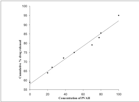
(3) www.ijpsonline.com. The release from the prepared inserts followed matrix diffusion kinetics in all cases, as evidenced by the regression coefficient (r2) values, which were always higher for Q vs. t1/2 (between 0.917 and 0.997) than for Q vs. t (0.774 to 0.936). Similar release kinetics was reported for flurbiprofen19 and indomethacin20 from PVA gels prepared by freeze-thawing and PVA disks respectively. However, the calculated diffusion coefficient (n) values ranged between 0.737 and 0.851, indicating the prevalence of an anomalous release behaviour / mechanism, which seems to be dependent on both matrix swelling and erosion21. The minimal initial burst effect seen from the inserts (fig. 1) containing both PVAL and PVAH in varying proportions could be attributed to the more soluble nature of PVAH in comparison to PVAL. Further, the amount of drug released increased with an increase in the proportion of PVAH in the inserts. Perhaps, the presence of higher proportions of PVAH could have resulted in more pronounced matrix swelling and erosion. This was further confirmed by the fact that the drug release from batches containing PVAH alone (BPC1 to BPC3) was significantly (P < 0.05, t-test) higher than the corresponding batches containing only PVAL (fig. 2). Similar results were reported for indomethacin ocular inserts containing different proportions of PVAH and PVAL15. The effects of three different drug loadings were studied from inserts containing either PVAH or PVAL. The release profiles of the inserts containing PVAH indicated that there was no significant difference between the amounts of drug released at the end of 24 h. However, during the initial phases, a marked decrease in the amount of released drug was observed with decrease in drug loading, presumably due to the. presence of lesser amount of the drug at the periphery with decrease in drug loading. Similar behaviour was observed from the batches containing PVAL (fig. 1). The rate of release was however proportional to the drug loading, showing a significant increase with increase in drug loading. Since not much difference was observed in the release profiles from either of the inserts (PVAL and PVAH), the matrix residue was weighed, after drying on tissue paper, at the end of the dissolution studies. The PVAH inserts showed 35-38% reduction from their original weight, while the PVAL inserts showed a reduction of 22-25%. This was again indicative of the fact that the drug release was controlled by swelling /erosion of the matrix along with diffusion, which explained the independence of drug release to drug loading.. m rf o d ns a lo tio n amost important component that may The plasticizer is the w c i o mechanical l properties of the films as it lowers affect the d b the glass-transition of the polymer. Hence, ufromtemperature e release . drug the plasticized insert (BPC ) was P e ) corresponding unplasticized rcompared f with the wIn theseomtwo batches, both PVAL and PVAHinsert r o (BPC ). are c fo present n . in equal proportions, and any difference in the k pattern e drelease w could be directly related to the presence l o b e absence plasticizer. PVA could be effectively n byof athevariety la M orplasticized k i of plasticizers like PEG 200 and d propylene glycol, glycerol, a by e400, etc. In this study, glycerol v a d .m at 10% concentration was selected as it gave sufficiently is te w pliable films to allow for uniform subdivision into inserts s w F without breaking the film. The amount of drug released o D from BPC was significantly higher than the amount w P te h ( is si h T a. 100. 22. 7. 8. 7. 100 95. 80. 90 85 Cumulative % drug released. Cumulative % derug release. 60. 40. 20. 80 75 70 65 60. 0 0. 5. 10. 15. 20. 25. 55 0. Time (h). Fig. 1: Drug release from inserts containing only PVAL and PVAH In vitro release studies were performed on BPC1-- (-�-), BPC2 (�-), BPC 3 (-�-), BPC 4- (-�-), BPC5 (-�-) and BPC6 (-�-) 628. 20. 40 60 Concentration of PVAH. 80. 100. Fig. 2: Effect of increasing PVAH concentration on drug release at the end of 24 h. Indian Journal of Pharmaceutical Sciences. September - October 2006.
(4) www.ijpsonline.com. released from the unplasticized insert (BPC8). The presence of glycerol in BPC 7 enhanced the hydrophilicity of the PVA matrix. This in turn resulted in enhanced permeability of the inserts, resulting in enhanced swelling and the consequent increase in the porosity of the matrix, thus accounting for higher drug release. S. aureus ATCC 25923, the most common ocular pathogen; and P. aeruginosa ATCC 27853, the most opportunistic ocular pathogen, were used as model organisms in the study. The 0.5 McFarland standard suspensions of S. aureus and P. aeruginosa revealed a count of 1.02×10⁶ and 1.43×10⁶ organisms/ml respectively. The antimicrobial effectiveness of the inserts was tested by varying the volume of the inoculum. The inserts were more effective in inhibiting the growth of P. aeruginosa when compared to S. aureus (Table 2, data shown for 30 and 48 h only). Challenging was done with 20% v/v of the initial inoculum size after 3 h to simulate the number of growing organisms.. When S. aureus was the organism, batches BPC1 and BPC 4 containing 0.5 mg of CPH were effective in inhibiting the growth in all the tubes for 24 h, beyond which growth was observed in two tubes each at 30 and 48 h for batch BPC1 at inoculum volume of 2 and 3 ml. In case of batch BPC4, growth was observed in one tube each at 48 h at an inoculum volume of 2 and 3 ml, indicating that the batch BPC4 composed of only PVAL sustained the drug release more effectively than BPC1 containing the same amount of PVAH. Decrease in the drug loading in the inserts composed of either of the PVA did not produce any remarkable change in the effectiveness of the inserts against the two microorganisms.. m rf o d (BPC ) was unable to inhibit the The unplasticized insert stubes a n growth of S. aureus in all the from the 24 hour (at o o l i 3 ml inoculum level) tonwards, whereas in contrast the n plasticized a insert (BPC ) inhibited the corresponding w c i l in all the tubes for 30 h. This growthoof S. aureus d ubpropensity indicated the of the plasticized insert to e . drug due to the channelling P amount) of theresulting e ofhigher rrelease f effect w the plasticizer, in faster and higher mrelease. r o amount of drug o fo kn .c le edOverall,owwhen S. aureus was the organism, the inserts b nable to inhibit the growth for the entire duration of la M were i thekstudy (48 h) when the inoculum volume was 1 ml. At d inoculum volumes (2 and 3 ml), the inserts were a by ehigher v a to inhibit the growth in all the tubes, showing an si ted w.m unable efficiency of 60-80% from the 24 hour onwards. s w Introduction of one more insert at the 24 hour would F o D have resulted in the inhibition of the growth in all the w P te h ( tubes beyond the 24 hour. In other words, and by s si i implication, the developed inserts have the potential of once-a-day application in the treatment of ocular Th a infections, the various ocular barriers in the ocular 8. th. 7. TABLE 2: IN VITRO MICROBIOLOGICAL EFFICACIES OF THE INSERTS AGAINST P. AERUGINOSA AND S. AUREUS Batch code BPC1 BPC2 BPC3 BPC4 BPC5 BPC6 BPC7 BPC8 BPC9 BPC10 BPC11 BPC12 BPC13 BPC14. Inoculum volume (ml) 2 3 2 3 2 3 2 3 2 3 2 3 2 3 2 3 2 3 2 3 2 3 2 3 2 3 2 3. Time (h) P. aeruginosa S. aureus 30 48 30 48. -5 -5 -4 (+1) -5 -5 -5 -5 -5 -5 -5 -5 -5 -5 -5 -5 -5 -5 -5 -5 -5 -5 -5 -5 -4 (+1) -5 -5 -5 -5. -5 -5 -5 -5 -5 -4 (+1) -5 -5 -5 -5 -5 -5 -5 -5 -5 -5 -5 -5 -5 -5 -4 (+1) -4 (+1) -5 -3 (+2) -5 -5 -5 -5. -3 -3 -4 -4 -4 -3 -4 -4. (+2) (+2) (+1) (+1) (+1) (+2) (+1) (+!) -5 -5 -4 (+1) -3 (+2) -5 -5 -4 (+1) +5 -5 -4 (+1) -4 (+1) -3 (+2) -4 (+1) -4 (+1) -4 (+1) -4 (+1) -3 (+2) -4 (+1) -5 -2 (+3). -3 -3 -3 -3 -3 -3 -4 -4 -4 -4 -4 -3 -4 -4 -3. (+2) (+2) (+2) (+2) (+2) (+2) (+1) (+1) (+1) (+1) (+1) (+2) (+1) (+1) (+2) +5 -5 -4 (+1) -4 (+1) -3 (+2) -4 (+1) -3 (+2) -4 (+1) -4 (+1) -3 (+2) -3 (+2) -4 (+1) -2 (+3). At 1 ml of inoculum level, inserts were able to inhibit test organism. ± indicates presence/absence of growth and the number indicates the number of tubes in which growth was present or absent. (n = 5). September - October 2006. th. th. th. domain notwithstanding. This study describes the complete methodology for the development of the CPH ocular insert, including the in vitro release studies on a flow-through apparatus and analysis of drug release kinetics. The studies also demonstrated the effectiveness of the prepared inserts against two of the most common ocular pathogens (S. aureus and P. aeruginosa). Further, the in vitro release data correlated well with the microbiological efficacy data without having to simultaneously use the rabbit eye model, which, in our opinion, is a substantial improvement over an earlier reported method23. These inserts have the potential to form the basis of a once-daily therapy of. Indian Journal of Pharmaceutical Sciences. 629.
(5) www.ijpsonline.com. ocular infections. However, validation of this model with other categories of antibacterials using other ocular pathogens and correlation with in vivo models are required before it could be viewed as a potential alternative to the in vivo models.. REFERENCES 1. 2.. Schoenwald, R.D. Clin. Pharmacokinet., 1990, 18, 255. Hume, L.R., Lee, H.K., Benedetti, L., Sanzgiri, Y.D., Topp, E.M. and Stella, V.J., Int. J. Pharm., 1994, 111, 295. 3. DiColo, G., Burgalassi, S., Chetoni, P., Fiashi, M.P., Zambito, Y. and Saettone, M.F., Int. J. Pharm., 2001, 215, 101. 4. Kawakami, S., Nishida, K., Mukai, T., Yamamura, K., Nakamura, J., Sakeda, T., Nakashima, M. and Sasaki, H., J. Control. Release, 2001, 76, 255. 5. Sasaki, H., Tie, C., Nishida, K. and Nakamura, J., J. Control. Release, 1993, 27, 127. 6. Baleens, V., Catalos, V., Boisrame, B., Varesio, E. and Gurny, R., J. Control. Release, 1998, 52, 215. 7. Lee, V.H.K. and Robinson, J.R., Int. J. Pharm., 1989, 53, 219. 8. Lee, Y.C., Simamaora, P. and Yalkowsky, S.H., J. Pharm. Sci., 1997, 86, 430. 9. Wood, R., W. Lee, V.H.K., Kreiuter, J. and Robinson, J.R., Int. J. Pharm., 1985, 23, 175. 10. Badenoch, P.B., Hay, G.J., McDonald, P.J. and Coster, D.J.A., Arch. Ophthalmol., 1985, 103, 718. 11. Hobden, J.A., Reidy, J.J., O’Callaghan, R.J. and Hill, J.M., Arch. Ophthalmol., 1988, 106, 1605. 12. Aswad, M.I., Barza, M. and Baum, J., Arch. Ophthalmol., 1989, 107, 1667.. 13. Pavan-Langston, D., Langston, R.H.S. and Geary, P.A., Arch. Ophthalmol., 1975, 93, 1349. 14. Troudale, M.D., Barlow, W.E. and McGuigan, L.J.B., Arch. Ophthalmol., 1989, 107, 1664. 15. Kim, H., Robinson, M.R., Lizak, J.M., Tansey, G., Lutz, R.J., Yuan, P., Wang, N.S. and Csaky, K.G., Inves. Ophtalmol. Vis. Sci., 2004, 45, 2722. 16. Pandit, J.K., Hari Kumar, S.L., Mishra, D.N. and Balasubramaniam, J., Indian J. Pharm. Sci., 2003, 65, 146. 17. Jorgensen, J.H., Turnidge, J.D. and Washington, J.A. In: Murray, P.R., Pfaller, M.A., Tenover, F.C., Baron, E.J. and Yolken, R.H., Eds., Mannual of Clinical Microbiology, 7th Edn., ASM Press, Washington, DC, 1999, 1526. 18. Balasubramaniam, J., ThilekKumar, M., Pandit, J.K. and Kant, S., Pharmazie., 2001, 56, 793. 19. Takamura, A., Ishii, F. and Hikada, S., J. Control. Rel., 1992, 20, 21. 20. Thanoo, B.C., Sunny, M.C. and Jayakrishnan, A., J. Pharm. Pharmacol., 1993, 45, 16. 21. Vazquez, M.J., Perez Macros, B., Gomez-Amoza, J.L., MartinezPacheo, R., Souto, C. and Conchrio, A., Drug. Dev. Ind. Pharm., 1992, 18, 1355. 22. Deamin, R.D., In; Seymour, R.B. Eds., Additives of Plasticizers, Academic Press, New York, 1978, 203. 23. Devarajan, P.V., Bhogte, C.P., Majali, A.B., and Sabharwal, S., Drug Develop. Ind. Pharm., 1999, 25, 781.. m rf o d ns a lo tio n a w c i do ubl e P ). e fr w m r fo kno .co le ed ow b la M dkn i a by e v a si ted w.m s w F o PD te h (w is si h T a. 630. Accepted 13 September 2006 Revised 27 February 2006 Received 16 June 2005 Indian J. Pharm. Sci., 2006, 68 (5): 626-630. Indian Journal of Pharmaceutical Sciences. September - October 2006.
(6)
Figure

Related documents
The layers were separated and the aqueous layer was extracted with ethyl acetate (3×50 mL), the combined organic layers were washed with saturated aqueous sodium
For the field arithmetic we investigate polynomial and normal basis arithmetic for these specific fields; in particular for the challenges on Koblitz curves normal bases become
The attack is performed using only the visible features of encrypted packets exchanged over the network Since web applications use client–server architecture, the
“Reproductive” cloning is defined in order to be outlawed but this term inadvertently reminds us of the position of human cloning within a spectrum of technologies of assisted
On backlighting of the gel mould, both fractures appeared to have a spiral pattern commencing on the medial cortex 5 cm distal to the bullet tract (junction of middle and distal
In this inaugural issue of International Journal of Quality Innovation , the Editor-in-Chief reports the evolution of quality management and the need for innovative research
