Transcriptional Activation by the Mouse Oestrogen Receptor
Paul Shemavon Danielian
Thesis presented in partial fulfilment of the degree of Doctor of Philosophy at the University of London
June 1993
Molecular Endocrinology Laboratory Biochemistry Department Imperial Cancer Research Fund University College London
Lincoln's Inn Fields Gower Street
ProQuest Number: 10018667
All rights reserved
INFORMATION TO ALL USERS
The quality of this reproduction is dependent upon the quality of the copy submitted.
In the unlikely event that the author did not send a complete manuscript and there are missing pages, these will be noted. Also, if material had to be removed,
a note will indicate the deletion.
uest.
ProQuest 10018667
Published by ProQuest LLC(2016). Copyright of the Dissertation is held by the Author.
All rights reserved.
This work is protected against unauthorized copying under Title 17, United States Code. Microform Edition © ProQuest LLC.
ProQuest LLC
789 East Eisenhower Parkway P.O. Box 1346
Abstract
The oestrogen receptor is a ligand inducible transcription factor that belongs to a family of nuclear receptor proteins. Previous studies have shown that in transient transfection experiments two regions of the oestrogen receptor protein are capable of stimulating transcription. One, located N-terminal to the DNA binding domain called transcriptional activation function 1, TAF-1, can stimulate transcription constitutively whilst TAF-2, located in the C-terminal hormone binding domain requires oestrogen binding for its activity. Analysis of mutant mouse oestrogen receptors has identiAed a region of the hormone binding domain important for TAF-2. The amino acid
sequence in this region is conserved in other members of the nuclear receptor family. Point mutation of conserved hydrophobic residues practically abolished hormone dependent transcriptional stimulation by TAF-2 without affecting oestradiol or DNA binding. The ability of TAF-2 and TAF-1 to cooperate in stimulating transcription in the full-length receptor was also abolished by mutation of the conserved hydrophobic residues but not conserved acidic residues. Mutagenesis of the corresponding residues in the glucocorticoid receptor showed similar results indicating that these residues may be important for ligand inducible transcription by other members of the nuclear receptor family.
A yeast genetic screen was established to identify target(s) for TAF-2 that were important for its ability to stimulate transcription. No candidate targets were identified but analysis of the TAF-2 defective mutants suggested that the mechanism whereby TAF-2 stimulates transcription in mammalian cells is not conserved in yeast.
Finally the effects of mutations in the steroid binding domain upon
Acknowledgements.
Contents
Title page. 1
Preface. 2
Abstract. 4
Acknowledgements. 5
Contents. 6
Abbreviations. 12
Chapter 1: Introduction.
Intracellular steroid hormone receptors. 15
Steroid hormone regulated genes. 17
Glucocorticoid responsive genes. 18
Oestrogen responsive genes. 20
Retinoic acid, thyroid hormone and vitamin D 21
response elements.
The nuclear receptor family. 24
Functional analysis of oestrogen and glucocorticoid receptors. 25
DNA binding. 26
Hormone binding. 28
Dimérisation. 29
Transcriptional activation. 30
Identification of transcriptional activation domains. 30 Synergistic transcriptional activation by steroid 33 hormone receptors.
Gene regulation by specific steroid hormones. 35
Eukaryotic transcription factors. 37
Transcription by RNA polymerase H. 38
Transcriptional activation. 40
Chromatin and transcriptional activation. 42
In vitro analysis of the role of chromatin in transcription. 42 Genetic analysis of the relationship between chromatin and 43 transcription in yeast.
Chapter 2: Materials and Methods. Materials.
Chemicals. 47
Radiochemicals. 48
Enzymes. 48
Plasmids. 49
Miscellaneous. 49
Buffers. 50
Bacterial media and plates. 52
Yeast media and plates. 52
Cell culture media. 52
Methods.
Bacterial Transformation. 53
Storage of bacteria. 53
Preparation of competent bacteria. 53
Transformation. 53
Preparation of plasmid DNA. 53
Small scale plasmid preparation (mini-prep). 53
Large scale plasmid preparation. 54
DNA manipulation and subcloning. 55
Restriction endonuclease digestion. 55
Agarose gel electrophoresis. 55
Purification of restriction fragments. 55
Preparation of vectors. 56
Oligonucleotide kinasing and annealing. 56
Ligations. 56
Polymerase chain reactions. 56
DNA sequencing. 57
Preparation of DNA and sequencing reactions. 57
Electrophoresis of sequencing reactions. 57
In vitro protein analysis. 57
Complementary RNA synthesis. 57
In vitro translation. 58
SDS polyacrylamide gel electrophoresis. 58
Western blotting. 59
Ligand binding. 60
Band shift assay. 60
Yeast transformation and library screening. 61
Yeast transformation. 61
Library screening. 62
Colony colour assay. 62
Plasmid curing. 63
Liquid assay for P-galactosidase activity. 63
Yeast protein extract preparation. 64
Cell culture methods. 64
Maintenance of cell stocks. 64
Charcoal treatment of serum. 65
Transient transfection. 65
Calcium phosphate precipitation. 65
Electroporation. 66
DEAE-dextran. 67
Whole cell steroid binding assay. 67
Harvesting cell monolayers. 68
Assay of cytoplasmic luciferase activity. 68
Assay of cytoplasmic CAT activity. 68
Whole cell protein extract. 69
Indirect immunofluoresence. 69
Chapter 3: Functional analysis of a conserved region required for hormone dependent transcriptional activation by the mouse oestrogen and glucocorticoid receptors.
Introduction. 71
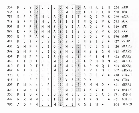
IdentiEcation of a conserved sequence. 71
Functional analysis of the conserved region. 73
Mutagenesis strategy. 73
Transient transfection assays. 74
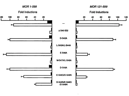
Transcriptional activation by mutant receptors. 74
Mutations in the conserved region appear not to affect other 77 receptor functions.
Assessment of wild-type and mutant receptor protein levels 77 in transfected cells.
Expression of receptors in COS-1 cells. 77
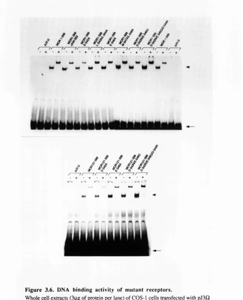
Examination of the mutant receptor proteins in the band shift assay. 83 Analysis of the ability of the mutant receptor proteins to 83 bind oestradiol.
Mutations in the conserved region affect TAF-2 when attached 85 to a heterologous DNA binding domain.
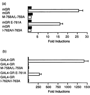
Analysis of the corresponding glucocorticoid receptor mutants. 86 Analysis of GAL4-glucocorticoid receptor chimeras. 87 Mutation of the conserved amino acids appear not to affect other 87 glucocorticoid receptor functions.
Analysis of the cooperation between the N-and C-terminus of the 91 mouse oestrogen receptor in stimulating gene transcription.
Assessment of transcriptional activation by TAP-1. 91
Cooperation in the wild-type receptor. 93
Mapping the region of the N-terminus important for cooperation. 95
Conclusions. 100
Chapter 4: A yeast genetic screen for proteins that interact with TAF-2 of the mouse oestrogen receptor.
Introduction. 102
Choice of strategy. 102
Indirect immunoprécipitation and affinity column 103 chromatography.
Screening A,gtl 1 libraries with labelled protein. 104
A genetic screen in yeast. 104
The genetic screen in yeast. 105
Analysis of SRF-ER in yeast 107
Screening the library. I l l
Testing SRF-ER in mammalian cells. 113
Conclusions. 117
Chapter 5: Examination of m utant mouse oestrogen receptors with altered ligand specificity.
Introduction. 119
Functional analysis of receptors containing mutations in the 119 hormone binding domain.
Analysis of oestradiol binding by the mutant receptors. 119 Transcriptional activation by the mutant receptors in NIH 3T3 cells. 120 Transcriptional activation by the mutant receptors in chicken embryo 123 fibroblast cells.
Oestradiol binding by H-520, A, M-521 appears to be temperature 126 sensitive.
Analysis of DNA binding by receptors in the presence of ligands. 129
Conclusions. 132
Chapter 6: Discussion.
The hormone dependent transcriptional activation function in 134 nuclear receptors.
A "TAF-2 like" activity in steroid hormone receptors. 134 Thyroid hormone, retinoic acid and retinoid X receptors. 137 The deletion of the conserved region in v-erbA abolishes 138 hormone dependent transcriptional activation.
The ecdysone receptor and orphan receptors. 139
Cooperation between the N-and C-terminal activation domains 142 in stimulating transcription.
Ligand and the activity of TAF-2. 146
Ligand binding and receptor function. 149
The requirement of ligand for DNA binding. 151
The genetic screen in yeast. 155
TAF-2 of the human oestrogen receptor in yeast. 157
Possible target(s) for the glucocorticoid receptor in yeast. 159 Possible target(s) for nuclear receptors identified in vitro. 159
Appendix. 162
Bibliography. 175
List of Figures and Tables.
Chapter 1.
1.1 DNA binding sites for nuclear receptors. 22
Chapter 3.
3.1 Sequence alignment of nuclear receptor proteins. 72 3.2 Transcriptional activation by mutant oestrogen receptors. 75
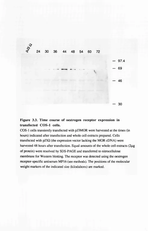
3.3 Time course of oestrogen receptor expression in 79
transfected COS-1 cells.
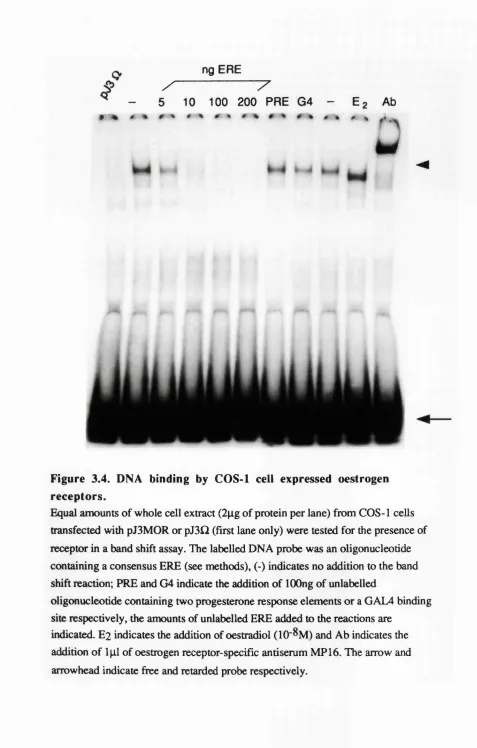
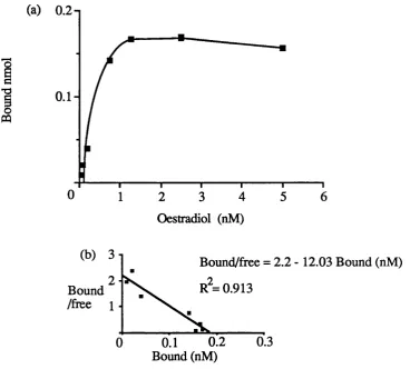
3.4 DNA binding by COS-1 cell expressed oestrogen receptors. 80 3.5 Oestradiol binding by the wild-type oestrogen receptor. 81
3.7 Transcriptional activation by the wild-type and mutant mouse glucocorticoid receptors.
88
3.8 In vivo interference assay. 90
3.9 Analysis of TAF-1 activity in NIH 3T3 cells. 92
3.10 Cooperation between the N-and C-terminal domains in transcriptional activation.
94
3.11 Mapping the region in the N-terminus important for cooperation. 96
3.12 DNA binding by N-terminal deletion mutants. 97
3.13 Analysis of the requirements for cooperation between the N-and C-terminal activation domains.
99
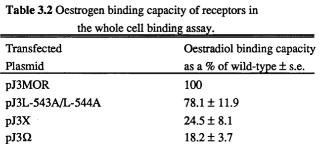
Table 3.1 Oestradiol binding by wild-type and mutant receptors. 84 Table 3.2 Oestradiol binding capacity of receptors in the whole cell 84
binding assay.
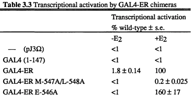
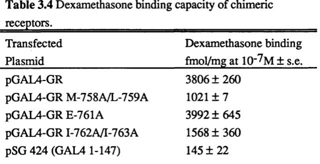
Table 3.3 Transcriptional activation by GAL4-ER chimeras. 86 Table 3.4 Dexamethasone binding capacity of chimeric receptors. 89
Chapter 4.
4.1 Yeast screen strategy. 106
4.2 Transcriptional activation by SRF-ER chimeric proteins in yeast. 109 4.3 Transcriptional activation by SRF-ER in yeast is oestradiol dependent. 110
4.4 Expression of SRF-ER chimeras in yeast. 112
4.5 Transcriptional activation by SRF-ER chimeras in NIH 3T3 cells. 115
4.6 DNA binding activity of SRF-ER chimeras. 116
Chapter 5.
5.1 Transcriptional activation by mutant oestrogen receptors. 121 5.2 Analysis of transcriptional stimulation by G-525R in the presence 125
of oestradiol and 4-hydroxytamoxifen.
5.3 Analysis of the relative levels of transiently expressed wild-type 128 and mutant receptors.
5.4 DNA binding activity of the wild-type and mutant receptors 130 expressed in COS-1 cells.
5.5 The effects of ligand on the ability of the wild-type and mutant 131 receptors to bind DNA.
Table 5.1 Oestradiol binding by wild-type and mutant receptors. 120
Chapter 6.
Abbreviations. ATP bp BSA CAT cDNA CEF cpm CTD C-terminal C Y C 1 dATP dCTP dGTP dTTP DCC DEAE DEPC DMEM DMSO DNA DNase DTT EDTA ER ERE PCS GRE GTP HEPES hsp kb Kd l a c Z MMTV MOPS MGR mRNA adenosine 5'-triphosphate base pair
bovine serum albumin
chloramphenicol acetyltransferase complementary DNA
chicken embryo fibroblast counts per minute
carboxyl-terminal domain carboxyl-terminal
iso-1-cytochrome C gene
2'-deoxyadenosine-5’-triphosphate 2’-deoxycytidine-5’-triphosphate 2'-deoxyguanosine-5’-triphosphate 2'-deoxythymidine-5'-triphosphate dextran coated charcoal
diethylaminoethyl diethyl pyrocarbonate
Dulbecco's modified Eagle's medium dimethyl sulphoxide deoxyribonucleic acid deoxyribonuclease 1,4-dithiothreitol ethylenediaminetetraacetic acid oestrogen receptor
oestrogen response element foetal calf serum
glucocorticoid response element guanosine 5'-triphosphate
N-2-hydroxyethylpiperazine-N'-2-ethanesulphonic acid heat shock protein
kilobase
dissociation constant p-galactosidase gene
mouse mammary tumour vims morpholinopropanesulphonic acid mouse oestrogen receptor
NP40 N-terminal
ODx
ONPG PAGE PBSA PCR PEG PMSF PRE RAR RNA RNase RXR S SDS SRE SRF SV40 T3 TAP TBP TEMED tk Tris Triton X-100 Tween 20 X-gal nonidet P40 amino-terminaloptical density at a wavelength of x nm o-nitrophenyl-p-D-galactopyranoside polyacrylamide gel electrophoresis phosphate buffered saline A polymerase chain reaction polyethylene glycol
phenylmethylsulphonylfluoride progesterone response element retinoic acid receptor
ribonucleic acid ribonuclease retinoid X receptor
Svedberg units
sodium dodecyl sulphate serum response element serum response factor simian virus 40
3, 5, 3'-triiodo-L-thyronine
transcriptional activation function TATA binding protein
N'NW'N'-tetramethylethylenediamine thymidine kinase
Chapter 1
The survival of most multicellular organisms relies on the ability of cells to respond to intercellular signalling molecules. In many cases these signalling
molecules bind to receptors that cause changes in the expression of appropriate genes. Receptors for intercellular signalling molecules located at the cell surface induce changes in gene expression indirectly through other molecules that "transmit" the signal to the nucleus. However, some signalling molecules, that are able to diffuse across cell membranes, bind to intracellular receptors that function as transcription factors, regulating gene expression directly. This thesis describes a study of one intracellular receptor, that for the steroid hormone oestradiol.
The oestrogen receptor belongs to a large family of nuclear hormone receptors that contains receptors for steroid hormones, vitamin D, retinoids and thyroid
hormones that play important roles in growth, development and homeostasis. The receptors for the gonadal steroid hormones, oestrogens, progestins and androgens are essential for the development and function of mammalian reproductive systems (King and Mainwaring, 1974; Cunha et al.^ 1991). The adrenal steroid hormones,
glucocorticoids, regulate carbohydrate and protein metabolism and the
mineralocorticoids control water and electrolyte metabolism whilst vitamin D is important for bone formation (Hughes and O' Malley, 1991) and the absorption of calcium ions and inorganic phosphate from the intestine. The insect steroid hormone ecdysone also binds a member of the nuclear hormone receptor family and initiates changes in development during moulting and metamorphosis (Andres and Thummel,
1992). The affects of retinoids and thyroid hormones, that are important for the growth and development of mammals are also mediated by members of the nuclear receptor family (reviewed in Ragsdale and Brockes, 1991; Chaterjee and Tata, 1992; Maden and Holder, 1992).
Intracellular steroid hormone receptors.
Although steroid hormones are capable of entering most cells physiological responses are restricted to certain tissues. By following the fate of injected
Jensen et al., 1968). These observations gave rise to the two-step model which proposed that receptor in the cytoplasm (the 9 S form) bound oestradiol and then migrated to the nucleus where it was detected, transformed to a 5 S form. Similar observations were also made of the progesterone, androgen and glucocorticoid receptors and further studies suggested that the 4-5 S form of the receptor bound to DNA or chromatin (reviewed in King and Mainwaring, 1974).
Biochemical analysis indicated that the 8-9 S forms of the steroid receptors contained a common component (Joab et j/., 1984; Schuh et al., 1985) that was subsequently identified as the heat shock protein hsp90 (for examples see Catelli
etal.y 1985; Snachez et at., 1985; Denis et at., 1987; Redeuilh etal., 1987; Sanchez
et al.y 1987). Ligand binding and/or thermally induced transformation of the receptor to the 5 S form resulted in the dissociation of hsp90 (Sanchez et at., 1985; Denis
etal.y 1987; Redeuilh etal., 1987; Sanchez et a l, 1987; Denis et al., 1988; Howard and Distelhorst, 1988; DeMarzo et a i, 1991). This transformation process also appeared to involve receptor dimérisation (Miller et al., 1985b; DeMarzo et al.,
1991). Pulse-chase labelling experiments using methionine (Howard and
Distelhorst, 1988) showed that labelled hsp90 could be co-immunoprecipitated with the glucocorticoid receptor from extracts of untreated cells but not from cells treated with dexamethasone, a synthetic glucocorticoid. Rexin etal., (1988,1992) showed that the glucocorticoid receptor could be cross-linked to hsp90 when the cross-linking was performed using intact cells. These two reports suggest that the glucocorticoid receptor associated with hsp90 in vivo rather than, non-specifically, during the extraction procedure.
A major role of hsp90 appears to be to maintain the receptor in an inactive state in the absence of ligand. On the basis of other studies hsp90 may also be important for folding of the receptor protein and/or transport across membranes (reviewed in Smith and Toft, 1993). In the case of the glucocorticoid receptor the binding of hsp90 appears to be important for high affinity ligand binding (Bresnick
et al.y1989; Nemoto et al., 1990). All these possibilities are supported by studies of yeast containing exogenous glucocorticoid and oestrogen receptors where lowering the level of hsp90 reduces the ability of the receptor to stimulate transcription (Picard
Although cell fractionation experiments suggested that steroid receptors were located in the cytoplasm and migrated to the nucleus upon the binding of ligand immunocytochemical studies have indicated that the oestrogen (King and Greene,
1984; Picard et al.y 1990b) and progesterone receptors are predominantly nuclear in the absence or presence of ligand (for examples see Perrot-Applanat, 1985;
Guiochon-Mantel et al.y 1989). In contrast, the glucocorticoid receptor is
predominantly cytoplasmic and translocates to the nucleus in the presence of ligand (Govindan, 1980; Papamichail et al.y 1980; Picard and Yamamoto, 1987; Wikstrom et al.y 1987). In the cases of the oestrogen and progesterone receptor the cell
fractionation results may reflect "tight nuclear binding" in the presence of ligand. Nuclear localisation signals have been identified in the glucocorticoid (Picard and Yamamoto, 1987), progesterone (Guiochon-Mantel e t a l . y 1989) and oestrogen receptors (Picard et al.y 1990b; Ylikomi et al.y 1992). One, that is conserved in many members of the nuclear receptor family, located near the C-terminus of the DNA binding domain is composed of groups of basic amino acids (reviewed in Dingwall and Laskey, 1991) whilst a second may exist in the hormone binding domain and require the binding of hormone for its activity. Recent studies indicate that the progesterone, glucocorticoid and oestrogen receptors cycle between the nucleus and the cytoplasm (Guiochon-Mantel e t al.y 1991; Madan and DeFranco, 1993;
S. Dauvois, ICRF, personal communication). The differences between the localisation of receptors may be related to the ability of the hormone binding domain to repress (in the absence of ligand) the function of the nuclear localisation signal near the DNA binding domain (see Ylikomi e t al.y 1992) thus altering the efficiency of receptor import into the nucleus.
Steroid hormone regulated genes.
Initial biochemical analysis of steroid hormone induced proteins indicated that one of the first changes induced by the hormone was an increase in the amount of RNA and that the induction could be prevented by inhibitors of transcription such as actinomycin D (for examples see Walters e t al.y 1985). These data along with
evidence that indicated that the transformed receptor was able to bind DNA suggested that steroid receptors may be able to stimulate transcription of specific genes.
Following the examples of prokaryotic and viral transcription factors the possibility that high affinity DNA binding sites for receptors were located near responsive genes was examined.
Using partially purified glucocorticoid receptor Payvar e t al.y (1981) showed, by filter binding assay, that the receptor bound specifically to a cloned DNA fragment of the mouse mammary tumour virus (MMTV) that was known to be hormone
caused an increase in the amount of mRNA from several MMTV genes by increasing the efficiency of initiation of transcription (Ringold et al., 1977; Ucker and
Yamamoto, 1984). The initial report of Payvar et al., (1981) was followed by the analysis of mainly glucocorticoid and oestrogen responsive genes that will be reviewed in the following sections.
Glucocorticoid responsive genes.
As indicated above some of the initial analyses were performed using cloned MMTV DNA that contained genes whose transcription was increased by
glucocortcoids in vivo (Ringold etal., 1977; Young etal., 1977; Grove etal., 1980). High affinity binding sites for the glucocorticoid receptor were found in the long terminal repeat (LTR) sequences upstream of the promoter as well as positions far from the promoter (Geisse et al., 1982; Govindan et al., 1982; Pfahl et al., 1982; Payvar et al., 1983). Scheidereit et al., (1983) mapped some of these binding sites in the LTR to between positions -202 and -50 relative to the transcription initiation site. The ability of these sequences to confer hormone induced transcription upon other genes was confirmed in transfection experiments (Buetti and Diggelmann, 1981; Lee
et al., 1981; Ponta et al., 1985) and deletion analysis identified a DNA fragment of a few hundred base pairs 5' of the MMTV promoter that conferred glucocorticoid induction of gene transcription upon the Herpes simplex virus thymidine kinase gene promoter (Chandler et al., 1983; Hynes etal., 1983; Majors and Varmus, 1983). This sequence conferred hormone responsive transcriptional activation when joined to the promoter in either orientation or when placed over 1 kb upstream (Chandler et al.,
1983) thus functioning as a classical enhancer (Muller et al., 1988).
The positions of the glucocorticoid receptor binding sites in responsive genes were determined by DNase I footprinting and méthylation interference in the mouse mammary tumour virus DNA (Payvar et al., 1983; Scheideriet et al., 1983;
Scheideriet and Beato, 1984), sequences upstream of the human metallothionein-IlA
gene (Karin et al., 1984), the lysozyme (Renkawitz et al., 1984) and uteroglobin genes (Cato et al., 1984) and in the first intron of the growth hormone gene (Moore
et al., 1985). Based on these studies the binding site for the glucocorticoid receptor was suggested to be 15 base pairs that contained an imperfect inverted repeat of TGTTCT with a three nucleotide gap between the repeats (Scheidereit et al., 1986). Subsequent studies showed that a 15 bp glucocorticoid response element, GRE, was sufficient to confer glucocorticoid induction upon a heterologous promoter (Klock
MMTV LTR between positions -201 and -58 were important (Buetti and Kuhnel, 1986; Kuhnel et al.j 1986; Chalepakis et a/., 1988).
This region of the MMTV LTR contains four potential binding sites for the glucocorticoid receptor one distal to the promoter, centred at position -177 and three proximal, centred at positions -120, -100 and -85. The distal sequence is an imperfect inverted repeat of the TGTTCT sequence whilst the proximal sites lack an inverted repeat in the correct position also the number of receptors that bind to the proximal sites is unclear (Chalepakis e to/., 1988). The deletion and linker scanning
mutagenesis described above also showed that mutation of the binding site of another transcription factor, nuclear factor I (CTF/NFI), located at position -70 reduced the level of induction approximately 10-fold. The importance of the CTF/NFI binding site was confirmed by mutagenesis and transient transfection experiments using cells with low CTF/NFI levels (Cato e t al.y 1988a; Bruggemeier e t al.y 1990). Two binding sites for another transcription factor Oct-1 have also been found in the MMTV
promoter between positions -57 and -37 (note that these were not present in the sequences tested by some groups) and the mutation of the distal site reduces the level of glucocorticoid induction 4-to 5-fold whilst mutation of the proximal site has little affect (Bruggemeier e t a l . y 1991). Mutation of the CTF/NFI and Oct-1 binding sites virtually abolishes induction in response to glucocorticoids (Bruggemeier e t al.y
1991).
Glucocorticoid response elements have also been found near other genes whose transcription is increased by glucocorticoids. In the tryptophan oxygenase gene response elements were found 450 and 1,200 base pairs upstream of the site of initiation of transcription (Danesch e t al.y 1987). The DNA sequence near position -450 also contains a binding site for the CACCC box binding factor that is able to cooperate with the glucocorticoid receptor to stimulate transcription (Danesch e t al.y
1987; Schule e t al.y 1988b). In the case of the tyrosine aminotransferase (TAT) gene two GREs were found approximately 2.5 kb upstream of the cap site (Becker e t al.y
expression (reviewed in Nitsch et al.y 1991).
Further studies indicated that a GRE and a TATA box were sufficient to obtain hormone induced stimulation of gene transcription only if the GRE was close to the TATA box (Bradshaw et a l . , 1988; Ham et al.y 1988; Strahle et al.y 1988). The progesterone (Thomson et al.y 1990; Meyer et al.y 1992) and oestrogen receptors (Tora et al.y 1989b) can also stimulate transcription from promoters containing the receptor binding site and a TATA box only. Hormone dependent transcriptional activation when binding sites were located far from the TATA box could be obtained using two or more GREs or a GRE in combination with a binding site for another transcription factor (Schule et al.y 1988a, b; Strahle et al.y 1988). These results suggested that the glucocortcoid receptor could cooperate with other transcription factors to stimulate transcription.
Surprisingly the same DNA sequences identified as GREs could also mediate an increase in gene transcription in response to progestins, androgens and
mineralocorticoids, for example those in the MMTV LTR were shown to be responsible for induction by all three of these steroids (for examples see Cato et a l.,
1986; Darbre et al.y 1986; Arriza et al.y 1987; Chalepakis et al.y 1988; Ham et al.y 1988) whilst a GRE alone was shown to be sufficient to confer progesterone and glucocorticoid and androgen induction of transcription upon a heterologous promoter (Strahle et al.y 1987; Ham et al.y 1988). Although the DNA sequence of the binding sites for these receptors are similar DNase I footprinting and méthylation protection studies have shown differences between the binding of the progesterone and
glucocorticoid receptors to the MMTV LTR (von der Ahe et al.y 1986; Chalepakis et al.y 1988). Chalepakis et al.y (1988) and Cato et al.y (1988a) also identified point mutations in the MMTV LTR that did not significantly alter the affinity of the receptors for the binding sites but affected the induction in response to progestins, glucocorticoids and possibly androgens differently.
Oestrogen responsive genes.
In oviparous vertebrates the synthesis of yolk proteins such as vitellogenin and ovalbumin is induced by oestradiol (for a review see Whali, 1988). Following the cloning of the vitellogenin genes A l, A2, B1 and B2 from Xenopus laevis and the chicken vitellogenin II and apo-very low density lipoprotein (VLDL) II genes, Walker
et al.y (1984) derived a putative sequence for an oestrogen response element (ERE) by searching for similar sequences in the 5' flanking regions of these genes.
The authenticity of the sequence suggested by Walker et al.y (1984) was verified by subsequent functional analysis of the Xenopus vitellogenin A2 gene that identified 35 base pairs containing the sequence 5 -GGTCACAGTGACC-3' that conferred
Functional analysis of the 5’ flanking region of the Xenopus vitellogenin B 1 gene identified a 33 base pair sequence that was able to confer oestrogen inducible transcription when placed in either orientation upstream of a heterologous promoter or when placed 1.7 kb downstream of the reporter gene (Seiler et al.y 1986; Martinez et al.y 1987; Martinez and Wahli, 1989). This sequence contained two 13 bp imperfect inverted repeats both of which were required for oestrogen induced stimulation of transcription. The ERE described by Klein-Hitpass et al.y (1986) contained a palindrome of the sequence TGACC similar to that of a GRE, in fact changing two base pairs in the palindrome of the ERE 5'-AGGTC ACAGTG ACCT-3* to
5 -AGAACACAGTGTTCT-3' (Figure 1.1, see later) is sufficient to convert the ERE into a GRE (Klock et al.y 1987; Martinez et al.y 1987). Further mutational analysis of the ERE from the vitellogenin A2 gene identified base pairs important for receptor binding in vitro and oestrogen receptor induced transcription in vivo based on these results the sequence GGTCANNNTGACC was proposed as a consensus ERE where N indicates any nucleotide (Klein-Hitpass et al.y 1988).
Oestrogen response elements have also been found in sequences flanking other oestrogen responsive genes such as the chicken ovalbumin gene (Tora et al.y
1988a) and the human pS2 gene (Berry et al.y 1989). These EREs, however, are not perfect inverted repeats. In the case of the ovalbumin gene two regions 5' of the start of transcription have been identified as being important for oestrogen regulation. One is located close to the transcription start site and contains two GGTCA motifs (Tora
et al.y 1988a) whilst the second is located 3.3 kb from the transcription start site contains four half-palindromes (TGACC sequences) in a region of 675 bp (Kato
et al.y 1992). Combining these two elements results in a highly oestrogen inducible promoter (~80-fold induction). In combination with the distal element that functions as an ERE the proximal element can be replaced by the binding site for other
transcription factors suggesting that the oestrogen receptor can cooperate with other transcription factors to stimulate transcription (Kato et al.y 1992). The proximal ERE in the pS2 gene can confer oestrogen induction upon a heterologous promoter
although the level of induction is 3-4 times lower than that conferred by a consensus ERE (Berry et al.y 1989). This may be a reflection of the fact that the affinity of the oestrogen receptor for this ERE, as judged in vitroy is approximately 5 times lower than that for a consensus ERE (Kumar and Chambon, 1988; Berry et a/., 1989).
Retinoic acid, thvroid hormone and vitamin D response elements.
DNA binding site sequences Receptors
AGAACAnnnTGTTCT GR, PR, AR, MR.
AGGTCAnnnTGACCT ER.
AGGTCAnAGGTCA RXR, COUP-TF/RXR
RAR/RXR, RXR/PPAR.
AGGTCAnnAGGTCA RAR/RXR.
AGGTCAnnnAGGTCA VDR/RXR.
AGGTCAnnnnAGGTCA TR/RXR.
AGGTCAnnnnnAGGTCA RAR/RXR.
AAAAGGTCA NGFI-B.
Figure 1.1. DNA binding sites for nuclear receptors.
Idealised DNA binding sites for the following nuclear receptors are shown:
glucocorticoid receptor (GR), progesterone receptor (PR), androgen receptor (AR), mineralocorticoid receptor (MR), oestrogen receptor (ER), retinoid X receptor (RXR), chicken ovalbumin upstream transcription factor (COUP-TF), retinoic acid receptor (RAR), peroxisome proliferator-activated receptor (PPAR), vitamin D receptor (VDR), thyroid hormone receptor (TR), nerve growth factor induced clone B
spaced by 3 ,4 or 5 nucleotides respectively (note that EREs are also composed of similar sequence arranged as an inverted repeat with a three nucleotide gap, see Figure 1.1). Further studies, however, have shown that this is not always the case and is further complicated by the fact that vitamin D, thyroid hormone and retinoic acid receptors can bind DNA as heterodimers with the retinoid X receptor, RXR (for examples see Yu et al.y 1991; Bugge et al.y 1992; Kliewer et al.y 1992b; Leid et al.y 1992b; Marks et al.y 1992; Rosen et al.y 1992; Zhang et al.y 1992). For example the studies of Carlberg et al.y (1993) showed that maximal transcriptional activation by the vitamin D receptor from a response element (from the osteopontin gene) occurred in the presence of RXR, suggesting that the presence of heterodimers, whilst maximal transcriptional activation from a different response element (found in the osteocalcin gene) occurred in the presence of the vitamin D receptor only suggesting that this element was bound as a homodimer. The retinoid X receptors bind a naturally occurring isomer of aH-trans retinoic acid, 9-cis retinoic acid (Levin et al.y 1992; Heyman et al.y 1992) whilst the retinoic acid receptor binds both these molecules.
Response elements found in retinoic acid regulated genes are often composed of direct repeats of PuGGTCA as well as PuGTTCA sequences separated by 1, 2 ,4 or 5 nucleotides. Pu indicates a purine nucleotide (for examples see Leid e t al., 1992a;
Mader e t al.y 1993a). In transfection experiments the highest levels of transcriptional activation are often observed from direct repeats separated by 1,2 or 5 nucleotides (Durand e t al.y 1992; Mader e t al.y 1993a). In these cases it is possible that the retinoic
acid receptor (RAR) is bound to DNA as a heterodimer with RXR. RXR alone appears to preferentially act through to direct repeats spaced by only 1 or 2
nucleotides (Manglesdorf e t al.y 1991; Mader e t al.y 1993a). In the case of the thyroid
hormone receptor natural response elements are often composed of direct imperfect repeats of AGGTCA or AGGTAA sequences spaced by 4 nucleotides but other arrangements do exist (Umesono etal.y 1991; Kim e t a l . , 1992; Williams e t al.y 1992).
For example in the rat growth hormone gene a response element 5' to the gene is composed of a direct repeat (spaced by 4 nucleotides) as well as an inverted repeat with no nucleotide gap (described in Kim e t al.y 1992) whilst a response element in the third intron is composed of two direct repeats with no nucleotide gap (Sap e t al.y
1990).
Functional analysis of orphan receptors (members of the nuclear receptor family for which no ligand has been identified) has indicated that some also bind repeats of sequences similar to AGGTCA. For example COUP-TF (chicken
1992; Kliewer et al., 1992a; Tran et al., 1992) and the orphan receptor HNF-4 (Ladias
et al., 1992; Mietus-Snyder et al., 1992). COUP-TF and ARP-1 can also heterodimerise with RXR and therefore may repress transcription by direct
competition for the DNA binding site or possibly by competing for RXR (Berrodin
et al., 1992; Kliewer et al., 1992a; Widom et al., 1992). The orphan peroxisome proliferator-activated receptors appear to bind imperfect direct repeats of AGGTCA sequences spaced by one nucleotide (Dryer et al., 1992; Kliewer et al., 1992c; Tugwood et al., 1992) possibly as heterodimers with RXR (Kliewer et al., 1992c; Gearing et al., 1993; Keller et al., 1993). In regulated genes response elements composed of direct repeats can be found in either DNA strand, whether this influences the response is as yet unclear.
Interestingly some orphan receptors bind to slightly different response
elements. For example NGFI-B and Rev-ErbAa bind elements composed of a single GGTCA sequence preceded by a short A/T rich sequence (Wilson et al., 1991; Harding and Lazar, 1993) whilst FTZ-Fl and ELP/SF-1 bind PyCAAGGPyC based sequences and Ad4BP binds this and PuPuAGGTCA sequences. Pu and Py indicate purine and pyrimidine nucleotides respectively (described in Lala et al., 1992; Tsukiyama et al., 1992; Honda et al., 1993). Some idealised DNA binding sites for some nuclear receptors are shown in figure 1.1.
The nuclear receptor family.
A comparison of the predicted protein sequences of the glucocorticoid, oestrogen and thyroid hormone receptors identified several regions of similar sequence suggesting that these proteins may be members of a superfamily of ligand inducible transcription factors. On the basis of this sequence homology other cDNA clones were isolated that encoded similar proteins. These included retinoic acid receptors and a number of orphan receptors for which, as yet, a ligand has not been identified (for reviews see Evans, 1988; Green and Chambon, 1988; Beato, 1989; Ham and Parker, 1989; Oro et al., 1992). To date over 30 members of the nuclear receptor family have been cloned that may have evolved from a single common ancestor (Amero et al., 1992; Laudet et al., 1992).
All members of the nuclear receptor family, on the basis of sequence
homology, have a similar DNA binding domain composed of two zinc finger motifs and in most cases contain a C-terminal ligand binding domain. The high sequence homology in the DNA binding domain is reflected in the similarity of the sequences bound by these receptors (described above). Functional analysis of the members of the nuclear receptor family has also indicated similarities in their mechanisms of action, these studies are described elsewhere in this chapter and chapter 6. In contrast, however, the region N-terminal to the DNA binding domain is, in general, poorly conserved in sequence or size between members of the nuclear receptor family. For example in the vitamin D receptor this region is composed of only 24 amino acids in comparison to 603 amino acids in the mineralocortcoid receptor (Evans, 1988). This region may therefore be important for receptor, tissue and/or gene specific regulation of transcription by nuclear receptors.
Functional analysis of oestrogen and glucocorticoid receptors.
The first members of the nuclear receptor family for which cDNA clones were isolated included the human glucocorticoid (Hollenberg et al., 1985) and oestrogen receptors (Green et al., 1986). The aim of this section is to review the subsequent analyses of mainly these receptors to provide a general idea of nuclear receptor structure and function.
A cDNA encoding the human glucocorticoid receptor was isolated in 1985 using specific antiserum to screen an expression library made from a lymphoid cell line (Hollenberg et al., 1985). The protein encoded by this cDNA, called hORa, was synthesised in vitro and shown to bind glucocorticoids with high affinity and
specificity. Hollenberg et al., (1985) also isolated a second clone (hORp) that encoded a protein that had a different C-terminus firom hORa. This protein failed to bind glucocorticoids suggesting that the hormone binding domain was located in the C-terminus of the molecule. Analysis of the predicted protein sequence indicated that the C-terminal half of the glucocorticoid receptor was related to the product of the
v-erbA gene found in avian erythroblastosis virus (see Krust et al., 1986).
The human oestrogen receptor cDNA, cloned by Green et aL, (1986) also encoded a protein that was related to the product of the v-erbA gene. Comparison of the protein sequences of the human and chicken oestrogen receptors, v-erbA and the glucocorticoid receptor revealed that the cysteine, lysine and arginine rich region was highly conserved between all these proteins and that a slightly less well conserved region, suggested to be a ligand binding domain, was located C-terminal to the
putative DNA binding domain (see Krust et a/., 1986). In the former region 9 cysteine residues were conserved in all of these proteins and these were proposed to form two zinc Angers.
On the basis of the homology between the chicken and human oestrogen receptors the protein sequences were divided into six regions, A to F (Krust et a/.,
1986). The clones for the cellular homologues of v-erbA were also isolated and found to encode receptors for thyroid hormones (Sap et al., 1986; Weinberger et al., 1986). Following the successful expression of functional receptors from these cDNA clones in heterologous cells and in vitro mutant cDNA clones were generated to assess the role of different regions of the receptor proteins.
DNA binding.
The highly conserved cysteine rich region, described as region C by Krust
et al., (1986), was first shown to contain the protein sequences necessary for target gene recognition by "domain swapping" experiments (Green and Chambon, 1987). In these experiments region C of the oestrogen receptor was replaced with that of the glucocorticoid receptor. This chimeric receptor, in the presence of oestradiol,
stimulated the transcription from a glucocorticoid but not oestrogen responsive reporter plasmid. The converse replacement was subsequently shown to confer dexamethasone inducible transcription from an oestrogen responsive element upon a chimeric glucocorticoid receptor (Kumar et al., 1987). Green and Chambon, (1987) also showed that the replacement of two highly conserved cysteines with other amino acids in region C of the oestrogen receptor abolished the stimulation of gene
transcription but not oestradiol binding.
By analysing the ability of rat glucocorticoid receptor mutants to co-immunoprecipitate a fragment of the MMTV-LTR DNA containing GREs Rusconi and Yamamoto, (1987) showed that approximately 140 amino acids encompassing the cysteine rich region was sufficient to bind specifically to MMTV LTR DNA with a similar efficiency to that of the wild-type receptor. One mutation that replaced four amino acids, including one of the highly conserved cysteines, abolished DNA binding verifying the importance of the cysteine rich region.
Structural analysis of a fragment of the rat glucocorticoid receptor containing the DNA binding domain using visible light spectroscopy and extended X-ray absorption fine structure spectroscopy suggested that this polypeptide coordinated two zinc ions each of which was bound by four cysteine residues (Freedman et al.j
1988). The binding of zinc was also shown to be required for the folding of this polypeptide into a DNA binding form. The replacement of 8 of the 9 conserved cysteine residues with serine and/or alanine abolished receptor function whilst replacement of some of the critical cysteines residues with histidine residues, that are able to coordinate zinc ions, did not abolish function (Seveme et a/., 1988).
Mutational analysis of region C in the oestrogen receptor indicated that both putative zinc fingers were required for the ability of the receptor to stimulate
transcription in transfected cells and that up to 6 amino acids could be inserted between the zinc fingers without reducing the ability of the receptor to stimulate transcription more than 3-fold (Green et al., 1988). This report also showed, using "swapping" experiments, that the N-terminal zinc fingers of the oestrogen and glucocorticoid receptors were sufficient to confer transcriptional activation from oestrogen and glucocorticoid responsive elements respectively.
The subsequent point mutagenesis of the N-terminal zinc finger identified amino acids important for discriminating between an ERE and a ORE (Danielsen
et al., 1989; Mader et al., 1989; Umesono and Evans, 1989). The replacement of the two amino acids between the third and forth cysteine residues of the N-terminal zinc finger of glucocorticoid receptor DNA binding domain with the corresponding amino acids of the oestrogen receptor resulted in a mutant that stimulated transcription poorly from a glucocorticoid responsive gene but efficiently from a oestrogen responsive gene (Danielsen et al., 1989; Umesono and Evans, 1989). In the case of the oestrogen receptor the corresponding amino acids and one located between the zinc fingers (these amino acids constitute the P box) were identified as being
important for discriminating between oestrogen and glucocorticoid response elements (Mader et al., 1989). Discrimination between these binding sites in the glucocorticoid receptor appears to rely upon both positive contacts with specific base pairs and inhibitory (negative) contacts with base pairs composing EREs (Zilliacus et al.,
1992). Interestingly in thyroid hormone and retinoic acid receptors two amino acids in the P box are the same as those in the oestrogen receptor and these receptors bind similar response elements based on repeated AGGTCA sequences (described above) whilst Ad4BP, ELP and FTZ-Fl that can bind different DNA sequences have
different amino acids in the P box (Tsukiyama et al., 1992; Ueda et al., 1992; Honda
et al., 1993).
The replacement of sequences in the C-terminal zinc finger of the
receptor suggested that residues between the first and second cysteine residues (called the D box) are important for determining the recognition of response elements having none (some thyroid response elements) or three nucleotides (as in EREs and GREs) between the palindromic sequences (Umesono and Evans, 1989). In the oestrogen receptor these amino acids have been shown to be important for dimérisation of the DNA binding domain and their replacement with the corresponding residues in the thyroid hormone or retinoic acid receptor abolishes dimérisation, of the DNA binding domain alone, on an ERE (Hirst et al.y 1992; Mader et al., 1993b). These residues may therefore also be involved in discriminating between EREs and other binding sites. Mader et al., (1993b) also showed that the 30 amino acids C-terminal to the zinc fingers in the oestrogen receptor were important for DNA binding especially to
imperfect palindromes. Amino acids in this region had also been shown to be important for DNA binding by NGFI-B and H-2RIIBP (retinoid X receptor type P) (Wilson et al., 1992) and FTZ-Fl (Ueda et al., 1992).
The structure of the DNA binding domain of the glucocorticoid receptor bound to DNA has been determined by X-ray crystallographic analysis (Luisi et al.,
1991) and two-dimensional nuclear magnetic resonance spectroscopy has been used to assess the structures of the DNA binding domains of the oestrogen (Schwabe et al.,
1990) and glucocorticoid receptors (Hard etal., 1990). These studies indicate that the DNA binding domain contains two amphipathic a-helices that contain the C-terminal pair of zinc binding cysteines of each "finger". The N-terminal helix, that contains the amino acids identified as being important for discriminating between GREs and EREs, lies in the major groove of the DNA helix containing the response element. The X-ray crystallographic analysis shows that each DNA binding domain binds to half of the palindrome and that residues in the D box are part of a dimérisation interface.
Hormone binding.
As indicated above analysis of a mutant glucocorticoid receptor, hGRP, indicated that sequences important for hormone binding were located in the
C-terminus of the molecule (Hollenberg et al., 1985). By analysing the consequences of deletions or insertions throughout the oestrogen and glucocorticoid receptors it was shown that a region of ~250 amino acids (region E in Krust et al., 1986), located C-terminal to the DNA binding domain was sufficient for high affinity hormone binding (Giguére et al., 1986; Kumar et al., 1986; Godowski et al., 1987; Hollenberg
covalently linked to cysteine residues such as ketonoestrol aziridine (an agonist) and tamoxifen aziridine (an antagonist) Harlow et al., (1989) showed that the cysteine residue 530 of the human oestrogen receptor could be labelled suggesting that it was close to the ligand in the hormone binding site. This amino acid, however, does not play a direct role in ligand binding since its replacement with an alanine or serine does not alter the affinity of the receptor for oestradiol significantly (Fawell et al.,
1990a; Reese and Katzenellenbogen, 1991b).
The C-terminal boundary of the ligand binding domain of the mouse oestrogen receptor has been located by deletion analysis to be between 61 and 77 amino acids from the C-terminus (Lees et al., 1989; Fawell et al., 1990a). Subsequent mutagenesis of this region identified amino acids important for oestradiol binding (Fawell et al., 1990a). In the case of the glucocorticoid receptor, however, deletion of only 5 amino acids from the C-terminus reduces the affinity for dexamethasone approximately 25-fold whilst binding is essentially abolished by deletion of 29 amino acids (Rusconi and Yamamoto, 1987). Analysis of mutant glucocorticoid receptors has identified amino acids throughout the hormone binding domain that are important for ligand binding. (Danielsen etal., 1986; Byravan etal., 1991; Garabedian and Yamamoto, 1992).
Dimérisation.
Early biochemical studies suggesting that the oestrogen receptor existed as a homodimer (Notides et al., 1981; Miller et al., 1985b) were verified by subsequent experiments that also demonstrated that steroid hormone receptors bound response elements as homodimers (Kumar and Chambon, 1988; Tsai et al., 1988). The functional analysis of mutant receptors has indicated that the DNA binding domain contains a weak dimérisation domain (Kumar and Chambon, 1988; Tsai et al., 1988; Dahlman-Wright et al., 1991) whilst the major dimérisation domain, at least in the oestrogen receptor, is located in the hormone binding domain (Kumar and Chambon,
1988; Guiochon-Mantel et al., 1989; Fawell et al., 1990a). Fawell et al., (1990a) identified a region within the hormone binding domain of the mouse oestrogen receptor that was essential for receptor DNA binding in vitro. This region contains a sequence of amino acids conserved in other members of the nuclear receptor family and the mutagenesis of some of these conserved amino acids prevented receptor dimérisation and DNA binding in vitro without abolishing ligand binding (Fawell
hormone, retinoic acid and retinoid X receptors (for examples see Forman et al.y
1989; Glass et al.y 1989; Selmi and Samuels, 1991; Spanjaard et al.y 1991; Leid et al.y
1992b; Marks et al.y 1992) that, in contrast to the steroid hormone receptors, can bind to response elements as heterodimers.
The association of a dimérisation function with the hormone binding domain is supported by some studies using the antioestrogen, ICI 164, 384 that show it prevents receptor dimérisation and DNA binding in vitro (Fawell et al.y 1990b; Arbuckle et al.y 1992). In vivo and in cultured cells this antioestrogen appears to promote degradation of receptor protein but whether this is a consequence of
preventing receptor dimérisation or another property of this ligand is unclear (Gibson
et al.y 1991; Dauvois et a/., 1992; Reese and Katzenellenbogen, 1992b).
The dimérisation function located in the hormone binding domain may also be important in determining the transcriptional response once a receptor has bound DNA. In the presence of hormone nuclear receptors in some cases, rather than stimulate gene transcription, can repress or not stimulate transcription significantly. For example the thyroid hormone receptor fails to stimulate transcription when bound to an oestrogen response element and the oestrogen receptor does not stimulate transcription when bound to a thyroid hormone response element (Holloway et al.y 1990). This study also showed that a chimeric receptor containing the N-terminus and DNA binding domain of the thyroid hormone receptor and the hormone binding domain of the oestrogen receptor only stimulated transcription when bound to the oestrogen response element. Similarly the reciprocal chimera only stimulated transcription from the thyroid hormone response element. These results suggest that dimérisation of the hormone binding domain directs upon which elements the receptor is transcriptionally active.
Transcriptional activation.
Identification of transcriptional activation domains.
The regions of the glucocorticoid and oestrogen receptor proteins important for transcriptional activation have been identified by analysing the ability of mutant receptors to stimulate transcription in transient transfection experiments. The initial studies of the human oestrogen receptor indicated that removal of the region
N-terminal to the DNA binding domain had little effect on the ability of the receptor to stimulate transcription in a hormone dependent manner when using the promoter from the Xenopus vitellogenin A2 gene (vit-tk-CAT) whilst mutants lacking the hormone binding domain (in the C-terminus) stimulated transcription, constitutively, to only 5% the level of the wild-type receptor in the presence of oestradiol (Kumar
et al.y 1987; Bouquel et al.y 1989). In contrast the deletion of the region N-or
significantly when the reporter plasmid containing promoter sequences from the pS2 gene (pS2-CAT) was used (Kumar et al., 1987). These results suggested that the region of the protein that overlapped the hormone binding domain, in the C-terminus of the receptor, was essential for efficient hormone dependent transcriptional
activation and that the requirement for the region N-terminal to the DNA binding domain for maximal transcriptional activation was promoter specific.
Subsequent studies showed that the region N-terminal to the DNA binding domain of the oestrogen receptor that stimulated transcription constitutively, at a low level, in HeLa cells but stimulated transcription efficiently in chicken embryo
fibroblast (CEF) cells (Tora et aL, 1989b; Berry et al., 1990) and GC2 cells (Waterman et al., 1988) suggesting that it may contain a cell specific activation domain. The N-terminal activation domain is called transcriptional activation function 1 (TAF-1) whilst the hormone dependent transcriptional activation domain located in the C-terminus was named transcriptional activation function 2 (TAF-2) (Tora et al.,
1989b; Berry et al., 1990). In HeLa cells, on some promoters, TAF-1 and TAF-2 in the oestrogen receptor appear to synergise (or cooperate) to stimulate transcription in the full-length receptor (Tora et al., 1989b). Analysis of the protein sequence of the oestrogen receptor suggested that these activation domains are not composed of a high proportion of acidic amino acids or glutamine residues as found in some
transcription factors. TAF-1, however, is slightly proline rich whilst TAF-2 does not fall into any of these categories. Additionally based on their ability to cooperate with or inhibit (by squelching, see later) transcriptional activation by themselves and transcription factors that contained acidic activation domains TAF-1 and TAF-2 were suggested to be functionally distinct from each other and from acidic activation domains (Tora et al., 1989b; Tasset et al., 1990; Oehler and Angel, 1992).
(Webster et al., 1988b). In an attempt to locate the region of the hormone binding domain of the oestrogen receptor important for TAF-2 each of the exons that encoded the hormone binding domain were tested for their ability to stimulate transcription (Webster et ah, 1989). As none of the exons stimulated transcription significantly the authors suggested that elements throughout the hormone binding domain formed TAF-2 upon the binding of oestradiol.
Analysis of the mouse oestrogen receptor also showed that the region N-terminal to the DNA binding domain stimulated transcription constitutively to a low level and that the region C-terminal to the DNA binding domain contained a ligand dependent transcriptional activation domain (Lees et ah, 1989). The
examination of C-terminal deletion mutants of the mouse receptor showed that TAF-2 activity could be reduced without significantly affecting high affinity oestradiol and DNA binding (Lees et ah, 1989). In these studies as well as those described in Kumar
et al., (1987) and Webster et ah, (1989) the DNA binding domain failed to stimulate transcription significantly.
The analysis of the human glucocorticoid receptor mutants identified two activation domains one N-terminal to the DNA binding domain (called Tl) and one located in the N-terminal region of the hormone binding domain (called x2) (Giguére
et ah, 1986; Hollenberg and Evans, 1988). Both of these activation domains, in contrast to those in the oestrogen receptor, were reported to be acidic activation domains. Further studies of the human, mouse and rat receptors suggested that the activation domain located N-terminal to the DNA binding domain may be responsible for the majority of the transcriptional activation stimulated by the wild-type receptor since deletion of the hormone binding domain could result in a constitutively active mutant that stimulated transcription to nearly wild-type levels (Danielsen et ah, 1987; Godowski et ah, 1987; Hollenberg et ah, 1987). The study of Godowski et ah, (1988) using chimeric proteins containing the DNA binding domain of the bacterial repressor lexA and the N-or C-terminal domains of the glucocorticoid receptor also indicated that an activation domain in the N-terminal region was responsible for the majority of the transcriptional activation by the receptor.
Two reports suggested that the DNA binding domain contains an activation domain, Miesfeld et ah, (1987) tested this directly by examining a mutant that contained the DNA binding domain only whilst Hollenberg et ah, (1987) concluded this from the fact that deletion of either the N-or C-terminal domains did not
domain to stimulate transcription in mammalian cells (Schena et a l., 1989; Zandi et
al.y 1993). A hormone dependent transcriptional activation domain in the
glucocorticoid receptor has been identiûed by joining the region C-terminal to the DNA binding domain to heterologous DNA binding domains (Hollenberg and Evans,
1988; Webster et al., 1988b).
In summary the oestrogen and glucocorticoid receptors contain at least two transcriptional activation domains, one located N-terminal to the DNA binding domain that can function without hormone and a second located in the hormone binding domain that requires the binding of hormone for its activity. The contribution of these domains to transcriptional activation by the wild-type receptor appears to be receptor, promoter and cell specific (Bouquel et a l., 1989 and see above). This is also true for other nuclear receptors that have been examined and will be discussed in chapter 6. For example, the naturally occurring A form of the progesterone receptor can stimulate transcription from the MMTV promoter and the promoter from the chicken ovalbumin gene whilst the B form (that has a slightly longer N-terminus than the A form) only stimulates transcription efficiently from the MMTV promoter (Tora
et al.y 1988b). The region N-terminal to the DNA binding domains of the retinoic and retinoid X receptor proteins are also important for promoter specific transcriptional activation (Nagpal et a i, 1992).
Svnergistic transcriptional activation bv steroid hormone receptors.
Analysis of steroid hormone responsive genes has indicated that, in many cases, the hormone induced gene transcription requires several transcription factor binding sites. In these cases transcription factors synergise (or cooperate) to stimulate gene transcription. Synergy is said to occur when the level of transcription observed in the presence of two (or more) transcription factors is greater than the sum of the levels of transcription stimulated by the factors when tested individually (Ptashne,
1988). Cooperation can occur at two levels, firstly transcription factors may bind to DNA in a cooperative manner and secondly the transcription factors may cooperate to stimulate gene transcription.
Oestradiol induced transcription of the vitellogenin B1 gene (Martinez et al.,
1987; Martinez and Wahli, 1989) requires the presence of two imperfect EREs. The mutation of one response element virtually abolishes hormone dependent
In the case of the vitellogenin B1 gene studies have shown that the binding of
oestrogen receptors to the imperfect EREs in the promoter is cooperative and this may account for the level of synergy observed in transcriptional activation (Martinez and Wahli, 1989).
Steroid receptors also cooperate with other transcription factors in stimulating gene transcription. As described previously the glucocorticoid receptor appears to cooperate with CACCC and possibly CCAAT box binding factors to stimulate transcription from the tryptophan oxygenase and tyrosine aminotransferase genes respectively (Danesch er fl/., 1987; Schule etal.^ 1988b; Strahie etal.^ 1988). Further analysis has indicated that cooperation between the CACCC box binding factor and the glucocorticoid receptor was optimal when the binding sites were aligned on the same side of the DNA helix raising the possibility that the cooperation may depend upon protein-protein interactions between these factors (Schule etal.^ 1988b). This study also showed that both of these transcription factors bound DNA efficiently in the absence of the other suggesting that the cooperation was not at the level of DNA binding. The glucocorticoid receptor can also cooperate with the transcription factor Oct-1 to stimulate transcription from the MMTV promoter, this cooperation appears to be at the level of DNA binding, (Bruggemeier et al.y 1991).
The ability of steroid receptors to cooperate with themselves and other transcription factors in stimulating transcription has also been examined using artificial DNA constructs (for examples see Kakidani and Ptashne, 1988; Schule
et al.y 1988a; Strahie et al.y 1988). Schule et al.y(1988a) reported that the
glucocorticoid receptor and the progesterone receptor cooperated with transcription factors that bound to Spl, NH or octamer sites and CCAAT or CACCC-boxes. In contrast to the results of Martinez and Wahli, (1989) described above
Ponglikitmongkol et al.y (1990) did not observe cooperative DNA binding of oestrogen receptors and suggested that cooperation between oestrogen receptors bound to pairs of perfect or imperfect EREs occurs at the level of transcriptional activation. This report also showed that maximal cooperation occurred when the EREs were aligned on the same face of the DNA helix suggesting a requirement for protein-protein interactions either between the receptor dimers or between the
receptors and factors bound at the promoter. Synergy between the oestrogen receptor and a chimeric protein containing the DNA binding domain of GAL4 joined to transcriptional activation domain of CTF/NFl also appears to occur at the level of transcriptional activation (Martinez et al.y 1991). The analysis of deletion mutants of steroid hormone receptors have suggested that in some cases the cooperation may depend upon different activation domains depending upon the cooperating
Gene reflatio n bv specific steroid hormones.
The receptors for glucocorticoids, androgens, progestins and
mineralocorticoids, as described previously, can stimulate transcription by binding to identical response elements, whilst other nuclear receptors appear to bind similar response elements. These observations raise the question of how a single ligand regulates the transcription of only a subset of genes that contain these response
elements as enhancers? This may be accomplished by controlling the secretion and/or metabolism of hormones or by the regulation of the types of functional receptors present in cells. Additionally differences between response elements and/or
surrounding DNA sequences could result in an element being bound preferentially by a particular receptor. Many of the studies, described above, showing that several receptors can stimulate transcription from the same response element used transient transfection experiments where the DNA is probably not, or incorrectly, organised into chromatin (Bresnick et al., 1990; Archer etal., 1992, see later). These
experiments may be misleading since Hager and Archer, (1991) have reported that transcription from the MMTV promoter in stable minichromosomes was refractory to stimulation by progesterone but responsive to glucocorticoids whilst the transiently transfected template was responsive to both these hormones. This would provide a mechanism whereby specific genes may be regulated by specific hormones although how this occurs is not clear. However, this mechanism may not occur universally since El-Ashry et al., (1989) reported that stably transfected MMTV is responsive to progestins and Strahie et al., (1989) demonstrated that the progesterone receptor, introduced into a cell line that lacked these receptors, could stimulate transcription of endogenous genes that were normally glucocorticoid regulated.
In transfection experiments transcriptional activation by different steroid receptors can be altered by the presence of components of the AP I transcription factor (reviewed in Miner and Yamamoto, 1991; Schule and Evans, 1991). AP I was originally identified as a complex that bound to phorbol ester-response elements and can be composed of heterodimers between members of the Fos and Jun transcription factor families (reviewed in Angel and Karin, 1991). This raises the possibility that the presence of differing amounts of Fos and Jun family members in cells could modulate steroid hormone responses to allow transcriptional regulation of a gene by only one type of receptor. For example Jun appears to inhibit transcriptional
activation by the glucocorticoid receptor but stimulate transcription by the androgen receptor (Shemshedini et al., 1991). These effects were also cell and promoter specific implicating additional regulatory mechanisms (Shemshedini et al., 1991).
In the case of the proliferin gene a response element (plfG) has been identified that when attached to a heterologous promoter can mediate repression or stimulation of transcription in response to glucocorticoids depending upon the cell type (Diamond
et al., 1990). This response element can be bound by AP I and the glucocorticoid receptor and further studies indicated that altering the ratio of Fos to Jun switches the transcriptional response from repression to stimulation (Diamond et al., 1990). These and several other studies have proposed that this event relies on a direct interaction between members of the nuclear receptor family and members of the Fos and Jun families (reviewed in Miner and Yamamoto, 1991; Schule and Evans, 1991). In contrast to the glucocorticoid receptor the mineralocorticoid receptor is unable to repress proliferin gene (pflG) transcription under similar conditions (Pearce and Yamamoto, 1993). Although the report did not show that these two receptors bound this element with similar affinity the functional analysis of chimeric proteins showed that the region N-terminal to the DNA binding domain of the glucocorticoid receptor was sufficient to confer regulation upon the mineralocorticoid receptor DNA and hormone binding domains suggesting that it did bind to the plfG element. The authors suggested that the N-terminal region of the glucocorticoid receptor was important for interaction with a component of AP I that caused the alterations in gene transcription. Members of the Jun and Fos families also differ in their ability to regulate
transcription by glucocrticoid receptors (Lucibello et al., 1990; Miner and Yamamoto, 1992).