COLLOID MEDIATED DRUG TRANSPORT ACROSS DERMAL ROUTE: A CRITICAL REVIEW
7
0
0
Full text
(2) Tripathy Surendra et al. Int. Res. J. Pharm. 2014, 5 (4) 12,13. skin . This refractiveness and fine structure of skin both vary between body sites, gender and species. It is higher in human and pigs than rodents. Diffusion barrier function The anatomical features suggested that the lateral overlapping of the quasi-columnar stacks of corneocytes are sealed tightly by densely packed lipid multi-layers, which are chemically attached to corneocyte envelope membranes. The in between space is hence filled up with the non-crystalline lipid domain. These are the major targets of the chemical skin permeation enhancers, which solubilise and partially extract the organized lipid in the skin there by increasing the numerosity of hydrophobic binding sites and regional lipid fluidity. As a consequence molecular partitioning and diffusion along the tortuous epidermal lipid layers become easier and permeation size limitation is overcome. The main factors for molecular permeation or diffusion across the horny layer of skin are molecular hydrophobicity, size and ability to interact with the other molecules, e.g. via H- bonding14-16. According to Cronin et al.17 correlation between permeation coefficient, octanol-water partition coefficient and molecular weight of permeant suggested that the effective width of skin permeation pathway cannot be spoken strictly correct, due to the strong relative hydrophobicity effect, but the limit molecular weight is 328 and KO/W is 1, roughly corresponds to a pore size of approximately 0.5 nm. It can be drawn from the above description that no much larger colloids and colloidal components can enter the skin in a practically meaningful quantity, except through transport shunts. Nanoporous barrier function The focussed force or pressure techniques including iontophoresis, diffuse mechanical stress like Ultrasound, hypodermic needle, electroporation and local thermal and ballistic treatment are very often used to break the barrier properties of the skin over spots with a diameter, which is typically in the range of 50-100 µm [Table 1]. Apart from that the natural hydrophilic pores in the stratum corneum, like the hair follicles and glands in the skin are arguable shunts for colloid entry into skin. The size distribution and numerosity of artificially created nanoporous pathways through the skin is sparse [Table 2]. On the basis of reports of Anguilella18, Pikal 19 and Ruddy and Hadzija,20 the estimated hydrophilic pores width in intact mammalian skin treated with colloidal penetrants to be 20-30 nm on an average. However, the trans epidermal pathway may be a series of short, discontinuous channels possessing various diameters and the critical factors for hydrophilic entity penetration is molecular size rather than molecular weight. Moreover, the hydrophilic pore density in the stratum corneum is taken to be between 108 -109 cm-1. This pore density corresponds to an effective porosity of approximately 0.5 % of total skin area assuming 25 nm path width. The funnel like geometry of inter-cellular junctions close to the skin surface weakens the impact of small porosity value. The nature and location of hydrophilic pathways The pathways with the lowest transport resistance lead between clusters of corneocytes in the stratum corneum. The cluster boundary parts do not have lateral overlapping and are surrounded by relatively weak packed and organized lipids. Such inter cluster space is therefore comparatively well accessible to the different colloid penetrants. Intercorneocyte pathways have a relatively high resistance to transport as it. leads between the intercellular lipid lamellae and these are predominantly found near all edges. Corneocyte packing and the average number of open pathways through the horny layer of the skin are affected by organ hydration21,22. Water is hence an efficient skin permeation and possibly penetration enhancer. Additionally, binding of water to polar substances in the skin like proteins and natural humidifiers contributes to the permeation enhancement too. Colloidal Binary Phases for Transdermal Application Before we think of any carrier system for transdermal drug delivery, the goal or primary objective is to implement minimal invasiveness or non-invasiveness techniques, to improve the drug pharmacokinetic and to achieve a more target specificity. Solids or nearly incompressible particles consisting of stiff molecules cross linked or crystalline materials, all have a simple arrangement in a fluid forming a colloidal system [Figure 2]. Such colloids including dendrimers, latex or metallic particles either float or sediment, form a suspension or else organise in a colloidal crystal, if having sufficient monodispersity. Soft and complex colloids comprising of flexible molecules exhibit several polymorphic forms, which are generally allowed by molecular rearrangement and conformation adaptation. Oils and amphipats in polar solvents including water spontaneously form a long range colloid order like micelles, cubic phases, micro emulsions, hexagonal phases, vesicles etc1. Some of these groups have a potential to improve transdermal drug delivery, or even serve as drug carriers. Changing water concentration in a blend with one or more fatty molecules (oils, amphipats) yields different mesophases, among which only some are of nano- or submicron range. It should be therefore observed the structural changes followed by application on skin surface due to drying23. The ambient skin temperature must be kept in consideration as it varies between 25-30oC in cold as well as warm climate. The structural changes in each phase region can be influenced by available water volume, presence of salt, temperature etc. Most lipid layers become thinner with increase in hydration due to imbibitions of more water into lipid lamellae, up to a certain characteristic limiting hydration value. The hydration value again depends upon the lipid polarity, hydrogen bonding ability, charge etc. Hydrophilic i.e. charged and flexible lipid bilayers swell more. Packed, uncharged, relatively unpolar and crystalline lipid multi-lamellae in the stratum corneum are therefore not suitable for ambient hydration. The lipid multi-lamellar phase can swell and break into individual lipid vesicles by means of mechanical or chemical energy, which may be produced by high pressure homogenization, agitation, pH jump or osmotic shock. Higher the lipid bi layer elasticity, lesser the energy demands for vesiculation. Addition of an unpolar fluid component (oil / a fluid drug) to a water-amphipat mixture can generate a microemulsion. Here, an oily and an aqueous sub-phase are separated by the extended lipid mono layers. Oil droplets of nano size range are produced from the micro emulsion after mechanical disruption or by thermal agitation in some rare cases24,25. Variation of concentration of the major system components relatively or absolutely, causes an isothermal phase transition and may also cause colloidal collapse of the system, which subsequently may hamper the drug diffusion from a colloidal system. This disturbance is caused by the coalesced material. It may be also possible that the effective permeant accumulation in one of the phase following phase segregation can subsequently increase the drug transport. Page 233.
(3) Tripathy Surendra et al. Int. Res. J. Pharm. 2014, 5 (4) This enhancement of transport following colloidal collapse may be proved to be a danger or an opportunity, which is strong in case of an open application of complex colloid formulations on the skin. Though, solid particle suspensions have simple colloidal nature, the opportunity case is seldom found. Therefore, transdermal carriers must be small and discrete and must not cross any formulation phase boundary on skin in an uncontrolled pattern26.. original globular structure transforms into bicontinuous emulsion system generating a turbidity subsequently31,32. Hence, the composition and temperature play an important role in maintaining the characteristic structural length. Micro emulsion systems are now incorporated in conventional dermal preparations and are preferred in transdermal drug delivery. The popularity of micro emulsion for dermal application is second only to liposomes33-35.. Penetration/Permeation of Basic Nanosized Colloidal Aggregates across Skin Permeation/ penetration behaviour of some basic colloidal nano-aggregates are summarized below [Figure 3].. Nanoemulsions The formerly called submicron emulsions have a droplet size range of 20-500 nm and the larger droplets are stabilized by surfactants. These differ from microemulsions in that, these are non-equilibrium structures and energetic input methods are utilized which makes nanoemulsion a metastable system36. Nanoemulsion survives for a longer period before being changed to coalesced form. Concentrated nanoemulsions on shearing deteriorate. Therefore, concentrated nanoemulsions when are squeezed through nanoporous structures of skin may create problem37,38. However, nanoemulsions are currently of great interest in transdermal drug delivery.. Liposomes These are the most popular drug carrier aggregates well known for transdermal use. These are vesicular in nature comprising one or several lipid bilayers enclosing an aqueous core. The lipid bi layers are kept stiff in pharmaceutical liposomes to prevent drug leakage. The bilayer elastic energy keeps the shape of liposomes nearly spherical. To provide good encapsulation volume, liposomes have an average diameter above 75 nm. Conventional fluid bi layer liposomes hence cannot penetrate pores less than 30 nm and can be forced through wide pores by applying high pressure sufficient for vesicle fragmentation. The bi-layer stiffness in some cases in vesicles made up of the ordered chains lipid creates problem in trespassing through pores of size greater than 50 nm, even at a high pressure. Such vesicles are heated above their chains melting phase transition temperature before extrusion27,28. Micelles Micelle formation is a spontaneous process and starts near and above the amphipaths solubility limit. Polar surfactants in water or non polar surfactants in oil produce variety of shapes, but all the shapes must be at least in one direction in the nano size range. The different shapes involve cylindrical, disc like, spheroidal, spherical etc. Micelles possess a size always smaller than liposomes. If micelles have fatty core separated from aqueous phase by the polar heads, the micelles are said to be normal and where aqueous core separated by the polar heads from a fatty solvent, it is called inverse micelle. Constant size micelles are incompressible, but long shape structures are having flexibility. Lower aggregation number micelles can be formed by either compressing or by more diluted surfaces, but such micellar suspension upon application on skin cause the aggregates to grow and micelles transform into vesicular structures1. Nanoparticles Pharmaceutical nanoparticles range from the size and shape of a micelle to 1 µm. These are longer lived than micelles in suspension phase, due to polymeric conjugation or poor solubility of the components that form the core of nanoparticles. Metallic nanoparticles are naturally long lived and are having sound monodispersity and mechanical strength. So these particles cannot cross pores of comparable or smaller pore diameter than their own29,30. Micro emulsion These are optically clear and contain droplets of size less than 300 nm. These droplets are structurally similar to micelles, except that they contain extra oily component. With an increase in oil content (decrease in water content), the. Dendrimers These are nanosized, monodisperse, macromolecular structures proven to be effective for transdermal drug delivery. Lower generation cationic dendrimers were established to have better skin permeability than higher generation dendrimers. These can be designed to improve drug solubility in their core part or also the surface of dendrimers can be decorated with drug molecules as a surface attachment39. These are having viscosity imparting property which is helpful for easy handling. The dendrimers behave as unimolecular micelles. TDDS of different groups of drugs have been studied successfully by using PAMAM dendrimers40. However, molecular transport through intact skin is a function of molecular size, hence, these macromolecules sometimes face problem as a drug carrier to penetrate through skin. Concept of Colloid Migration through Skin Barrier Colloidal migration involves permeation and molecular penetration by a colloid. Permeability corresponds to the diffusion of a permeant through a barrier, whether penetrability corresponds to the molecular barrier penetration by a colloid. Colloidal permeation/penetration The driving force which drives the molecule across a semipermeable and porous membrane/ barrier is strictly depends upon the permeant properties as well as concentration. For simple solutions the permeant activity is complementary to solvent/ water activity. The trans-barrier gradient in hydration free energy drives the permeant diffusion through barrier. The hydration free energy can be calculated from hydration potential, which is again a function of water activity. The hydration free energy is directly proportional to the number of water molecules associated with a permeant, average free energy of such solvent molecule and trans-barrier water activity difference41. This concept also resembles the electrostatic energy difference, which is proportional to the number of charges, unit charge and electrical potential difference. The trans-barrier flux value is proportional to the trans-barrier permeant concentration or activity difference. The flux decreases exponentially with time, from the Page 234.
(4) Tripathy Surendra et al. Int. Res. J. Pharm. 2014, 5 (4) maximum initial value, with a time constant which is inversely proportional to the barrier permeability26. Hence, the transport process ceases when a steady state is achieved, where trans-barrier concentration and activity gradient are zero. Maintenance of the initial high trans-barrier flux can be achieved by applying an additional pressure on the donor or reservoir site. To ensure the gradient to be constant, the applied pressure must gradually increase with time. Practically this can be achieved by creating an external activity difference, which must rightly contribute to make up the disappearing trans-barrier drug and water activity difference. The concept is somewhat related to the transport enhancement achieved by drug super-saturated solution. The gradient matching option discussed in previous paragraph is proved to be of no use, when trans-barrier permeant concentration difference is nearer to zero always27. This particular case is encountered in permeant aggregation or encapsulation42-45. Higher number of drug molecule in an aggregate or capsule brings the trans-barrier diffusion to a standstill value. Externally created water activity difference is required to induce the transport. Hence, the rate of barrier penetration by permeant aggregates or capsule is proportional to external water activity difference, as no other flux driving gradients are there. The sensitivity of flux towards the water activity gradient in this case is such that, it is proportional to the drug cluster or carrier hydrophilicity, which linearly increases with the number of molecules in each aggregate. The process may go complicated when permeant aggregation is due to change in water activity or driving pressure, as seen in case of lipid vesicles that aggregate and fuse upon the application of external stress. To prevent such coalescence by resisting the external stress the colloids must have strong repellent property, which can be achieved by high surface hydrophilicity or charge. Inter-colloid repulsion must not hamper the colloid deformation during passage through a barrier from high stress zone to low stress zone. Any large aggregate must be highly adaptable to cross a barrier with pores smaller than the aggregate size. Such aggregates are insensitive towards the mismatch between vesicles and pore size46-49. Colloidal deformation and driving forces for transport Incorporation of components that contribute towards the softness and surface fluidity of the colloid can be added, which enable the colloid to flow across the barrier. Hence, higher the particle adaptability, more will be the maximum trans-barrier suspension flow. A proper blend of components with different solubility and amphiphilicity complementing each other is must required to ensure a best aggregate adaptability. Upon the application of a non-uniform local stress the arrangement of the different component in the deformed system is such that the more soluble components accumulate at the site of greatest local deformation and less soluble ingredients remain in abundance in the less deformed parts50 [Figure 4]. Here the role of membrane softener is pretty good to achieve optimum required deformability keeping the aggregate stability as such. Excessive use of softening agents (like surfactants, co-surfactants) leads to aggregate disintegration to mixed amphipat micelles. High surfactant or co-surfactant concentration on skin may damage the barrier too. Again, decrease in relative concentration of more soluble amphiphile decreases the deformability. Partially solubilised colloids and colloid stiffness over the skin greatly decreases the particle ability to cross nanoporous barriers on the skin, in contrast to stable and softer lipid. aggregates51,52. The reported driving forces for barrier penetration are the hydration gradient or hydration potential difference across stratum corneum and by generating transepidermal electrical potential. For the former case, the colloid must be superficially hydrophilic in nature and the skin should be non-occluded. Penetration by electrical driving gradients needs the colloids to be charged, deformable and stable enough. The minimum required electrical potential difference depends on relative colloid size and more strongly on colloid deformability, which is again dependent on aggregate composition as discussed earlier. The transport of hydrophilic colloids through non-occluded stratum corneum is a kind of reverse osmosis, powered by the water activity gradient as well as penetrant deformability. Water evaporation from the non-occluded skin is a continuous process which pertains to the water activity gradient. But, the transdermal colloid flux is more influenced by the aggregate adapatability than the water activity gradient in case of skin and artificial nano-porous barriers. Transferosomes are the best examples for this phenomenon, which are more stable deformable vesicle and have sound transdermal flux27,53. Interactions between the Skin and Colloidal Systems Epicutaneously applied aggregate colloids generally reduce the height of skin barrier by deteriorating inter-corneocyte lipid thickness by their aggregate components or by organ occlusion. This phenomenon significantly influences the drug partitioning or diffusivity or both in the stratum corneum. If the colloid dependent change of solvent activity exists on the skin surface, this can cause negative influence on drug diffusion, when superficial drug solubility consequently decreases due to the same54. When the colloid based drug formulation is applied on non-occlusive skin surface, the transdermal flux simply varies with a change in formulation composition. This phenomenon is commonly encountered in hydro-alcoholic drug carriers, where it is very difficult to maintain a constant formulation composition for a prolonged period on a non-occlusive skin surface53. In case of simple and highly adaptable vesicles, it has been reported that, the rate of highly adaptable colloid transport through the stratum corneum exceeds the rate of diffusion of the individual colloid components through the barrier. The flow of highly deformable aggregates across the stratum corneum is around 3 orders below in magnitude than the corresponding particle flux through an artificial semi-permeable barrier with 108 fixed size pores55,56. Hence, the path way for colloidal transport is created by the aggregates themselves and they penetrate as a whole entity when subjected to an external driving gradient. Moreover, the colloid mediated drug transport does not result from direct interactions of barrier components and colloid components, because such cases are never encountered, where, individual components or their clusters move across the barrier better than the whole mixed colloid. It is also improbable that the individualized, parallel, uncoupled action of more than one colloid component synergistically crack the skin barrier. If this were the case, the diffusion of each component must follow in a super additive manner according to individual component concentration gradient and partitioning behaviour27. Current Position and Future of Colloid Mediated Transdermal Drug Delivery Three decades ago L’ Oreal has filed the first patents on the preparation and usage of vesicles those were made from nonPage 235.
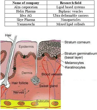
(5) Tripathy Surendra et al. Int. Res. J. Pharm. 2014, 5 (4) ionic surfactants for application on skin. In 1980s, a number of patents covering specific use of vesicles were filed. Since then, the patenting in this field is running constantly covering various colloid drug combinations, preparation methodology or a specific application related with transdermal drug delivery. A major breakthrough in patenting was brought by IDEA AG and since 1991; it holds several patents covering all preparations based on mixed amphipat aggregates and formulations containing alcohols. Currently companies involved in developing transdermal nanocarriers are given in Table 3. Intact skin, as a largest drug delivery site in body has tremendously attracted researchers for drug delivery and also for cosmetics. The standard established pharmaceutical formulations including hydrogels, ointments, creams as well as the novel nanocarriers like liposomes, niosomes, micro emulsions are still not efficient enough to be used across the skin barrier for a systemic delivery. Ultra deformable hydrophilic colloids like transferosomes have succeeded in overcoming certain problems without compromising the barrier properties. The best transdermally given formulations to date are achieved with lipid and membrane softeners. mixtures. The softeners may be a charged bio surfactant, a quasi-biological or synthetic surfactant or else an alcohol or a mixture. Some formulations include ethosomes, some niosomes, novosomes etc56. Ultra deformable, self-regulating vesicles are challenging carriers for transcutaneous delivery of highly potent drugs for a sustained action or also can be used for systemic delivery which usually needs a 100-1000 times higher dose than transcutaneous case. Ultra deformable carriers with high adaptability, self-regulating complex in form of mixed lipid vesicles do not obey simple concentration dependency relationship. Therefore, such drug carriers cannot be evaluated by conventional bio analytical methods like diffusion cell test and skin stripping, rather novel methods as well as open minded testing should be performed. Most of the research groups still rely on oldest or invasive transdermal drugs delivery and a few groups are involved in developing colloidal systems as tabulate above. A lot of opportunities are still waiting to be discovered in this area of transdermal drug delivery. However, this novel highly adaptable ultra deformable colloid mediated transdermal drug delivery systems are hoped to be in market in recent future.. Table 1: Commonly used techniques for enhancing Transdermal drug transport Possible options Regional Stratum corneum elimination Regional Stratum corneum perforation. Methods Abrasion (with sand paper), Ablation (RF or Ultrasonic field treatment) Cutaneous micro needles, Sonoporation by sonic pulses, Electroporation by high voltage pulse, High velocity impact of particles/ballistic droplets, Optoporation by Laser beam Hard Ballistic carriers, Soft Microinjected colloid carriers Penetration enhancer, Highly adjustable, soft carriers. Stratum corneum perforation and Cutaneous depot formation Skin penetration without/ with depot formation. RF = Radio Frequency Table 2: Natural hydrophilic Transdermal penetration shunts Pathway type Sweat duct Sebaceous duct Hair follicle Clusters junction Inter-corneocytes. Average path width (nm) ˃ 5 × 10⁴ 5-15 × 104 5-70 × 104 3-8 × 10⁴ 0.5-7 (normal), 20-30 (widened). Table 3: List of some leading pharmaceutical companies involved in development of colloid mediated TDDS Name of company Alza corporation Helix Pharma Idea AG Skye Pharma Yamanouchi. Research field Lipid based systems Biphasic vesicles Ultra deformable carriers Nanoparticles Mixed lipid colloids. Figure 1: Different layers of skin along with the skin appendages. Page 236.
(6) Tripathy Surendra et al. Int. Res. J. Pharm. 2014, 5 (4). Figure 2: Sequential molecular arrangement in a Lipid-Water binary phase (% water content increases from left to right). From left to right: Inverse micelle, inverse hexagonal phase, Lamellar phase (can swell and break into individual lipid vesicles), normal hexagonal phase, normal micellar phase. Figure 3: Pictorial representation of some colloidal systems showing Liposomes (a), Micelles (b), Nanoparticles (c), Globules of Nanoemulsion system (d) and Dendrimer (e). Figure 4: Distribution pattern of more soluble (Red part), Less soluble (Blue part) and Hydrophobic part (Yellow part) during barrier penetration, 26 upon the administration of an external stress. CONCLUSION This paper highlights the sparse distribution of drug transport pathways in dermal route and the potential of some major colloidal systems to penetrate through this route. The skin barrier is generally breached by the use of substantially high forces, as in case of micro needles, ballistic treatments and local abrasion. The use of energetic and ultra deformable colloidal systems is the only left option for transdermal application, apart from the invasive methods. Transferosomes are the newer colloidal systems with better deformability are having appreciable spontaneous barrier penetration. Colloidal nano-devices with particle size below 150 nm with no local dehydration on skin surface are requisite for a system to achieve an enhanced transdermal penetration. REFERENCES 1. Roth Y, Opatowski E, Lichtenberg D, Kozlov MM. Phase behaviour of dilute aqueous solutions of lipid–surfactant mixtures: effects of finite size of micelles. Langmuir 2000; 16 (5): 2052–2061. http://dx.doi. org/10.1021/la990984+ 2. Maghraby GME, Barry BW, Williams AC. Liposomes and skin: From drug delivery to model membranes. Eur. J. Pharm. Sci 2008; 34 (4-5): 203-222. http://dx.doi.org/10.1016/j.ejps.2008.05.002 3. Domínguez Delgado CL, Rodríguez Cruz IM, Escobar Chávez JJ, Calderón Lojero IO, Quintanar Guerrero D, Ganem A. Preparation and characterization of triclosan nanoparticles intended to be used for the treatment of acne. Eur. J. Pharm. and Bio pharm 2011; 79(1): 102-7. http://dx.doi.org/10.1016/j.ejpb.2011.01.017 4. Almeida AJ, Souto E. Solid lipid nanoparticles as a drug delivery system for peptides and proteins. Adv. Drug Delivery Rev 2007; 59(6): 478490. http://dx.doi.org/10.1016/j.addr.2007.04.007 5. Menjoge AR, Kannan RM, Tomalia DA. Dendrimer-based drug and imaging conjugates: design considerations for nanomedical applications. Drug Disc. Today 2010; 15: 171-185. http://dx.doi.org/10.1016 /j.drudis.2010.01.009. 6.. 7. 8. 9.. 10.. 11. 12. 13.. 14.. 15.. 16. 17.. Schroeter A, Engelbrecht T, Neubert RH, Goebel AS. New nanosized technologies for dermal and transdermal drug delivery: A review. J. Biomed. Nanotech 2010; 6(5): 511-28. http://dx.doi.org/10.1166 /jbn.2010.1149 Neubert RH. Potentials of new nanocarriers for dermal and transdermal drug delivery. Eur. J. Pharm. and Bio pharm 2011; 77(1): 1-2. http://dx.doi.org/10.1016/j.ejpb.2010.11.003 Chourasia R, Jain SK. Drug targeting through pilosebaceous route. Curr. Drug Targets 2009; 10(10): 950-67. http://dx.doi.org/10.2174/1389 45009789577918 Cevc G, Schatzlein A, Richardson H, Vierl U. Overcoming semi permeable barriers, such as the skin, with ultra deformable mixed lipid vesicles, transferosomes, liposomes, or mixed lipid micelles, Langmuir 2003; 19: 10753–10763. http://dx.doi.org/10.1021/la026585n Cevc G, Vierl U. Spatial distribution of cutaneous microvasculature and local drug clearance after drug application on the skin. J. Controlled Release 2007; 118(1): 18–26. http://dx.doi.org/10.1016 /j.jconrel.2006.10.022 Hajime I. Epidermal turnover time. J. Dermatol. Sci 1994; 8(3): 215– 217. http://dx.doi.org/10.1016/0923-1811(94)90057-4 Hadgraft J. Skin, the final frontier. Int. J. Pharm 2001; 224 (1-2): 1-18. http://dx.doi.org/10.1016/S0378-5173(01)00731-1 Pirot F, Kalia YN, Berardesca E, Singh M, Maibach HI, Guy RH. Stratum corneum thickness and apparent water diffusivity: facile and non invasive quantitation in vivo. Pharm. Res 1998; 15(3): 490–492. http://dx.doi.org/10.1023/A:1011996903513 Fartasch M, Bassukas ID, Diepgen TL. Structural relationship between epidermal lipid lamellae, lammelar bodies and desmosomes in human epidermis: an ultra structural study. Br. J. Dermatol 1993; 128(1): 1–9. http://dx.doi.org/10.1111/j.1365-2133.1993.tb00138.x Ponec M, Boelsma E, Weerheim A, Mulder A, Bouwstra J, Mommaas M. Lipid and ultra structural characterization of reconstructed skin models. Int. J. Pharm 2000; 203(1-2): 211 – 225. http://dx.doi.org/ 10.1016/S0378-5173(00)00459-2 Schaefer P, Bewick Sonntag C, Capri MG, Berardesca E. Physiological changes in skin barrier function. Skin Pharmacol. Appl. Skin Physiol 2002; 15(1): 7–19. Cronin MTD, Dearden JC, Moss GP, Murray Dickson G. Investigation of the mechanism of flux across human skin in vitro by quantitative. Page 237.
(7) Tripathy Surendra et al. Int. Res. J. Pharm. 2014, 5 (4) 18. 19.. 20.. 21.. 22.. 23. 24. 25. 26. 27. 28. 29.. 30.. 31.. 32.. 33. 34. 35. 36.. 37.. structure– permeability relationships. Eur. J. Pharm. Sci 1999; 7(4): 325–330. http://dx.doi.org/10.1016/S0928-0987(98)00041-4 Aguilella V, Kontturi K, Murtoma L, Ramirez P. Estimation of the pore size and charge density in human cadaver skin. J. Controlled Release 1994; 32(3): 249–257. http://dx.doi.org/10.1016/0168-3659(94)90235-6 Pikal MJ. Transport mechanisms in iontophoresis. I. A theoretical model for the effect of electro osmotic flow on flux enhancement in transdermal iontophoresis, Pharm. Res 1990; 7(2): 118–126. http://dx .doi.org/10.1023/A:1015816532532 Ruddy SB, Hadzija BW. Iontophoretic permeability of polyethylene glycols through hairless rat skin: application of hydrodynamic theory for hindered transport through liquid filled pores. Drug Des. Discov 1992; 8(3): 207–224. Fesq HP, Richardsen H, Cevc G, Ring J, Abeck D. Detection of transdermal inter cluster gaps in human epidermis with the help of highly deformable lipid vesicles (Transfersomes R) and confocal laser scanning microscopy. Arch. Derm. Res 1999; 291: 130. Tezel A, Sens A, Mitragotri S. Description of transdermal transport of hydrophilic solutes during low-frequency sonophoresis based on a modified porous pathway model. J. Pharm. Sci 2003; 92(2): 381–393. http://dx.doi.org/10.1002/jps.10299 Mak VHW, Potts RO, Guy RH. Does hydration affect intercellular lipid organization in the stratum corneum? Pharm. Res 1991; 8(8): 1064– 1065. http://dx.doi.org/10.1023/A:1015873511692 Dubey V, Mishra D, Nahar M, Jain NK. Vesicles as tools for the modulation of skin permeability. Expert Opin. Drug Deliv 2007; 4(6): 579–593. http://dx.doi.org/10.1517/17425247.4.6.579 Peter U, Roux D, Sood AK. Observation of a topological relaxation mode in micro emulsions. Phys. Rev. Lett 2001; 86: 3340–3343. http://dx.doi.org/10.1103/PhysRevLett.86.3340 Cevc G, Vierl U. Nanotechnology and the transdermal route A state of the art review and critical appraisal. J. Controlled Release 2010; 141: 277-299. http://dx.doi.org/10.1016/j.jconrel.2009.10.016 Maghraby GM, Williams AC, Barry BW. Can drug-bearing liposomes penetrate intact skin? J. Pharm. Pharmacol 2006; 58(4): 415–429. http://dx.doi.org/10.1211/jpp.58.4.0001 Fang JY, Hwang TL, Huang YL. Liposomes as vehicles for enhancing drug delivery via skin routes. Curr. Nanosci 2006; 2(1): 55–70. http://dx.doi.org/10.2174/157341306775473791 Huang X, Du Y, Yuan H, Hu F. Preparation and pharmacodynamics of low molecular- weight chitosan nanoparticles containing insulin. Carbohyd. Polym 2009; 76: 368-373. http://dx.doi.org/10.1016 /j.carbpol.2008.10.025 Goswami S, Bajpai J, Bajpai AK. Designing Gelatin Nanocarriers as a Swellable System for Controlled Release of Insulin: An In-Vitro Kinetic Study. J. Macromol. Sci 2010; 47: 119-130. http://dx.doi.org/10.1080 /10601320903458556 Osborne DU, Ward AJ, O Neill KJ. Micro emulsions as topical drug delivery vehicles I. Characterization of a model system. Drug Dev. Ind. Pharm 1988; 14(9): 1203–1219. http://dx.doi.org/10.3109 /03639048809151929 Osborne DU, Ward AJ, O Neill KJ. Micro emulsions as topical drug delivery vehicles: in-vitro transdermal studies of a model hydrophilic drug, J. Pharm. Pharmacol 1991; 43(6): 450–454. http://dx.doi. org/10.1111/j.2042-7158.1991.tb03511.x Kogan A, Garti N. Micro emulsions as transdermal drug delivery vehicles. Adv. Colloid Interface Sci; 2006. p. 123–126, 369–385. Trommer H, Neubert RH. Overcoming the stratum corneum: the modulation of skin penetration. A review. Skin Pharmacol. Physiol 2006; 19: 106–121. http://dx.doi.org/10.1159/000091978 Santos P, Watkinson AC, Hadgraft J, Lane ME. Application of micro emulsions in dermal and transdermal drug delivery. Skin Pharmacol. Physiol 2008; 21: 246–259. http://dx.doi.org/10.1159/000140228 Shakeel F, Baboota S, Ahuja A, Ali J, Aqil M, Shafiq S. Celecoxib nanoemulsion: skin permeation mechanism and bioavailability assessment, J. Drug Target 2008; 16(10): 733–740. http://dx.doi.org/ 10.1080/10611860802473402 Shakeel F, Ramadan W, Ahmed MA. Investigation of true nanoemulsions for transdermal potential of indomethacin: characterization, rheological characteristics, and ex vivo skin permeation studies. J. Drug Target 2009; 17(6): 435–441. http://dx.doi.org/ 10.1080/10611860902963021. 38. Kumar D, Aqil M, Rizwan M, Sultana Y, Ali M. Investigation of a nanoemulsion as vehicle for transdermal delivery of amlodipine. Pharmazie 2009; 64(2): 80–85. 39. Svenson S, Tomalia DA. Dendrimers in biomedical applications – reflections on the field. Advanced Drug Delivery Reviews 2005; 57(15): 2106-2129. http://dx.doi.org/10.1016/j.addr.2005.09.018 40. Tripathy S, Das MK. Dendrimers and their applications as novel drug delivery carriers. J App Pharm Sci 2013; 3(9): 142-149. 41. Davis AF, Hadgraft J. Effect of super saturation on membrane transport: 1. Hydrocortisone acetate. Int. J. Pharm 1991; 76(1-2): 1–8. http://dx .doi.org/10.1016/0378-5173(91)90337-N 42. Alvarez Román R, Naik A, Kalia YN, Guy RH, Fessi H. Skin penetration and distribution of polymeric nanoparticles. J. Controlled Release 2004; 99: 53–62. http://dx.doi.org/10.1016 /j.jconrel.2004.06.015 43. Alvarez Román R, Naik A, Kalia YN, Fessi H, Guy RH. Visualization of skin penetration using confocal laser scanning microscopy. Eur. J. Pharm. and Bio pharm 2004; 58(2): 301–316. http://dx.doi.org /10.1016/j.ejpb.2004.03.027 44. Larese FF, Agostin DF, Crosera M, Adami G, Renzi N, Bovenzi M, Maina G. Human skin penetration of silver nanoparticles through intact and damaged skin. Toxicology 2009; 255 (1-2): 33–37. http://dx.doi. org/10.1016/j.tox.2008.09.025 45. Baroli B, Ennas MG, Loffredo F, Isola M, Pinna R, Quintela MAL. Penetration of metallic nanoparticles in human full-thickness skin. J. Invest. Dermatol 2007; 127(7): 1701–1712. 46. Sonavane G, Tomoda K, Sano A, Ohshima H, Terada H, Makino K. In vitro permeation of gold nanoparticles through rat skin and rat intestine: effect of particle size. Colloids Surf., B Bio interfaces 2008; 65: 1–10. http://dx.doi.org/10.1016/j.colsurfb.2008.02.013 47. Yilmaz E, Borchert HH. Affect of lipid-containing, positively charged nanoemulsions on skin hydration, elasticity and erythema—an in vivo study. Int. J. Pharm 2006; 307(2): 232–238. http://dx.doi.org/10.1016 /j.ijpharm.2005.10.002 48. Anton N, Benoit JP, Saulnier P. Design and production of nanoparticles formulated from nano-emulsion templates—a review. J. Controlled Release 2008; 128(3): 185–199. http://dx.doi.org/10.1016/j.jconrel. 2008.02.007 49. Cevc G, Vierl U, Mazgareanu S. Functional characterisation of novel analgesic product based on self-regulating drug carriers. Int. J. Pharm 2008; 360: 18–28. http://dx.doi.org/10.1016/j.ijpharm.2008.04.002 50. Cevc G, Gebauer D. Hydration-driven transport of deformable lipid vesicles through fine pores and the skin barrier. Bio phys. J 2003; 84(2): 1010–1024. http://dx.doi.org/10.1016/S0006-3495(03)74917-0 51. Van den Bergh BAI, Bouwstra JA, Junginger HE, Wertz PW. Elasticity of vesicles affects hairless mouse skin structure and permeability. J. Controlled Release 1999; 62(3): 367–379. http://dx.doi.org /10.1016/S0168-3659(99)00168-6 52. Van der Merwe D, Riviere JE. Comparative studies on the effects of water, ethanol and water/ethanol mixtures on chemical partitioning into porcine stratum corneum and silastic membrane. Toxicol. In Vitro 2005; 19(1): 69–77. http://dx.doi.org/10.1016/j.tiv.2004.06.002 53. Cevc G, Mazagareanu S, Rother M, Vierl U. Occlusion effect on transcutaneous NSAID delivery from conventional and carrier-based formulations. Int. J. Pharm 2008; 359(1-2): 190–197. http://dx.doi.org /10.1016/j.ijpharm.2008.04.005 54. Maghraby GME, Williams AC, Barry BW. Oestradiol skin delivery from ultra deformable liposomes: refinement of surfactant concentration. Int. J. Pharm 2000; 196(1): 63–74. http://dx.doi.org/10.1016/S03785173(99)00441-X 55. Wachter C, Vierl U, Cevc G. Adaptability and elasticity of the mixed lipid bi layer vesicles containing non-ionic surfactant designed for targeted drug delivery across the skin. J. Drug Target 2008; 16: 611– 625. http://dx.doi.org/10.1080/10611860802230158 56. Cevc G. Transdermal drug delivery of insulin with ultra deformable carriers, Transfersomes. Clin. Pharmacokinet 2003; 42(5): 461–474. http://dx.doi.org/10.2165/00003088-200342050-00004 Cite this article as: Tripathy Surendra, Palei Narahari Narayan, Das Malay Kumar. Colloid mediated drug transport across dermal route: A critical review. Int. Res. J. Pharm. 2014; 5(4):232-238 http://dx.doi.org/10.7897/2230-8407.050450. Source of support: Nil, Conflict of interest: None Declared. Page 238.
(8)
Figure

Related documents
The Cochrane Collaboration policy is that each review should be updated every two years, thus we chose a four year period to allow for as many reviews as possible to be
This study aims to assess viability of Trichoderma harzianum TH03 and Pleurotus ostreatus PO2 isolates that propagated in different growth mediums: rice, corn and sawdust
Recall the major premises of the traditional LFG treatment of constituent coordi- nation (Bresnan, Kaplan, et al. 1985; Kaplan and Maxwell 1988; Dalrymple and Kaplan 2000;
Major challenges facing the CIELO team included how to accommodate the use of a new softer thermal PFNS spectrum, and updated PFNS spectra at higher energies, as recommended by
However, it is being increasingly recognized generally that we need to go beyond to the family, group, village, community and social levels if we are to more fully understand what
This multidimensional structure is a comprehen- sive reflection of the sociopsychological context of school disciplinary climate, covering student discipline perception and
Until now, countless efforts have been made to understand and control this phenomenon using various methods; perforating far above the original OWC; keeping
A new 239 Pu evaluation based on data form BRC-2009 for the continuum energy range and from JEFF-3.1.1 and recent ORNL work [7] for the thermal and resonance energy range will
