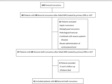
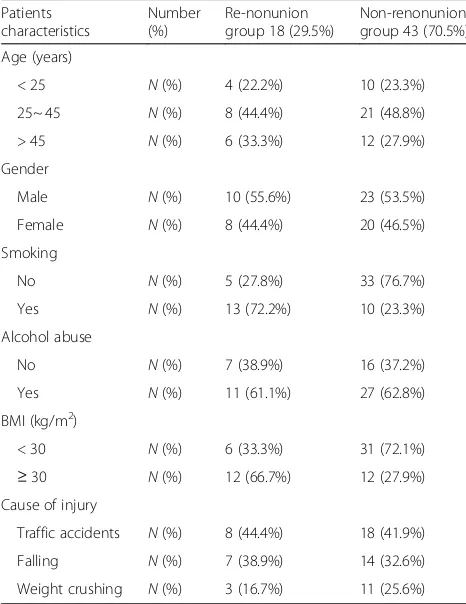
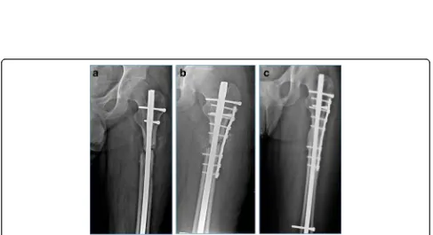
Factors associated with development of re-nonunion after primary revision in femoral shaft nonunion subsequent to failed intramedullary nailing
Full text
Figure


Related documents
In the present study, drug loaded transdermal patches of herbal extract were prepared by solvent evaporation method.. The extract was dissolved in mixture of
The study was a randomized controlled trial (RCT) in which one group (case) given prophylactic phenobarbital before occurrence of seizures compared to another group (control)
Therefore, this study aims to assess the prevalence of malaria and associated factors constituting traditional practices and mosquito net use in a pastoralist district of
IC1 Publication was published in the proceedings of the IEEE/RSJ International Conference on Intelligent Robots and Systems (IROS), IEEE International Conference on Robotics
8 Following Ariño 2003, we performed two tests to assess the reliability of measures of logistics partnership performance see Table 3: Table 3 Reliability tests for measures
Methods: All infants with respiratory distress born at 28 – 32 weeks ’ gestational age from January 2012 to December 2012 (n=90), who were eligible for exogenous pulmonary
These questions were primarily related to understanding the impact of contextual changes on service delivery and financial feasibility of the programs; coping strategies,
