INTRODUCTION
Quinine (Qu), a quinoline–methanol derivative from the bark of the cinchona tree has been used in the suppression and treatment of malaria for more than three centuries (Robert, et al., 2001). However there is a dearth of publications on the effects of Qu on the male reproductive system, and the few available lack details on how the effects could be reversed.
It has been reported that QU is effective in inhibiting sperm metabolism and motility (Trifunac and Berstein, 1982). In another study, it was reported that Qu immobilized 100% of human sperm within 20 seconds (Garg, et al., 1994).
Biosciences, Biotechnology Research Asia Vol. 4(1), 111-116 (2007)
The effects of quinine therapy on the
seminal fluid analysis and histology of testes of male rats
E.K. NWANGWA¹, J.C. IGWEH², U.E. UZUEGBU³ and E.C. ADEGOR*
¹Department of Physiology, College of Health, Sciences, Delta State University, Abraka (Nigeria) ²Department Physiology, College of Medicine, University of Nigeria, Enugu Campus, Enugu (Nigeria)
³Department Of Medical Biochemistry, College of Health Sciences, Delta State University, Abraka (Nigeria)
(Received: March 15, 2007; Accepted: March 25, 2007) ABSTRACT
The effects of intra muscular (IM) administration of quinine (Qu) on the seminal fluid analysis and testicular histology was studied. Thirty adult male Sprague–Dawley rats were randomly divided into five experimental groups (6 rats per group). Group 1 (control) were given clean drinking water and rat chow, group 2 were given intra-muscular (IM) quinine injection 10mg/kg body weight + water + rat chow. Group 3 and 4 were given, IM quinine 10mg/kg body weight + water + rat chow and graded proportions of extract of Telfairia occidentalis of 4.5g, and 6.0g, respectively. While Group 5 were given rat chow + water + 6.0g of Telfairia occidentalis. The results show a destruction in the testicular histology, decreased sperm count, activity and morphology of Group 2 administered with quinine only, which was statistically significant (P<0.05), and there was also a dose-dependent improvement in the parameters studied in the Group (3 and 4) given extract of Telfairia occidentalis. Group 5 did not show any Testicular destruction but showed an increase in sperm count, morphology and activity but was not statistically significant (P>0.05). This shows that Qu therapy has a destructive effect on the histology of testes and decreased the sperm count, activity and morphology but these effects could be reversed by the administration of extract of Telfairia occidentalis.
Key words: Testicular histology, quinine, seminal fluid analysis, Telfairia occidentalis.
The use of herbs in the treatment of different diseases is fast becoming revolutionalized. In some countries, it has been integrated into the health scheme despite advances in orthodox medicine.
to reduce plasma glucose, cholesterol, creatinine and triacylglycerol levels of alloxan–induced diabetic rabbits (Nwozo, et al., 2004) but had no effect on plasma protein level (Nwozo, et al., 2004). Its herbal preparation has been employed in the treatment of sudden attack of convulsion, malaria and anaemia (Gbile, 1986).
Little is known about the effect of Telfairia occidentalis on the male reproductive system except for some unverified claims by herbal practitioners that it increases sperm count and activity.
Therefore, this study is designed to verify some of these claims, confirm the effect of quinine on the histology of the testes and a possible testicular regenerative effect of Telfairia occidentalis extract. A positive result may encourage the consumption of Telfairia occidentalis leaves in our diets for a healthier living.
MATERIALS AND METHODS Test animals
Thirty adult male rats of the Sprague– Dawley strain were purchased from the Animal House Unit of the College of Medicine, Ambrose Alli University, Ekpoma. The rats weighed between 150 – 200g and were about 8 – 9 weeks old.
The rats were kept in standard room temperature and conditions for fourteen days to acclimatize. The rats were given unrestricted access to water and normal rat chow (Pfitzer Nig. Ltd., Ibadan). The animals were randomly divided into five experimental groups of six rats in each group. Group 1 (Control) were given clean drinking water ad libitum and rat chow.
Group 2 were given rat chow + intramuscular (IM) quinine 10mg/kg bwt.
Group 3 and 4 were given rat chow + 10 mg/kg b.wt. IM quinine + graded proportion of 4.5g and 6.0g respectively of aqueous extract of T. O. Group 5 were given rat chow + 6.0g T. O. extract. Preparation Of Telfairia occidentalis leaf extract
The Telfairia occidentalis leaves were purchased from a local market in Abraka. It was rinsed to remove sand and other debris. The
vegetable was authenticated by Prof JMO Eze of Botany Department, Delta State University, Abraka. The leaves were detached, weighed and dried on a laboratory bench to a constant weight. It was then milled.
The dried samples were extracted by cold percolation and maceration technique (Dhar, et al., 1968) and the quantity of dissolvable solid extract was observed to be 3g solid T. O/ ml.
Method of extract administration
The appropriate quantity of crude aqueous extract was given orally through an orogastric cannula into the stomach via the oesophagus (Prakash, 1981). The extract was administered once daily between 8-9 am for a continuous period of 30 days. The extracts were preserved in a refrigerator at 40C and the left over after 3 days
were discarded and new one reconstituted. The intragastric administration enabled us to determine the exact quantity that the rats received daily.
The quinine was given intra muscularly (lM) once daily for 5 days in a week continuously over a 2 weeks period.
Collection of samples
The rats were re-weighed and mean weight was obtained and recorded. After an overnight fast, the rats were sacrificed under chloroform anaesthesia.
Seminal fluid analysis
The r ight and left epididymis were dissected from the caput to the boundary between the cauda and the first part of vas deferens. For the sper m count, the right epididymis was homogenized in a physiological saline and examined under a microscope using a Neubauer hemocytometer. For the morphology and activity, the left epididymis was placed in a buffered normal saline and examined under microscope. The percentage of motile, sluggish and immotile sperm was calculated (Linder, et al., 1988, Liobet, et al., 1991).
The morphological appearance of abnormal sperm shape (Wyrobeck and Bruce, 1975) and their percentage (Wyrobeck and Bruce, 1975) were calculated.
Light microscopy and histology of testes The testes were carefully dissected out, and fat were trimmed off. The weight was then obtained and recorded. Each testis was fixed in 10% formal – saline and histological slide prepared.
Statistical analysis
Results were expressed as mean ± SEM. The evaluation of data for statistical significance between control and experimental groups was done using Students t-test. Statistical software, SPSS, was also used to analyze data. A value of P<0.05 was accepted as statistically significant.
RESULTS
The data obtained from the above study is as shown in the tables and figures.
Table - 1: The effects of quinine and extract of Telfairia occidentalis on the total body weight and testicular weight of Sprague–Dawley rats after 30 days.
GP 1: n = 6 Grp 2:n = 6 GP 3: n =6 GP 4: n = 6 GP 5:n=6 (control) Rat chow + Rat chow + Rat chow + IM Rat chow +
IM 10mg/kg IM 10mg/g 10mg /kg bwt 6.0g T. O bwt Qu. bwt Qu + Qu + 6.0g T. O extract
4.5g T. O. extract extract
Initial body weight (g) 165.90±17.07 164.80±1.37 165.20±12.35 165.20±12.35 163.00±10.44 Final body weight (g) 177.60±18.56 174.2 ± 2.58 180.00 ± 10.4 182.00 ± 11.57 191.60 ± 18.00 weight difference (g) 11.70 ± 12.31 9.40 ± 3.22 14.80 ± 6.45 16.80 ± 14.34 28.60 ± 7.63 * Testicular weight (g) 1.98 ± 0.005 1.52 ± 0.15 1.82 ± 0.64 1.86 ± 0.11 2.10 ± 0.007 Data is given as Mean ± SEM * Statistically significant (P< 0.05).
Qu = quinine. bwt = body weight and T.O. = Telfairia occidentalis
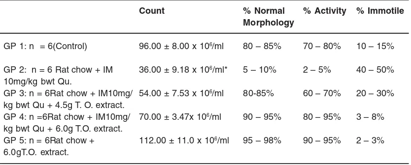
Table - 2: The effect of administration of quinine and extract of Telfairia occidentalis on seminal fluid analysis.
Count % Normal % Activity % Immotile
Morphology
GP 1: n = 6(Control) 96.00 ± 8.00 x 10⁶/ml 80 – 85% 70 – 80% 10 – 15%
GP 2: n = 6 Rat chow + IM 36.00 ± 9.18 x 10⁶/ml* 5 – 10% 2 – 5% 40 – 50%
10mg/kg bwt Qu.
GP 3: n = 6Rat chow + IM10mg/ 54.00 ± 7.53 x 10⁶/ml 80-85% 60 – 70% 20 – 30%
kg bwt Qu + 4.5g T. O. extract.
GP 4: n =6Rat chow + IM10mg/ 70.00 ± 3.47x 10⁶/ml 90 – 95% 80 – 95% 3 – 8%
kg bwt Qu + 6.0g T.O. extract.
GP 5: n = 6Rat chow + 112.00 ± 11.0 x 10⁶/ml 95 – 98% 90 – 95% 2 – 3%
6.0gT.O. extract.
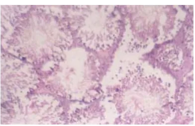
Fig. - 1-5 shows the effects of quinine and extract of Telfairia occidentalis on the histopathology of rats testes. Fig. -1: Photo micrography of cross section of
seminiferous tubules of normal (control) Sprague -Dawley rats (Group 1) stains:
haematoxylin & eosin. Magn. X 125.
Fig. -2: Photo micrography of cross section of seminiferous tubules of Sprague –Dawley rats
administered with 10mg/kg b.wt. QU for 2 weeks (Group 2) stains: haematoxylin & eosin.
Magn. X 125.
Fig. -3: Photo Micrography of Cross section of seminiferous tubules of Sprague – Dawley rats
administered with 10mg/kg b.wt. QU. For 2 weeks + 4.5g T. O. extract (Group 3) stains: haematoxylin & eosin. Mag. X 125.
Fig. -4: Photo micrography of cross section of seminiferous tubules of Sprague – Dawley rats
administered with 10mg/ kg b.wt. QU. For 2 weeks + 6.0g T. O. extract (Group 4) stains: haematoxylin & eosin. Magn. X 125.
DISCUSSION
Previous studies have demonstrated that Telfairia occidentalis extract significantly increase hematological parameters (Alada, 2000; Nwangwa, 2006). It is also reported that quinine induces, a general destruction of cells of the seminiferous tubules and testicular interstitium (Osinubi, et al., 2005) hence adversely affects spermatogenesis.
This study investigated the effect of Telfairia occidentalis on the seminal fluid analysis and the histology of the Testes. The results shows that the extract of Telfairia occidentialis increased the sperm count, morphology and activity of the rat though it was not statistically significant. This study also supported the previous report of quinine inhibiting sperm metabolism and motility (Trifunac and Berstein, 1982) as there was a statistical significant (P<0.05) decrease in all the sperm parameters studied following the administration of quinine.
The extract of Telfairia occidentialis showed a regenerative effect on the histology or the testes. Quinine is known to disrupt the tubular morphology and general destruction of testicular interstitium and
spermatogenesis (Osinubi, et al., 2005) but the extract of Telfairia occidentialis showed a near complete morphological regeneration and increased spermatogenesis. This work could not say whether there was an increased synthesis or secretion of testosterone from the leydig cells since radio immunoassays were not performed.
CONCLUSIONS
This present study has provided evidence that quinine therapy is capable of inducing degeneration in the testicular epithelium and that the administration of the extract of Telfairia occidentalis caused a regeneration of the destroyed epithelium and improved the quantity of sperm produced. This may become very impor tant especially this period of controversy over male contraceptive.
ACKNOWLEDGEMENTS
We wish to appreciate the technical assistance of Mr. Osadebe N. C. and Mr. Eghworo Ovocity Also to Udoh Patience A. for typing the manuscript.
1. Akabue PI., Kar A., Nnachetta FN. Toxicity of extracts of roots and leaves of Telfairia occidentalis Planta Med., 38(4): 339-343 (1980).
2. Alada A., The hematological effects of Telfairia occidentalis diet preparation. Afri. J. Biomed Res 3(3): 185- 186 (2000). 3. Dhar, MI., Dhar, MM., Dhawan BN.,
Mehrotra, B. Ray C. Screening of Indian Plants for biological activity: Part 1. Ind. J. Exp. Biol 16: 232 –236 (1968).
4. Garg, S., Doncel G, Chabra S, Upadhyay SN and Talwar GP. Synergistic Spermicidal activity of neem seed extract, reetha, saponins and quinine hydrochlor ide. Contraception 50: 185-190 (1994).
5. Gbile Z.O. Ethnobotany, taxonomy and conservation of medicinal plant. In: The State
REFERENCES
of Medicinal Plant Research in Nigeria A. O. Sofowara (Ed.) (1986).
6. Linder RE., Rehnberg GL., Strader LF., Diggs JP., Evaluation of reproductive parameters in adult male Wistar rats after subchronic exposure (gavage) to Benomyl. J. Toxicol Environ Health 25: 285-289 (1988). 7. Liobet JM., Irvent JJ., Ortega A., Domingo
JL., Influence of chronic exposure of uranium on male reproduction in mice Fundam. Appl. Toxicol. 6: 821-825 (1991).
8. Nwangwa EK., Effects of Aqueous extract of Telfairia occidentalis on hematological parameter of rats. J. App. Sci. (In press) (2006).
diabetic rats. Nig. J. Nat Prod Med 8: 45-47 (2004).
10. Olatunji, K., Health usefulness of vegetables. Health Monitor 3(4): 21 (2005).
11. Osinubi AA., Noronha CC., and Okanlawon, Morphometric and stereological assessment of the effects of long-term administration of quinine on the morphology of rat testes. West Afri. J. Med., 24(3): 200-205 (2005). 12. Prakash, O. Antifertility investigation on
embelin in rats – an oral contraceptive of plant origin part 1, Biological properties, Planta. 41: 259-266 (1981).
13. Robert A, Benoit–Vical F., Dechy-Cabaret O., and Meunier B. From classical antimalarial drugs to new compounds based on the mechanism of action of artemisinin. Pure Appl.Chem 73: 1173-1188 (2001).
14. Trifunac NP., and Berstein GS., Inhibition of the metabolism and motility of human sper matozoa by various alkaloids. Contraception 25: 69-87 (1982).