RESEARCH ARTICLE
EFFECT OF PROGRESSIVE EXERCISE PROGRAM ON THE BLEEDING FREQUENCY IN
HAEMOPHILIC PATIENTS
1, *
Maha HABIB,
2Hala EZZ EL DEIN,
3Talaat AL KEMARY and
4Gihan Samir, M.
1Department of Physical Therapy, Damanhour Medical National Institute, Cairo, Egypt 2Department of Physical Therapy for Cardiovascular/ Respiratory Disorders and Geriatrics,
Faculty of Physical Therapy, Cairo University, Cairo, Egypt
3Department of internal medicine and hematology, Damanhour Medical National Institute 4Department of Physical Therapy for Cardiovascular/ Respiratory Disorders and Geriatrics, Faculty of
Physical Therapy, Cairo University, Cairo, Egypt and Physical Therapy Department, Faculty of Applied Medical science, Umm Al Qura University, KSA
ARTICLE INFO ABSTRACT
Background and Objective: To examine the effect of progressive exercise program on the bleeding frequency in haemophilic patients.
Material and methods: Thirty male patients with haemophilia were assigned into two equal groups; group A was the study group and consisted of 15 haemophilic patients (type A and B) with mild and moderate severity received their medical treatment and on their normal activities and participated in progressed exercise program for eight weeks and group B was the control group and consisted of 15 haemophilic patients (type A and B) with mild and moderate severity received their medical treatment and on their normal activities not participated in the exercise program for eight weeks. All patients participated in the current study three times /week for eight weeks. All subjects in both groups were assessed for pain; clotting time and muscle test.
Results: Following treatment, the study group (A) demonstrated statistically highly significant improvement (P<0.001) in pain intensity measured by Borg scale for pain (P-Value was 0.000) with percentage of improvement 31.9 % and significant improvement (P<0.05) in pain (P-Value was 0.025) post treatment compared to the control group with percentage of improvement 8.38 %.
Following treatment, the study group (A) demonstrated statistically highly significant improvement (P<0.01) in bleeding measured by number of bleeding episodes (P-Value was 0.004) with percentage of improvement 41.83 % and significant improvement (P<0.05) in clotting time (P-Value was 0.042) post treatment compared to the control group with percentage of improvement47.03 %.
Following treatment, the study group (A) demonstrated statistically significant improvement in muscle strength measured by manual muscle test of right knee, left knee, right elbow, left elbow (P-Value was 0.005, 0.016, 0.003, 0.015) with percentage of improvement (16.78%, 19.15%, 33.19%,19.13%) respectively post treatment compared to the control group.
Conclusion: From these findings we can conclude that the designed therapy program consisted of electrical stimulation, stretching exercises and range of motion exercises when accompanied with the medical treatment can achieve much more improvement in pain reduction, increasing muscle strength and decrease bleeding in patients with haemophilia.
INTRODUCTION
Haemophilia is a congenital, recessive clotting disease which has two features. The first feature is cerebral hemorrhage which is considered the most dangerous and the second feature which is the musculoskeletal hemorrhage and is more common and disabling. Factor VIII (FVIII) deficiency results in haemophilia A while haemophilia B is caused by factor IX (FIX) deficiency (Felipe et al., 2011).
*Corresponding author: Maha HABIB,
Department of Physical Therapy, Damanhour Medical National Institute, Cairo, Egypt.
Although large number of severe haemophilic patients suffers from musculoskeletal injuries, they have similar life expectancy to that of the general population (Aznar et al.,
2009). The severity of haemophilia is determined by the level of VIII or factor IX clotting factors activity in the blood. There are three categories of the severity of haemophilia: in mild haemophilia the percentage of clotting factors activity in blood is from 5 % to 40% of normal while in moderate cases this percentage ranges from 1% to 5 % of normal and when the percentage of factor activity in blood is less than 1% of normal this is considered severe (Srivastava, 2005). Painful joint deformity is usually accompanied with severe haemophilia due
Article History:
Received 07th
May, 2018 Received in revised form 18th
June, 2018 Accepted 09th
July, 2018 Published online 30th
August, 2018
www.ijramr.com
International Journal of Recent Advances in Multidisciplinary Research
Vol. 05, Issue 08, pp.4026-4031, August, 2018
Keywords:
Clotting time, Exercises, Hemophilia, Pain.
Abbreviations
CM : Centimeter KG : Kilogram NS : Non Significant M : Meter P : Probability S : Significant SD : Standard Deviation
SPSS : Statistical Packaged For Social Science.
to chronic and recurrent bleeding (Green, 2006). Performing of resistance training exercises is beneficial either to children, adolescent, adult athletic or non-athletic individuals. Resistance training exercises have many benefits such as improving muscular strength, endurance and power, improving
motor performance and ergonomic tasks, increasing
cardiovascular fitness, lean body mass and tissue tensile strength including bone mineral density, improved blood lipid profiles, decreasing pain and reducing psychological stress (Callahan, 2009). As literatures reported that exercises may be of great benefits for haemophilic patients without investigating its progression effect. So, the aim of the study was to examine the effect of progressive exercise program on the bleeding frequency in haemophilic patients.
MATERIALS AND METHODS
The study was conducted from June 2016 to January 2018 at Damanhour Medical National Institute, Egypt. The study was designed as a prospective randomized clinical trial.
Inclusion criteria of the study group
This study was conducted on thirty males with haemophilia their age ranged from 20 to 40 years and referred from the out-patient clinic of internal medicine in Damanhour Medical National Institute in Damanhour in Egypt to participate in the present study. All patients participated in the current study three times /week for eight weeks.
Exclusion criteria of the study group
The patients with history of severe haemophilia, recent hemorrhage, skeletal deformities and Cardiac disease patients were excluded from the study.
Design: Pretest posttest design was used in this study. Thirty
patients with haemophilia were randomly allocated into two groups A and B equally in number:
Group (A): (study group)
Fifteen haemophilic patients (12 patients with type A and three patients with type B) with mild and moderate severity recieved their medical treatment and on their normal activities and participated in progressed exercise program for eight weeks.
Group (B): (control group)
Fifteen haemophilic patients (12 patients with type A and three patients with type B) with mild and moderate severity received their medical treatment and on their normal activities did not participate in the exercise program for eight weeks.
Procedure
All patients signed informed consent form about the purpose and nature of the study. A complete history was being taken and physical examination was being conducted for each patient before participation in the study.
Evaluation procedure:
1. Weight-height scale: The evaluation procedure had
been done for all patients in the two groups before starting the program.
Measurement of BMI was done by weight-height scale.
BMI= Weight (kg) / height2 (m2) Kg/ m2.
2. Blood tests: Measurement of clotting time which is
the time required for a sample of blood to coagulate in vitro under standard conditions is called "clotting time".
3. Procedure: Make a sufficient deep prick in the finger
tip with following all sterile precautions. Put a drop of the blood on the slide. The slide is placed on a warm plate 37 C, every 30 seconds, after putting a needle in the middle of the blood drop begin to assess it, blood coagulation start when fibrin threads appear attached to the needle. It takes from 3 to 10 minutes.
4. Borg pain Scale: The patient degree of pain was
measured by Borg pain Scale. The scale is based on a 0 10 range of pain, where 0 is no pain and 10 is unbearable pain.
5. Bleeding profile diary: Bleeding profile diary was
completed by each patient in both groups. Bleeding profile determined based on a diary completed by each patient for a period of one month before the study and after eight weeks from the study for both groups. For each bleeding incident, patients are requested to fill out the following information: site of the bleed; the exact joint, muscle or other place of the bleed and the cause of the bleed: traumatic or spontaneous. The percentage of traumatic bleeds per month was calculated as the traumatic bleeds for the month out of the total number of bleeds for the month multiplied by 100.
6. The Lafayette Manual Muscle Test System:
Eccentric muscle strength was objectively measured by The Lafayette hand-held device Manual Muscle Test System.
Treatment procedure:
Treatment had been carried out in Damanhour Medical National Institute. The program of the study was given for three times /week for eight weeks.
Study group A
Received their medical treatment and on their normal activities and participated in progressed exercise program for eight weeks.
1-Electrical stimulator
The quadriceps femoris and hamstring muscles of both legs were treated with electrical stimulation for 15 min a day, three days a week, for eight weeks. Placement of electrodes for quadriceps muscle one electrode was placed over the inside lower thigh area, and the other one over the front of the leg. Bipolar electrodes and an electro stimulator apparatus were used (Therapic 2000 modality stimulator was used). At any session frequency of 45 Hz and 300 μs pulse were used and a contraction and relaxing cycle of 12 s on and 8 s off. During application the intensity of the pulse was increased according to the patient sensation in successive sessions.
2-Free body weights
for each muscle group. It was done as warming up for 15 minutes and cooling down for 15 minutes .Frequency of stretching was three sessions/week. Strength exercises: were conducted three times /week for eight weeks (total of 24 treatments), as shown in Table 1.
Level 1 40% 1 RM, 1 X 10 reps Level 2 50% 1 RM, 1 X 10 reps Level 3 60% 1 RM, 1 X 10-20 reps
Control group B
Received their medical treatment and on their normal activities did not participate in the exercise program for eight weeks.
Data analysis
The data were coded, entered and processed on computer using A statistical package program Statistical Packaged for Social Science (IBM SPSS version 22, 2013). The significance level was determined by P ≤ 0.05. Descriptive statistical methods (proportion, mean, and standard deviation) were used in the evaluation of research data as well as the Kolmogorov– Smirnov distribution test for examining normal distribution. The unpaired samples t-test was used in intergroup comparison of quantitative data while, for intragroup comparisons the Paired samples t-test was used.
RESULTS
Baseline characteristics of the patients are shown in Table 2. The average age was 27.33 ± 3.15 years in the control group and 26.53 ± 4.34 years in the study group. The average height was 173.13 ± 11.64 cm in the control group and 175.87 ± 10.29 cm in the study group. The average weight was 75.13 ± 9.63 kg in the control group and 74.4 ± 12.66 kg in the study group. The average BMI was 24.11 ± 2.91 Kg/ m2in the control group and 23.73 ± 2.26 Kg/ m2 in the study group.
No side effects or complications were observed during the treatment.
No statistically significant difference was found between the 2 groups in terms of age and sex (P > 0.05).
The decrease in the Borg scale for pain in the study group at the end of the treatment was statistically significant in comparison to baseline (P < 0.05). The decrease in the Borg scale for pain in the control group at the end of the treatment was statistically significant in comparison to baseline (P < 0.05). The decrease in the borg scale for pain in the study group at the end of the treatment was significantly higher than in the control group (P < 0.05), as shown in Table 3. The decrease in the bleeding episodes for the study group at the end of the treatment was statistically significant in comparison to baseline (P < 0.05).The decrease in the bleeding episodes for the control group at the end of the treatment was statistically significant in comparison to baseline (P < 0.05). The decrease in the bleeding for the study group at the end of the treatment was significantly higher than in the control group (P < 0.05), as shown in Table 4. The decrease in the clotting time for the study group at the end of the treatment was statistically significant in comparison to baseline (P < 0.05). The decrease in the clotting time for the control group at the end of the treatment was statistically not significant in comparison to baseline (P > 0.05).
Table 1. Graduation of the study group treatment program during eight weeks
Time Program
First 2 weeks Electrical stimulation + Stretching exercises + Range of motion exercises.
Third and fourth weeks All of the above + level 1 of resistive exercise . At both Fifth and sixth
weeks
The same above, level 2 of resistive exercise was added.
Also at both Seventh and eighth weeks
The same above, level 3 of resistive exercise was added.
Table 2. Demographic characteristics of the patients
Variable study group
(n= 15)
control group
(n= 15) t-value P-value
Age (yrs.) 26.53 ± 4.34 27.33 ± 3.15 -0.577 0.568 (NS)
Height (cm) 175.87 ± 10.29 173.13 ± 11.64 0.681 0.501 (NS)
Weight (Kg) 74.4 ± 12.66 75.13 ± 9.63 -0.179 0.860 (NS)
BMI (Kg/ m2) 23.73 ± 2.26 24.11 ± 2.91 -0.392 0.698 (NS)
Data are presented as mean ± SD or number of patients.
Table 3. Comparative analysis of between study and control groups. borg scale of pain
Borg scale of pain Pre- treatment Post- treatment
Study group 7.93 ± 1.28 5.4 ± 1.24
Control group 8 ± 0.85 7.33 ± 0.72
t- value 0.868 0.000**
Level of significance (p- value) -0.168 -5.209
Significance NS S
Data are presented as mean ± SD, **P < 0.01.
Table 4. Comparative analysis of bleeding between study and control groups
Bleeding
Pre- treatment Post- treatment
Study group 4.47 ± 1.88 2.6 ± 1.24
Control group 4.07 ± 1.39 4.27 ± 1.62
t- value 0.662 -3.157
Level of significance (p- value) 0.513 0.004**
Significance NS S
Data are presented as mean ± SD, **P < 0.01.
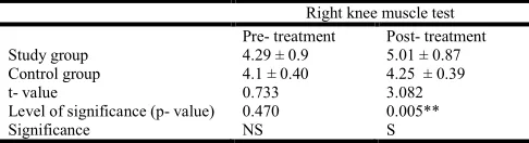
Table 5. Comparative analysis of right knee muscle test between study and control groups
Right knee muscle test Pre- treatment Post- treatment
Study group 4.29 ± 0.9 5.01 ± 0.87
Control group 4.1 ± 0.40 4.25 ± 0.39
t- value 0.733 3.082
Level of significance (p- value) 0.470 0.005**
Significance NS S
Data are presented as mean ± SD, **P < 0.01.
Table 6. Comparative analysis of left knee muscle test between study and control groups
Left knee muscle test Pre- treatment Post- treatment
Study group 4.02 ± 0.79 4.79 ± 0.76
Control group 4.02 ± 0.4 4.21 ± 0.45
t- value 0.000 2.552
Level of significance (p- value) 1.000 0.016*
Significance NS S
Data are presented as mean ± SD, *P < 0.05.
Table 7. Comparative analysis of right elbow muscle test between study and control groups
Right elbow muscle test Pre- treatment Post- treatment
Study group 3.45 ± 0.65 4.25 ± 0.6
Control group 3.43 ± 0.49 3.55 ± 0.58
t- value 0.126 3.204
Level of significance (p- value) 0.900 0.003**
Significance NS S
Data are presented as mean ± SD, **P < 0.01.
Table 8. Comparative analysis of left elbow muscle test between study and control groups
Left elbow muscle test Pre- treatment Post- treatment
Study group 3.66 ± 0.55 4.36 ± 0.49
Control group 3.62 ± 0.54 3.83 ± 0.63
t- value 0.168 2.587
Level of significance (p- value) 0.868 0.015*
Significance NS S
Data are presented as mean ± SD, *P < 0.05.
Table 9. Comparative analysis of clotting time between study and control groups
clotting time Pre- treatment Post- treatment
Study group 6.54 ± 0.84 5.92 ± 0.87
Control group 6.69 ± 0.86 6.58 ± 0.82
t- value -0.460 -2.134
Level of significance (p- value) 0.649 0.042*
Significance NS S
Data are presented as mean ± SD, *P < 0.05.
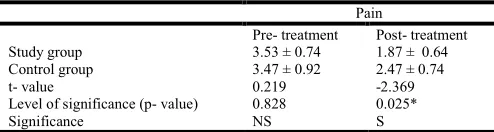
Table 10. Comparative analysis of pain between study and control groups
Pain
Pre- treatment Post- treatment
Study group 3.53 ± 0.74 1.87 ± 0.64
Control group 3.47 ± 0.92 2.47 ± 0.74
t- value 0.219 -2.369
Level of significance (p- value) 0.828 0.025*
Significance NS S
Data are presented as mean ± SD, *P < 0.05.
The increase in the left knee muscle strength for the study group at the end of the treatment was significantly higher than in the control group (P < 0.05), as shown in Table 7. The increase in the right elbow muscle test for the study group at the end of the treatment was statistically significant in comparison to baseline (P < 0.05). The increase in the right elbow muscle test for the control group at the end of the treatment was statistically significant in comparison to baseline (P < 0.05). The increase in the right elbow muscle strength for the study group at the end of the treatment was significantly higher than in the control group (P < 0.05), as shown in Table
8. The increase in the left elbow muscle test for the study group at the end of the treatment was statistically significant in comparison to baseline (P < 0.05). The increase in the left elbow muscle test for the control group at the end of the treatment was statistically significant in comparison to baseline (P < 0.05). The increase in the left elbow muscle strength for the study group at the end of the treatment was significantly higher than in the control group. (P < 0.05), as shown in Table 9. The decrease in pain for the study group at the end of the treatment was statistically significant in comparison to baseline (P < 0.05). The decrease in the pain for the control group at the end of the treatment was statistically significant in comparison to baseline (P < 0.05). The decrease in the pain for the study group at the end of the treatment was significantly higher than in the control group (P < 0.05), as shown in Table 10.
DISCUSSION
The results of the current study showed that there was a significant difference between study and control groups post treatment in pain, bleeding and muscle test values. The present study results agreed with Joyce et al., 2010 who mentioned that regular exercise program can significantly improve joint ROM in patients with haemophilic arthritis who had not previously participated in a regular exercise program .There was also increase of function that accompanies increased ROM. Improved the elbow ROM is accompanied by enhancing the independency in doing activities of daily living such as the ability of individuals to reach their head, neck, and face, helping them to wash their face and hair, shave, apply makeup, brush and floss teeth, feed themselves. Strength significantly improved for all muscle groups (Joyce et al.,
2010). The present study results agreed with Pelletier et al.,
1987 who reported that there was increased strength in the right hamstring and quadriceps muscle without reporting any side effects. The present study results agreed with Joyce et al.,
2010 who reported that it is safe and effective to do intensive isotonic strengthening program for haemophilic patients (Pelletier et al., 1987). Joyce et al., 2010 also mentioned that Haemophilia and bleeding disorders are rare and may result in chronic musculoskeletal problems and functional disabilities. Physical therapists have a big role in protecting people with bleeding disorders from episodes of bleeding and joint destruction and also promoting independency.
Although exercise is recommended for recovery from bleeding episodes, as well as for health, fitness, and enhanced quality of life, there are few reported evidence based studies reported that there are many safety guidelines should be followed to void occurrence of any dangerous situations with haemophilic patients (Pelletier et al., 1987). Haemophilic patients are characterized by reduced muscle strength (Falk et al., 2000), which in turn leading to joint instability resulting in decrease in their ability to withstand stress (Tiktinsky et al., 2002). When the muscles become weaker, the joint is less protected which results in increasing the frequency and severity of haemarthrosis and leads to chronic synovitis and joint arthropathy .Enhancement in muscle strength will increase joint stability, provide protection for the joint, and eventually result in reduced frequency and severity of spontaneous bleeds, thus delaying arthropathy (Tiktinsky et al., 2002). Tiktinsky et
al., 2002 also mentioned that resistance training has a great
general (e.g. elevated endorphin levels in response to aerobic exercise, and resistance training in particular (e.g. elevated levels of anabolic hormones, it is tempting to speculate that some of these effects may be related to the apparent reduction in pain (Tiktinsky et al., 2002). The results of the current study agreed with Tiktinsky et al., 2002 who mentioned that increased muscle strength and increased range of motion were accompanied by reduced bleeding frequency. Additionally, it was noted that recurrent haemarthrosis were evident when one of the subjects discontinued his training (Tiktinsky et al.,
2002). The results of the current study agreed with Tiktinsky et
al., 2002 who mentioned that resisted exercise training is of
great benefits in reducing the frequency of spontaneous bleeds so the joint damage which is accompanied with the bleeds in young haemophilic patients will decrease (Tiktinsky et al.,
2002). The results of the current study agreed with Pelletier et al. 1987 who mentioned that reported a 40–70% strength enhancement in a 12-year-old haemophilia patient suffering from chronic knee arthroses who trained for 3 weeks, performing 10 isometric repetitions of 10 s once daily. The authors noted that throughout the training period there was no increase in haemarthroses or muscle bleeds, although no data were provided (Tiktinsky et al., 2002). The results of the current study agreed with Tiktinsky et al., 2002 who mentioned that resistance training resulting in decreasing bleeding frequency. The reduction of bleeding was accompanied by obvious reduction in the associated pain. (Tiktinsky et al., 2002). The available results demonstrate an increase in activity behavior without negative influences on bleeding frequency and factor consumption (Herbsleb et al.,
2006). Frequency and severity of bleeding episodes in patients with haemophilia vary significantly, not only between patients with identical levels of factor deficiency, but also in the same individuals during different periods. Exercise is considered the only practical applicable method which can alter the coagulation parameters in patients with haemophilia (Koch et al., 1984).
The findings of the current study disagree with Koch et al.,
1984 who reported that although there was shortening of the prothrombin time after exercise in both mild and severe haemophilias, there were no significant changes in any component of the factor VIII molecule in the severe patients (Koch et al., 1984). The results of the present study disagree with Broderick et al., 2012 who reported a small risk of bleeding associated with vigorous physical activity especially with children and adolescents with haemophilia (Broderick et
al., 2012). The results of the present study disagree with Ross
et al., 2009 who did not report correlation between the
correlation physical activity and bleeding outcomes in 37 children with severe haemophilia in the two separate studies the author designed (Ross et al., 2009). The results of the present study disagree with Tiktinsky et al., 2009 who studied 44 children with haemophilia not receiving prophylaxis and found no correlation between number of bleeds and physical activity level, although the group that practiced strenuous exercise had a higher proportion of traumatic bleeds. These studies involved between-participant comparisons so are potentially exposed to selection bias (Tiktinsky et al., 2002). The results of the present study disagree with Collins et al.,
2009 who designed a cohort study on children and adults with hemophilia and reported that the increase in the risk of bleeding increased by 1.4% to 2.2% depending on their age for every additional hour per week as factor VIII levels were less than 1%. In this study pharmacokinetic measures were used to
measure clotting factor levels for each child. There are many factors which may interfere with the accuracy of the results such as the absence of the control group to make comparison with the study group and also the bias occurred in the sample selection (Collins et al., 2009).
Conclusion
On the findings of the present data, it is possible to conclude that the designed therapy program consisted of electrical stimulation, stretching exercises and range of motion exercises when accompanied with the medical treatment can achieve much more improvement in pain reduction, increasing muscle strength and decrease bleeding in patients with haemophilia. So, it is recommended to be added to the traditional medical treatment of patients with haemophilia.
Acknowledgement: The author would like to thank the study
participants for their time and effort.
Conflict of Interest Statement: The authors declare that there
is no conflict of interest in this study. The manuscript has not been published and is not under consideration for publication in any other journal. The manuscript has been read and approved by authors.
Funding statement: The authors whose names were listed
immediately above certify that they have no affiliations with or involvement in any organization or entity with any financial interest (such as participation in speakers’ bureaus; membership, employment, consultancies, stock ownership, or other equity interest or non-financial interest (such as personal or professional relationships, affiliations, knowledge or beliefs) in the subject matter or materials discussed in this manuscript.
REFERENCES
Aznar J.A., Lucia F., Abad-Franch L., Jimenez-Yuste V., Perez R., Batlle J. et al. 2009. Haemophilia in Spain. Haemophilia, 15:665–75.
Broderick C R., Herbert R D., Latimer J, Barnes C, Curtin J A., Mathieu E et al. 2012. Association Between Physical Activity and Risk of Bleeding in Children With Hemophilia JAMA, 308(14):1452-1459.
Callahan L.F. 2009. Physical activity programs for chronic arthritis. Curr Opin Rheumatol., 21: 177–82.
Collins PW, Blanchette VS and Fischer K. 2009. rAHFPFM Study Group. Break-through bleeding in relation to predicted factor VIII levels in patients receiving prophylactic treatment for severe hemophilia A. J Thromb
Haemost., 7(3):413- 420.
Falk B, Portal S, Tiktinsky R, Weinstein Y, Constantini N, Martinowitz U. 2000. Anaerobic power and muscle strength in young hemophilia patients. Med Sci Sports
Exerc., 32: 52–7.
Felipe Q., Sofia P., Joe E. et al. 2011. Hemophilia : exercises and sports. Apunts Med Esport., 46(169): 29-39 Global Report.
Green D. 2006. The management of acquired haemophilia. Haemophilia; 12: 32–36.
Joyce C, Janet Tuller, Jonathan M. Rose Ruth Mulvany, Audrey R. Zucker-Levin, Michael Jeng and Marion Dugdale, 2010. Effects of a 6-Week, Individualized, Supervised Exercise Program for People With Bleeding Disorders and Hemophilic Arthritis. PHYS THER.; 90:509-526.
Koch B, Galioto FM, Kelleher J. 1984. Physical fitness in children with hemophilia. Arch Phys Med Rehabil., 65: 324–6.
Pelletier JR, Findley TW, Gemma SA. 1987. Isometric exercise for an individual with hemophilic arthropathy.
Phys Ther., 67: 1359–1364.
Ross C, Goldenberg NA, Hund D, Manco-Johnson MJ. 2009. Athletic participation in severe hemophilia: bleeding and joint outcomes in children on prophylaxis. Pediatrics, 124(5):1267-1272.
Srivastava A. 2005. Guidelines for the management of hemophilia": World federation of hemophilia available at: http:// wfh.org/2/ docs /publications/ Diagnosis and treatment /Guidelines management hemophilia. PDF Tiktinsky R, Falk B, Heim M, Martinovitz U. 2002. The effect
of resistance training on the frequency of bleeding in haemophilia patients: a pilot study. Haemophilia, 8: 22–7.