Review Article
Diabetic Retinopathy: A Concise Review
Reeta Devi*,1 Savita Kumari,1 Ankit Verma,
Rubina Sharma1
Department of Pharmacology1, Department of
Pharmaceutics2, CT Institute of Pharmaceutical
Sciences, Shahpur, Jalandhar, India
Date Received: 12th June 2016; Date accepted:
June 2016; Date Published: 7th
E-mail:reetadevi7618@gmail.com
Abstract
Diabetic retinopathy is most important diabetic complication and remains the primary caus avoidable blindness in working-aged persons. As the global prevalence of diabetes mellitus conti ues to increase, diabetic retinopathy remains a principal cause of vision loss in several developed countries. Most favorable control of blood pre sure, blood glucose, and possibly blood lipids r mains the foundation for decrease of risk of retin pathy development. Novel approaches for DR treatment are intraocular steroid injection and anti vascular endothelial growth-factor (VEGF) agents, are less damaging to the retina than are older treatments. This article will summarize key dete tion and management approaches for the compl cations of diabetes with special prominence on retinopathic complications.
Keywords: Diabetic Retinopathy, Laser Treatment, Vitrectomy
INTRODUCTION
Diabetic retinopathy (DR) is a commo
multi-factorial disease of the retina with great s cial impact and complex pathogenesis which i cludes a variety of different molecules, cells and factors. Diabetic retinopathy results in ch
mediators such as neurotrophic factors, growth factors, vasoactive agents, cytokines/chemokine’s and adhesion molecules which results in vascular
Diabetic Retinopathy: A Concise Review
Ankit Verma,2
, Department of , CT Institute of Pharmaceutical
har, India
2016; Date accepted: 30th th July 2016
reetadevi7618@gmail.com
Diabetic retinopathy is most important diabetic complication and remains the primary cause of aged persons. As the global prevalence of diabetes mellitus contin-ues to increase, diabetic retinopathy remains a principal cause of vision loss in several developed countries. Most favorable control of blood
pres-lood glucose, and possibly bpres-lood lipids re-mains the foundation for decrease of risk of retino-pathy development. Novel approaches for DR treatment are intraocular steroid injection and
anti-factor (VEGF) agents, to the retina than are older treatments. This article will summarize key detec-tion and management approaches for the compli-cations of diabetes with special prominence on
Diabetic Retinopathy, Laser Treatment,
Diabetic retinopathy (DR) is a commonly known factorial disease of the retina with great so-cial impact and complex pathogenesis which in-cludes a variety of different molecules, cells and
nopathy results in change in trophic factors, growth tive agents, cytokines/chemokine’s and adhesion molecules which results in vascular
injuries and ultimately cell death.
past, diabetic retinopathy was commonly cons dered as vascular disorder of the retina which u timately leads to vascular damage. It has become evident from last few years that significant retinal neurons damage is also present in early stages of diabetic retinopathy. As the surveys continue and data are produced, it seems that d
neurons also plays a significant role in micro va cular. 5-9
SYMPTOMS OF DIABETIC RETIN
TYPES OF DIABETIC RETINOPATHY
1. ‘Non-Proliferative’ diabetic retinopathy (NPDR)
2. ‘proliferative’ diabetic retinop
Non-proliferative diabetic retinop
NPDR is characterized by micro infarcts, hemor hages, exudate, and micro aneu
can be classified into mild, moderate and severe depending on the extent of these changes (Table 1). Micro infarcts also known as cotton wool spots or delicate exudates which show up in cutting edge phases of NPDR because of vascular i
and they show up as white injuries with unclear edges when they heal they may frame a disco raged area because of tissue loss.
injuries and ultimately cell death.1-4 In the current
past, diabetic retinopathy was commonly consi-ed as vascular disorder of the retina which ul-cular damage. It has become evident from last few years that significant retinal neurons damage is also present in early stages of abetic retinopathy. As the surveys continue and produced, it seems that degeneration of neurons also plays a significant role in micro
vas-SYMPTOMS OF DIABETIC RETINOPATHY
TYPES OF DIABETIC RETINOPATHY
Proliferative’ diabetic retinopathy
‘proliferative’ diabetic retinopathy (PDR)
proliferative diabetic retinopathy.10
NPDR is characterized by micro infarcts, hemorr-urysm. This further can be classified into mild, moderate and severe depending on the extent of these changes (Table 1). nfarcts also known as cotton wool spots or delicate exudates which show up in cutting edge phases of NPDR because of vascular impediment and they show up as white injuries with unclear edges when they heal they may frame a
Hemorrhages happen because of break of wea-kened vessels. They can be little spots or bigger blot hemorrhages present inside the thickly pressed more profound layers of retina.
Hard exudates comprise of lipoproteins and differ-ent proteins spilling through strange retinal ves-sels. They show up as yellow lipid stores with a waxy or gleaming appearance and may shape a circinate example around foci of spilling vessels and micro aneurysms.
Micro aneurysms are out pouchings of vessels and are among the primary clinically distinguishable indications of retinopathy. They emerge because of expanding of debilitated narrow dividers or endo-thelial buds endeavoring to revascularize ischemic retina. They show up as modest red dots, ordinari-ly fleeting to the macula. Even though micro aneu-rysms are not settled components and may even disappear. Sudden appearance of various miniatu-rized scale aneurysms means that compounding
retinal ischemia
Proliferative diabetic retinopathy (PDR). 11 PDR is the highly developed phase of diabetic reti-nopathy. It is described by new vessel arrangement usually emerging on the optic disk (New vessels on the disk) or emerge on different parts of the retina (new vessel somewhere else or NVE) gener-ated by ischemic changes in the retina and an im-balance amongst angiogenic and antiangiogenic variables. The New vessel on the disk (NVD) con-veys the most noticeably bad visualization because of numerous variables including connection of the vitreous to the optic disk. Early phase of PDR be-gins as neovascularization and pre-retinal hemorr-hages (Table 1). This may advance to vitreous he-morrhages and in late stages it might bring about tractional retinal separation and neovascular glau-coma.
Table 1: The Early Treatment Diabetic Retinopathy Study (ETDRS) grading system. 12
Types Sub types Characteristics
Non-Proliferative Diabetic
Retinopathy (NPDR)
Mild to moderate Hemorrhages, intra-retinal, hard exudates, Macular edema, micro aneurysms,
Moderate to severe Extensive intra-retinal hemorrhages and/or micro aneurysms, intra retinal microvascular abnormalities (IRMA), venous beading, Cotton wool spots
Severe to very severe IRMA, venous beading, Plus Cotton wool spots. Sim-plified by 4:2:1 i.e. Intraretinal hemorrhages: Venous beading: IRMA.
Proliferative Diabetic Retinopathy (PDR)
Disk neovascularization Neovascularization somewhere else in the retina
Early PDR Pre-retinal hemorrhage
Proliferative Diabetic Retinopathy with high-risk criteria
High risk:- the occurrence of any of the following:
• Vitreous hemorrhage
• New vessels on the disk >1/3 DD
(most significant prognostic factor for severe visual loss)
• New vessels somewhere else >1/2 DD
Advanced eye diseased PDR
Neo vascularization of the iris, Tractional retinal de-tachment.
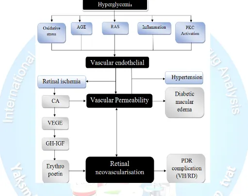
Pathophysiology And Current Strategies. 13-15
Our consideration of the pathophysiological mechanisms essential for the development of diabetic retinopathy is always evolving with new research.
AGE:- Advanced Glycation End-products; RAS:- Renin-Angiotensin System; PKC:- Protein Kinase C; CA:- Carbonic Anhydrase; VEGE:- Vascular Endothelial Growth Factor; GH-IGF:- Growth Factor–Insulin Growth Factor; PDR:- Proliferative Diabetic Retinopathy; VH:- Vitreous Haemorrhage; RD:- Retinal Detachment
Table 2: Current techniques of treatments in diabetic macular edema 16
Sr. no. Current strategies Treatment For
1 Systemic factor control Lipids, blood pressure and blood glucose is still the ’gold standard’ treatment for diabetic retinopathy.
2 Focal/grid laser non-centre-involving diabetic macular edema only
3
Anti-vascular endothelial growth factor (VEGF) intravi-treal injections
patients with centre-involving diabetic macular oedema
4 Steroids For poor responders to anti-VEGF therapies
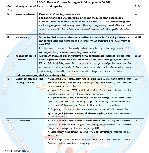
Table 3: Role of Current Strategies in Management Of DR Sr.
No.
Management of diabetic retinopathy Ref.
1 Laser treatment Perform PRP for high risk of PDR.
For earlystagesof PDR, start PRP after any maculopathy isStabilized Suppose PRP for severe NPDR, mainly if there is T2DM, impending cata-ract surgery,poor follow-up compliance, pregnancy, renal disease, and severe disease in the fellow eye or confirmation of retinopathy develop-ment.
17
2 Vitrectomy Consider that there is vitrectomy within 3 months for T1DM patients
hav-ing severe vitreous hemorrhage in eyes which suspected to have very se-vere PDR.
Furthermore, consider the early vitrectomy for eyes having severe PDR, not responding to extensiveandaggressive PRP.
18-22
3 Management of
Cataract
Carefully evaluate DR in patients with considerable cataract. Before cata-ract surgery made possible efforts to treat any DME with grid/focal laser. When DR is stable, consider that cataract surgery helps to improve the vision in diabetic patients. If the cataract is moderate to advanced, so con-sider surgery to sufficiently assess need or to permit laser treatment.
23-26
Role in managing diabetes retinopathy
4 Laser Treatment
(Pho-tocoagulation)
• Multiple RCT, including the ETDRS and DRS, have found that the pan-retinal photocoagulation (PRP) considerably reduces the risk of severe vision loss.
At least 50% from PDR and that grid or focal laser photocoagula-tion decreases the risk of moderate vision loss.
• Apply focal laser photocoagulation utilizing 100-micron laser burns to the areas of focal spillage (i.e. spilling microaneurysms) and areas of fine non-perfusion in the perimacular section.
• Apply grid laser photocoagulation utilizing 50-100 micron burn up in a grid pattern to areas of diffuse spillage and non-perfusion at the macula.
19
5 Vitrectomy • The Diabetic Retinopathy Vitrectomy Study (DRVS) was a
multi-focus RCT that assessed signs and timing of standards plana vitrec-tomy for management of cutting edge DR.
• Vitrectomy was found in little RCT to advantage chronic or dif-fuse DME.
OCT is significant to establish and evaluate DME, and to confirm footing and its reaction to surgery.
18
ABBREBIATIONS:-
CONCLUSION
Regardless of good control of systemic risk factors, a huge number of patients will in any case advance to create vision-undermining diabetic retinopathy (either macular oedema or proliferative retinopa-thy). The present standard for the management of DR are laser treatment, vitrectomy, and not all around powerful in inversion of visual loss. Along these lines, new methodologies have likewise de-veloped, for example, utilization of intraocular organization of hostile to VEGF specialists and corticosteroids in selected eyes. However, oph-thalmologists and physicians should be awake not only of the noticeable benefits but also of the poss-ible risks associated with these novel therapies.
REFERENCES
1. Abcouwer SF, Gardner TW. Diabetic retinopa-thy: loss of neuroretinal adaptation to the di-abetic metabolic environment. Annals of the New York Academy of Sciences. 2014; 1311: 174–190.
2. Ola MS, Nawaz MI, Siddiquei MM, Al-Amro S, Abu El-Asrar AM. Recent advances in un-derstanding the biochemical and molecular mechanism of diabetic retinopathy. Journal of Diabetes and Its Complications. 2012; 26(1): 56– 64.
3. Qian HH, Ripps. Neurovascular interaction and the pathophysiology of diabetic retinopa-thy. Experimental Diabetes Research.2011; 2011: 1-11.
4. Tarr JM, Kaul KM, Chopra EM, Kohner, Chib-ber R. Pathophysiology of diabetic retinopa-thy. ISRN Ophthalmology. 2013; 2013: 1-13. 5. Antonetti DA, Barber AJ, Bronson SK.
Diabet-ic retinopathy: seeing beyond glucose-induced microvascular disease. Diabetes.2006; 55(9): 2401–2411,
6. Hernandez C, Simo R, Neuro-protection in diabetic retinopathy. Current Diabetes Re-ports, 2012; 12(4): 329– 337,
7. Jindal V. Neurodegeneration as a primary change and role of neuroprotection in diabetic retinopathy. Molecular Neurobiology. 2015; 51(3): 878–884,.
8. Zhang X, Wang N, Barile GR, Bao S, Gillies M. Diabetic retinopathy: neuron protection as a therapeutic target. International Journal of
Bi-ochemistry and Cell Biology. 2013; 45(7): 1525– 1529,
9. Simo R, Hernandez C. Novel approaches for treating diabetic retinopathy based on recent pathogenic evidence. Progress in Retinal and Eye Research. 2015; 48: 160–180.
10. Diabetic Retinopathy Study Research Group. Preliminary report on effects of photocoagula-tion therapy. Am. J. Ophthalmol. 1976; 81: 383– 396.
11. Patz A, Smith RE. The ETDRS and diabetes 2000. Ophthalmology. 1991; 98: 730–740. 12. Abdulrahma A. Alghadyan M.D. Diabetic
re-tinopathy – An update. Saudi Journal of Oph-thalmology. 2011, 25, 99-111
13. Curtis TM, Gardiner TA, Stitt AW. Microvas-cular lesions of diabetic retinopathy: clues to-wards understanding pathogenesis? Eye.2009; 23: 1496–1508.
14. Antonetti DA, Barber AJ, Bronson SK. Diabetic retinopathy: seeing beyond glucose-induced microvascular disease.Diabetes.2006; 55: 2401– 11.
15. Ciulla TA, Amador AG, Zinman B. Diabetic retinopathy and diabetic macular edema: pa-thophysiology, screening, and novel therapies. Diabetes Care.2003; 26: 2653–64.
16. New treatments for diabetic retinopathy-Das1,2, S. Stroud1, A. Mehta1 & S. Rangasa-my3 Diabetes, Obesity and Metabolism.17: 219–230, 2015.
17. Early photocoagulation for diabetic retinopa-thy. ETDRS report number 9. Early Treatment Diabetic Retinopathy Study Research Group. Ophthalmology. 1991; 98: 766-785.
18. Early vitrectomy for severe vitreous hemorr-hage in diabetic retinopathy. Two-year results ofa randomized trial. Diabetic Retinopathy Vi-trectomy Study report 2. The Diabetic Retino-pathy Vitrectomy Study Research Group. Arch.Ophthalmol. 1985; 103: 1644-1652. 19. Early vitrectomy for severe vitreous
hemorr-hage in diabetic retinopathy. Four-year results of a randomized trial: Diabetic Retinopathy Vi-trectomy Study Report 5 Arch Ophthal-mol.1990; 108: 958-964.
21. Pendergast SD, Hassan TS, William GA. Vi-trectomy for diffuse diabetic macular ede-maassociated with a taut premacular posterior hyaloid. Am J Ophthalmol. 2000; 130: 178-186. 22. Kaiser PK, Riemann CD, Sears JE, Lewis H.
Macular traction detachment and diabetic ma-cular edema associated with posterior hya-loidal traction. Am J Ophthalmol. 2001;131:44- 49.
23. Wilson A, Baker R, Thompson J, Grimshaw G. Coverage in screening for diabetic retinopathy according to screening provision: results from a national survey in England and Wales. Di-abet. Med. 2004; 21: 271-278.
24. Sangha SS. Severe diabetic retinopathy after cataract surgery [letter]. Am. J. Ophthalmol. 1994; 118: 681-682.
25. Mittra RA, Borrillo JL, Dev S, Mieler WF, Koe-nig SB. Retinopathy progression and visual Outcomes after phacoemulsification in pa-tients with diabetes mellitus. Arch Ophthal-mol. 2000; 118: 912-917.