Structural Dynamics as a Contributor to Error-prone
Replication by an RNA-dependent RNA Polymerase
*
□SReceived for publication, October 2, 2014, and in revised form, October 30, 2014Published, JBC Papers in Press, November 6, 2014, DOI 10.1074/jbc.M114.616193
Ibrahim M. Moustafa‡, Victoria K. Korboukh‡1, Jamie J. Arnold‡, Eric D. Smidansky‡, Laura L. Marcotte§2, David W. Gohara§3, Xiaorong Yang¶, María Antonieta Sa´nchez-Farra´n储, David Filman§, Janna K. Maranas储, David D. Boehr¶, James M. Hogle§, Coray M. Colina**, and Craig E. Cameron‡4
From the‡Department of Biochemistry and Molecular Biology, the¶Department of Chemistry, the储Department of Chemical Engineering, and the**Department of Materials Science and Engineering, The Pennsylvania State University, University Park, Pennsylvania 16802 and the§Department of Biological Chemistry and Molecular Pharmacology, Harvard Medical School,
Boston, Massachusetts 02115
Background:The physical basis for polymerase fidelity remains unclear.
Results:Molecular dynamics simulations, NMR, and pre-steady-state kinetics of a mutator polymerase reveal conformational states of the active site that serve as fidelity checkpoints.
Conclusion:Protein dynamics, largely concealed by x-ray crystallography, govern incorporation fidelity. Significance:Strategies have been inspired for engineering (anti)mutator polymerases and attenuated viruses.
RNA viruses encoding high- or low-fidelity RNA-dependent RNA polymerases (RdRp) are attenuated. The ability to predict residues of the RdRp required for faithful incorporation of nucleotides represents an essential step in any pipeline intended to exploit perturbed fidelity as the basis for rational design of vaccine candidates. We used x-ray crystallography, molecular dynamics simulations, NMR spectroscopy, and pre-steady-state kinetics to compare a mutator (H273R) RdRp from poliovirus to the wild-type (WT) enzyme. We show that the nucleotide-bind-ing site toggles between the nucleotide bindnucleotide-bind-ing-occluded and nucleotide binding-competent states. The conformational dynamics between these states were enhanced by binding to primed template RNA. For the WT, the occluded conformation was favored; for H273R, the competent conformation was favored. The resonance for Met-187 in our NMR spectra reported on the ability of the enzyme to check the correctness of the bound nucleotide. Kinetic experiments were consistent with the conformational dynamics contributing to the established pre-incorporation conformational change and fidelity check-point. For H273R, residues comprising the active site spent more time in the catalytically competent conformation and were more positively correlated than the WT. We propose that by linking the equilibrium between the binding-occluded and binding-competent conformations of the nucleotide-binding pocket and other active-site dynamics to the correctness of the
bound nucleotide, faithful nucleotide incorporation is achieved. These studies underscore the need to apply multiple biophysical and biochemical approaches to the elucidation of the physical basis for polymerase fidelity.
The list of (re)emerging viruses continues to expand, and with this expansion comes an increased risk of the evolution of a highly virulent, highly transmissible strain and a correspond-ing pandemic. It is cost-prohibitive to treat every (re)emergcorrespond-ing agent as the etiologic agent of the next pandemic. However, when that pandemic comes, a rapid response with antiviral therapies and vaccines will be absolutely essential. With this circumstance in mind, the viral RNA-dependent RNA poly-merase (RdRp)5is a very attractive target for both the design of
broad spectrum, antiviral therapies and mechanism-based strategies for viral attenuation and vaccine development (1, 2). It is becoming increasingly clear that the ability of a virus population to be sustained in the presence of antiviral responses of the host and other bottlenecking events requires a genetically diverse virus population. In the presence of this genetic diversity, selection can act to evolve the most robust population for that place and time. For RNA viruses, an impor-tant source of this genetic diversity is the nucleotide misincor-poration frequency of the RdRp (3). For example, in poliovirus one or two errors are introduced into the viral genome during each replication cycle. Interestingly, in the coronaviruses (e.g.
severe acute respiratory syndrome virus), a proofreading exo-nuclease exists that can remove mistakes made by the RdRp (4). It is possible that both enzymatic (e.g.deamination) and oxida-tive (e.g. 8-oxo-G) modifications of RNA also contribute to genetic diversity (5).
*This work was supported by National Institutes of Health Grant R01 AI045818 from NIAID (to C. E. C.).
□S This article containssupplemental Movies S1–S5.
1Present address: AstraZeneca, Innovative Medicines, Discovery Sciences, 35 Gatehouse Dr., Waltham, MA 02451.
2Present address: Dept. of Biochemistry and Molecular Pharmacology, Uni-versity of Massachusetts Medical School, 364 Plantation St., Worcester, MA 01605.
3Present address: Dept. of Biochemistry and Molecular Biology, St. Louis Uni-versity School of Medicine, 1100 South Grand Ave., St. Louis, MO 63104. 4To whom correspondence should be addressed: Dept. of Biochemistry and
Molecular Biology, The Pennsylvania State University, 201 Althouse Labo-ratory, University Park, PA 16802. Tel.: 814-863-8705; Fax: 814-865-7927; E-mail: [email protected].
Numerous examples exist in the literature providing some evidence for sensitivity of viral populations to mutagens like the antiviral agent ribavirin. Resistance to this drug yields viruses harboring mutations in the RdRp that decrease genetic diver-sity while simultaneously decreasing virus fitness (6 –10). Active-site mutagenesis of the RdRp or the proofreading exo-nuclease can lead to increased genetic diversity but neverthe-less decreases virus fitness (11–14). Collectively, observations such as these lead to the suggestion that an optimal genetic diversity exists for maximal fitness. When tested, viruses exhib-iting perturbed genetic diversity are attenuated and serve as vaccine strains (15).
The ability to rationally design RdRp derivatives with increased or decreased nucleotide incorporation fidelity would be of great practical value. The RdRp from poliovirus (PV) is an ideal model system for elucidating physical mechanisms governing nucleotide incorporation fidelity because of the substantial tools that can be applied to this system. Of particular impor-tance to the question of the mechanistic basis of fidelity are the following: pre-steady-state kinetics (16, 17), crystallography (18, 19), NMR (20, 21), molecular dynamics (MD) simulations (22–25), as well as the existence of both low (H273R)- and high-fidelity (G64S) RdRp derivatives (5, 6, 8).
Several observations suggest that nucleotide incorporation fidelity of PV RdRp is governed by the conformational dynam-ics of the active site. First, residues implicated in nucleotide incorporation fidelity are remote from the active site, suggest-ing allosteric control of the conserved structural motifs in the active site involved in nucleotide binding and/or the nucleoti-dyl transfer reaction (Fig. 1A). It is now well established that dynamics and allostery are inextricably linked (26). Second, a conformational change step (Fig. 1B, ERnNTP^ *ERnNTP) observed kinetically has been implicated as the step affected in the high fidelity G64S RdRp (6). The structure of G64S RdRp is essentially identical to the wild-type enzyme (27). Finally, struc-tural studies show that a conformational rearrangement of con-served residues is required for stable binding of an incoming nucleotide (Fig. 1C) (18, 19).
Here we exploited the PV H273R and G64S RdRp to interro-gate the physical basis for nucleotide incorporation fidelity. Our data are consistent with a model in which equilibrium between two conformational states of the active site that inter-convert on the nanosecond timescale are linked to the fidelity checkpoint that occurs on the millisecond timescale. Perturba-tion of this equilibrium alters nucleotide incorporaPerturba-tion fidelity, as shown for the PV RdRp derivatives studied here. Impor-tantly, the active-site amino acid residues contributing to the two conformational states are highly conserved, leading us to propose these interactions as a starting point for the develop-ment of engineered RdRp with altered fidelity that can be extrapolated to other RNA virus systems.
MATERIALS AND METHODS
Construction of H273R Expression Vector
Mutation encoding substitution of His-273 by arginine was introduced into the WT PV RdRp using overlap extension PCR
and subcloning into the expression plasmid pET26-Ub as described previously (5, 28).
Expression and Purification of Proteins
and induced with 0.5 mMisopropyl--D-thiogalactopyranoside (IPTG). Cell growth continued for an additional 4 h at 25 °C before harvesting. Cell pellets were washed once with buffer containing 10 mMTris, pH 8.0, and 1 mMEDTA and stored at
⫺80 °C. Frozen cell pellets were thawed on ice, suspended in lysis buffer (100 mM potassium phosphate, pH 8.0, 0.5 mM EDTA, 20% glycerol, 1 mMdithiothreitol (DTT), 60MZnCl2,
2.8g/ml pepstatin A, and 2.0g/ml leupeptin), and disrupted by passage through a French pressure cell at 20,000 p.s.i. Phe-nylmethanesulfonyl fluoride (PMSF) and Nonidet P-40 were added immediately after lysis to final concentrations of 1.0 mM and 0.1%, respectively. To precipitate nucleic acid, polyethyl-eneimine was slowly added to the cell lysate at a concentration of 0.025%. The lysate was stirred for 30 min at 4 °C and then centrifuged at 25,000 rpm. Ammonium sulfate fraction-ation was done by slowly adding ammonium sulfate at 40% saturation to the clear solution from the polyethyleneimine precipitation step. The fraction of proteins containing RdRp was pelleted by centrifugation at 25,000 rpm for 30 min at 4 °C. The ammonium sulfate pellet was suspended in buffer A (50 mMTris, pH 8.0, 20% glycerol, 1 mMDTT, 0.1% Nonidet P-40, and 60 MZnCl2) and diluted to 50 mMNaCl. The protein sample was loaded onto a phosphocellulose column (What-man, P-11) that was pre-equilibrated with buffer A containing 50 mMNaCl (⬃1 ml bed volume/20 mg of total protein was used) at a flow rate of 1.0 ml/min. The column was washed to baseline with the equilibrating buffer and eluted with a linear gradient from 50 to 350 mMNaCl in buffer A. Fractions con-taining the protein (WT or H273R), checked by SDS-PAGE, were pooled and diluted to a final concentration of 50 mMNaCl and were further fractionated on an S-Sepharose column fol-lowed by a Q-Sepharose column (equilibrated, washed, and eluted in the same way as the phosphocellulose column). Pro-tein fractions eluted from the Q-Sepharose column were diluted to adjust the salt concentration to 50 mM NaCl and finally concentrated by passing over a Q-Sepharose column equilibrated with buffer B (50 mMHEPES, pH 7.5, 20% glycerol, 1 mMDTT, 0.1% Nonidet P-40, and 60MZnCl2) containing 50 mMNaCl. The protein was stripped from the Q-Sepharose col-umn using 500 mMNaCl in buffer B. The concentrated protein (⬃10 mg/ml) was aliquoted and stored at⫺80 °C.
Pulse-Chase and Pulse-Quench Experiments
Pulse-chase and pulse-quench experiments were performed in the reaction buffer (50 mMHEPES, pH 7.5, 10 mM 2-mercap-toethanol, 60MZnCl2, and 5 mMMnCl2) at 30 °C using a rapid chemical quench-flow instrument (KinTek Corp., Austin, TX). The enzyme solution (4M), WT or H273R, was incubated with a symmetrical self-complementary 10-nt RNA substrate con-taining uracil as the templating base for the first incorporation (S/S-U, 20 M) (GE Healthcare) and rapidly mixed with [␣-32P]ATP (100
M) (MB Biomedicals), and then reactions at various time points were either quenched or pulse-chased by adding a large excess of ATP (20 mM) (Sigma) to proceed for an additional 30 s. Reactions were quenched with HCl to a final concentration of 1.2N followed by immediate neutralization using 3.0MKOH. Samples were quantitated via sequencing gel analysis. Kinetic parameters were obtained by
simulation of the data using KinTek Explorer 2.03 (KinTek Corp.). All rate constants were determined experimentally, except where noted. The agreement between experimental data and kinetic simulations was determined by visual inspection.
Stopped-flow Nucleotide Incorporation Assays
Stopped-flow pre-steady-state nucleotide incorporation assays were performed using a model SF-2001 stopped-flow apparatus (KinTek Corp.) equipped with a water bath. The incorporation of the correct nucleotide (UTP) and nucleotide with an incorrect sugar (2⬘-dUTP) was achieved by using the symmetric RNA duplex S/S-UA substrate (Eurofins Genomics, Huntsville, AL) with the sequence 5⬘ -GpyrCAUGGGCCCA-3⬘), where pyrC or pyrrolo-C at the⫹1 templating position is a fluorescent analog of the cytidine nucleoside that retains its Watson-Crick base-pairing capacity with G. For incorporation of the correct nucleotide (UTP) next to the site of misincorpo-ration, we used the symmetric RNA duplex S/S-UG substrate (Eurofins Genomics) with the sequence 5⬘ -GpyrCAUGGGC-CCG-3⬘), containing pyrC at the⫹1 templating position and a G-U mismatch at the 3⬘-end. All reactions were conducted at 30 °C in the reaction buffer (50 mMHepes, pH 7.5, 10 mM 2-mercaptoethanol, 5 mMMgCl2, and 60MZnCl2). The
reac-tion condireac-tions for PV RdRp, WT or H273R, were optimized. For WT assays, the enzyme (0.5M) was incubated with pyrC-labeled S/S-UA primed template (0.25 M) or pyrC-labeled S/S-UG primed template (1.5M) in the reaction buffer for 3 min and then mixed rapidly with UTP or 2⬘-dUTP solutions of different concentrations. For H273R, similar assays were per-formed using H273R enzyme (2 M) and S/S-UA (1M) or S/S-UG (2.5M). The excitation wavelength was 350 nm, and fluorescence emission was monitored using a 440-nm cut-on filter (model HW440lp, Chroma Technology Corp., Rocking-ham, VT). For each experiment, at least four fluorescent traces were averaged. Relative fluorescence was plotted as a function of time and fit to a single exponential equation (Equation 1), yielding an observed rate constant,kobs,
F⫽A⫻exp共⫺kobst兲⫹C (Eq. 1)
whereAis the amplitude andCis the end point. Values forkobs
were plotted as a function of nucleotide concentration and fit to a hyperbolic equation (Equation 2), providing estimates forkpol,
the maximal rate constant for nucleotide incorporation, and
Kd,app, the apparent dissociation constant for nucleotide.
kobs⫽kpol[NTP]/Kd,app⫹[NTP] (Eq. 2)
Solvent Deuterium Kinetic Isotope Effect (SDKIE)
Experiments were performed essentially as described above for the stopped-flow nucleotide incorporation assays, except enzyme, substrates, and buffers were prepared in 100% D2O. The pD was used instead of pH for solutions in D2O and was
adjusted according to the relationship (pD⫽pH⫹0.4). The SDKIE was calculated as the quotientkpol(H2O)/kpol(D2O).
Crystallization, Data Collection, and Determination of H273R Structure
disrupt interface I of PV RdRp (27). Prior to crystallization, the H273R protein, purified as described above, was further frac-tionated on a HiLoad 26/60 Superdex 200 gel filtration column, equilibrated with buffer containing 5 mMTris, pH 7.5, 200 mM NaCl, 0.1 mMEDTA, and 2 mMDTT as a polishing step. Crys-tals of H273R were grown using vapor diffusion at 20 °C by mixing equal volumes of the protein (10 mg/ml) and the crys-tallization reservoir containing 2 Msodium acetate and 0.1M sodium cacodylate, pH 6.8. For data collection, crystals were soaked briefly in the crystallization solution, adjusted to con-tain 20% glycerol as a cryoprotectant, prior to flash-cooling in a nitrogen stream and use in the diffraction experiment. Diffrac-tion data were collected at the 19-ID beamline at the Advanced Photon Source (Argonne, IL). The data were integrated, merged, and scaled using DENZO and SCALEPACK (29). The structure was determined by molecular replacement in PHASER (CCP4) (30) using the structure of WT PV RdRp (PDB 1RA6) (18) as the search model. The model was built using Coot (31, 32) and refined with REFMAC5 (33, 34).
Molecular Dynamics Simulations
System Setup—The WT PV RdRp and the G64S mutant in their free forms were investigated by all-atom MD simulations (25 ns) in our previous study (23). Thus, simulations for the two systems described here (termed WT and G64S, respectively) started from the end of the previous 25-ns trajectories and extended to a total length of 150 ns. For the H273R mutant, the solved crystal structure (PDB code 4R0E) was used to prepare the initial model. The two surface residues 446 and Asp-455 introduced to help in crystallization were mutated back to the WT residues Leu-446 and Arg-455, respectively. It should be mentioned that the two surface mutations, L446D and R455D, have no impact on the observed dynamics of the WT protein.6The starting coordinates for the WT binary complex
were obtained from the crystal structure (PDB code 3OL6) (19). The primer-template RNA of the crystal structure was trimmed to include only nucleotides that are within 5 Å distance from protein atoms. The initial simulated structure (termed WT-RNA) included protein residues 1– 461 (chain A), a 13-mer RNA template strand (chain B), a 9-mer RNA primer strand (chain C), and 153 structural waters. Also, the two surface res-idues, Asp-446 and Asp-455, reported in the crystal structure were mutated back to the WT residues. For the binary com-plexes of the mutants (G64S and H273R), because there is no available crystal structures these complexes were constructed based on the WT-RNA complex by single residue replacement, leaving other protein residues and RNA base sequences unchanged. The steric clashes generated in the two mutant sys-tems (G64S-RNA and H273R-RNA) were removed by subse-quent energy minimization and equilibration.
MD Simulations Protocol—MD simulations were carried out using the program AMBER12 (35, 36) with AMBER99bsc0 (37), a version of AMBER99SB (38) with improved parameters for nucleic acid. In all simulations, an integration time step of 1 fs was used. Periodic boundary conditions using cutoff radii of 9 Å (in simulations of the free forms) and 12 Å (in simulations of the
RNA-bound forms) were applied in calculating nonbonded interactions. Electrostatic interactions were treated using the particle mesh Ewald method (39, 40). The SHAKE algorithm (41) was employed to constrain all bond lengths involving hydrogen atoms. All MD simulations were performed in explicit water (TIP3P model) (42), imposing a minimal distance of 20 Å between the edge of the solvent box and any protein atom. The simulations were conducted following a scheme that was essentially similar to our previously published work (23). Specifically, the coordinates of the simulated systems were first relaxed to remove any steric clashes between the atoms of pro-teins, RNA, waters, and ions in multiple steps using SANDER; each system was subjected to two cycles of energy minimization followed by short constrained dynamics (100 ps) under condi-tions of constant pressure and temperature (NPT). Subse-quently, the systems were energy-minimized by applying convergence criteria for the energy gradient DRMS (the root-mean-square of the Cartesian elements of the gradient) of 0.1 kcal/mol-Å. Next, each system was slowly heated to 300 K over a period of 150 ps under conditions of constant volume and temperature (NVT) and applying a Berendsen thermostat (43). This was followed by a 200-ps NVT dynamics before switching to NPT dynamics for another 150 ps, applying the Berendsen method with temperature and pressure coupling constants of 1 ps (weak coupling). The NPT dynamics was continued for the remainder of the MD simulations utilizing the parallel version of PMEMD in AMBER12. Analyses were performed using PTRAJ and CPPTRAJ programs (44) of the AMBER package on MD trajectories, which were generated from snapshots retained every 1 ps. All MD simulations were carried out on 128 Intel Xeon X5675 Six-Core processors managed by the High Performance Computing Group at The Pennsylvania State University and on 128 Cray XT3 processors (TeraGrid resourc-es; transitioned to XSEDE in 2011) managed by the Pittsburgh Supercomputing Center.
Cluster Analysis
To identify distinct conformations generated over the last 100 ns of MD simulations, we performed the cluster analysis. The refinement “means” algorithm (45) in PTRAJ was used to cluster the structures extracted at 20-ps intervals across the trajectories. Initially, the number of clusters was determined by carrying out the cluster analysis using the average-linkage (45) algorithm in PTRAJ.
Principal Component Analysis (PCA)
PCA (46, 47) was carried out to separate major motions from irrelevant noise sampled during MD simulations over the last 75 ns by diagonalizing the variance/covariance matrix (S) of the positional deviations of C␣atoms with respect to the average structure. The elements of this matrix are given by Equation 3,
Sij⫽cov共r兲⫽具共ri⫺具ri典t兲共rj⫺具rj典t兲典t (Eq. 3)
whereSij is the element of the covariance (cov) matrix,S; ri
represents the coordinatesx,y, andzof the C␣iatom;rj
repre-sents the coordinatesx,y, andzof the C␣jatom;冓ri冔represents the coordinatesx,y, andzof the C␣iatom in the average
ture;冓rj冔represents the coordinatesx,y, andzof the C␣jatom in
the average structure; andtdenotes the time average over the MD trajectory.
Diagonalization of the matrixSproduced an orthogonal set of eigenvectors (or modes) that describe the directions of max-imum variation in the observed conformational space sampled during simulations. The variance around an average structure represented atomic displacements or motion. In this analysis, our focus was to identify the regions that exhibited changes in the dynamics of the H273R-RNA complex relative to the WT-RNA complex. To do so, for each C␣atom, contributions to the total variance along the top 10 modes (contained⬃50% of the total variance) were summed and normalized to the average of the least contributing residues (5% of total residues). The PCA sum corresponding to WT-RNA was subtracted from that of H273R-RNA to obtain the⌬PCA values for each residue. The
⌬PCA values were mapped onto the structure as shown in Fig. 9.
Dynamic Cross-correlation Map (DCCM)
Information about correlated motions sampled during MD simulations was obtained from the DCCM analysis (48). In this analysis, the cross-correlation of the atomic displacements of C␣atoms was examined across the last 75 ns of the MD simu-lations. For the displacement vectors⌬riand⌬rjof atomsiand
j, respectively, the cross-correlation is given by Equation 4,
C共i,j兲⫽ 具⌬ri䡠⌬rj典
冑
具⌬ri典2具⌬rj典2(Eq. 4)
Matrix C, for which the elementscij are given by the above
equation, was calculated for C␣atoms in PTRAJ. The calcu-lated matrix was visualized and analyzed using MATLAB 2012b (MathWorks, Natick, MA). For completely correlated motions
c(i,j)⫽1, and for completely anti-correlated motionsc(i,j)⫽
⫺1. Complete correlation indicates that the motions have the same phase as well as the same period. Deviations from 1 (or
⫺1) imply either that the motions ofiandjare less correlated (or anti-correlated) or that they deviate from motion along a straight line.
Residence Time Analysis
This analysis was performed using C␣atoms and snapshots extracted from the last 50 ns of the MD trajectories. In this
analysis, we viewed the trajectory of C␣atoms as a collection of successive local cages. For a given C␣atom, the residence time was the average time that the atom spent in a cage. To identify a cage, the atomic coordinates over a 3-ps window were grouped, and a running average of the center of mass was cal-culated. A cage was identified when the distance between three consecutive centers of mass varied less than 5%. The cage iden-tified was characterized by its center of mass and radius; the latter was defined as the one that circumscribed 80% of the cage positions. We considered that an atom left its cage when it spent more than 6 consecutive ps outside of it. This analysis brings new insights to the dynamics of the relatively less mobile atoms, which exhibit longer residence time compared with the more mobile atoms. Further information and details of this analysis will be presented in a separate publication.
Protein NMR
NMR samples were prepared as described previously (20, 49). All NMR experiments were done at 293 K following previ-ously published procedures (20, 49) using a Bruker Advance III 600 MHz spectrometer equipped with a 5-mm inverse detec-tion triple resonance (1H/13C/15N) single axis gradient TCI
cryoprobe.
Figure Preparation
All structures in Figs. 1–14 were prepared with CHIMERA 1.9 (50).
Structure Deposition
The atomic coordinates and structure factors have been deposited in the PDB under ID code 4R0E.
RESULTS
PV H273R RdRp Is an Error-prone Polymerase
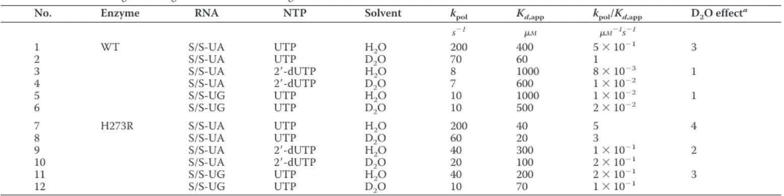
We recently described a PV mutant that exhibits a mutator phenotype in cells and that is attributable to a change of His-273 of the RdRp to Arg (HHis-273R) (5). In that study, we showed that the GMP misincorporation frequency was⬃3-fold higher than observed for the wild-type (WT) enzyme. To determine whether the error-prone activity of the H273R RdRp was lim-ited to base mispairing, we evaluated the ability of this enzyme to utilize a nucleotide with an incorrect sugar configuration (Table 1, 2⬘-dUTP) and the ability of this enzyme to extend a TABLE 1
Kinetic parameters for UTP and 2ⴕ-dUTP incorporation by WT and H273R enzymes in protonic and deuteronic solvents Values are rounded to 1 significant figure. Standard errors range from 10 to 20% of the measured values.
No. Enzyme RNA NTP Solvent kpol Kd,app kpol/Kd,app D2O effect
a
s⫺1
M M⫺1s⫺1
1 WT S/S-UA UTP H2O 200 400 5⫻10⫺
1 3
2 S/S-UA UTP D2O 70 60 1
3 S/S-UA 2⬘-dUTP H2O 8 1000 8⫻10⫺
3 1
4 S/S-UA 2⬘-dUTP D2O 7 600 1⫻10⫺
2
5 S/S-UG UTP H2O 10 1000 1⫻10⫺
2 1
6 S/S-UG UTP D2O 10 500 2⫻10⫺
2
7 H273R S/S-UA UTP H2O 200 40 5 4
8 S/S-UA UTP D2O 60 20 3
9 S/S-UA 2⬘-dUTP H2O 40 300 1⫻10⫺
1 2
10 S/S-UA 2⬘-dUTP D2O 20 100 2⫻10⫺
1
11 S/S-UG UTP H2O 40 200 2⫻10⫺
1 3
12 S/S-UG UTP D2O 10 70 1⫻10⫺
1
aThe D
mispaired primer terminus (Table 1, S/S-UG RNA). To do this, pre-steady-state kinetic methods were employed to determine the maximal rate constant for single nucleotide incorporation (kpol) and the apparent dissociation constant for nucleotide
(Kd,app). As shown previously (5), the catalytic efficiency (kpol/ Kd,app) of the H273R RdRp was 10-fold higher than the WT (Table 1, compare line 7 with line 1). In both experiments, H273R RdRp appeared more promiscuous than the WT, even after taking into account the differences in catalytic efficiency.
Crystal Structure of H273R RdRp Reveals No Significant Differences Relative to WT
Solution of the x-ray crystal structure of the high fidelity PV G64S RdRp was unable to show a significant difference relative to WT, and the altered fidelity was attributed to differences related to dynamics (23). Although we did not expect substan-tial structural perturbations with this enzyme, we pursued the x-ray crystal structure of the H273R RdRp to make sure that we did not miss any significant structural difference that could explain the biochemical and biological differences observed for the H273R mutant. Data collection and refinement statistics are shown in Table 2. Superposition of the C␣atoms of H273R and the WT RdRp structures showed a root-mean-square devi-ation (r.m.s.d.) of 0.33 Å, which was essentially no change (Fig. 2A). Most of the changes were localized to the site of the H273R substitution; these changes accommodated the arginine side chain very well (Fig. 2B).
MD Reveal a Nucleotide Binding-occluded State and
Nucleotide Binding-competent State That Interconvert on the Nanosecond Timescale
MD Design—Previously, we performed 25-ns all-atom and 1
s coarse-grained MD simulations of four picornaviral RdRp (23, 24). These studies revealed conserved and correlated motions of the conserved structural/functional motifs compris-ing the RdRp active site. Analysis of the high fidelity G64S RdRp revealed a substantial change in these motions, which we inter-preted as the basis for the change in fidelity relative to WT (23). In those studies, only the free enzymes were analyzed. In this study, we performed a 150-ns all-atom MD simulation for WT, H273R, and G64S RdRp in the absence and presence of primed template RNA. The primer was 9 nt in length, and the template was 13 nt in length (see “Materials and Methods”). The RdRp-RNA complexes were derived from the structure of the WT RdRp-RNA complex (PDB code 3OL6) (19) (see Materials and Methods”). Analysis of the r.m.s.d. of the backbone of the stable core of the protein (palm subdomain) revealed that all trajecto-ries had reached stability by 50 ns (Fig. 3). We used only the last 100 ns of the trajectory for data analysis. The average r.m.s.d. value decreased when RNA was bound (compare each enzyme
FIGURE 2.Crystal structure of H273R RdRp.A, structural model of H273R RdRp (green) is superimposed onto the structure of WT RdRp (gray), indicating very little difference between these structures (r.m.s.d.⬃0.33 Å). The site of mutation is indicated.B, the 2Fo⫺Fcelectron density map contoured at the
1.4level is shown at the site of mutation. The larger Arg side chain is accom-modated without any steric issues.
FIGURE 3.Analysis of the r.m.s.d. for trajectories of MD simulations.To determine the duration of time to use for analysis, the r.m.s.d. for the backbone atoms of the palm subdomain of the free (WT, G64S, and H273R) and binary complexes (WT-RNA, G64S-RNA, and H273R-RNA) were plotted (black line). In addition, the r.m.s.d. values for the sugar-phosphate backbone of the primer RNA (cyan) and template RNA (blue) of the RNA substrate were plotted. The proteins exhibited no dramatic fluctuations after 50 ns; therefore, the last 100 ns of the trajectories were used for analysis. Over this range of time, the amplitude of motion of the primed template was larger but regular.
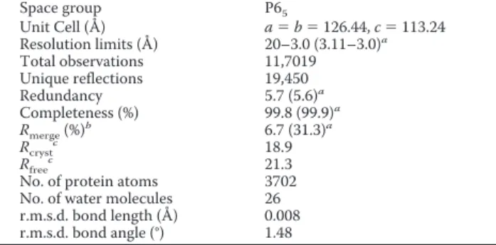
TABLE 2
Data collection and refinement statistics for the H273R structure
Space group P65
Unit Cell (Å) a⫽b⫽126.44,c⫽113.24 Resolution limits (Å) 20–3.0 (3.11–3.0)a
Total observations 11,7019 Unique reflections 19,450 Redundancy 5.7 (5.6)a
Completeness (%) 99.8 (99.9)a
Rmerge(%)
b
6.7 (31.3)a
Rcryst
c
18.9 Rfree
c
21.3 No. of protein atoms 3702 No. of water molecules 26 r.m.s.d. bond length (Å) 0.008 r.m.s.d. bond angle (°) 1.48
aValues in parentheses are for the highest resolution shell. bR
merge⫽ 兺兩Ih⫺ 具Ih典兩/兺Ih, over allhwhereIhis the intensity of reflectionh.
cR
crystand Rfree⫽ 兺储Fo兩⫺兩Fc储/兺兩Fo兩, whereFoandFcare the observed and
calcu-lated amplitudes, respectively.Rfreewas calculated using the 5% of data excluded
in the absence and presence of RNA shown in Fig. 3), consistent with RNA binding reducing the overall flexibility of the protein. The sugar-phosphate backbone of the primer, which is in a stable duplex with the template and has substantial interactions with the enzyme (19), was least flexible (Fig. 3). In contrast, the template was much more flexible (Fig. 3); most of this flexibility derived from then⫹2 ton⫹4 regions of template that inter-act with the fingers subdomain away from the inter-active site (19).
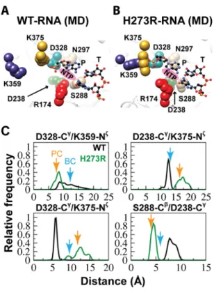
Identification of an Equilibrium between the Nucleotide Binding-occluded and Nucleotide Binding-competent States—A comparison of the crystal structures of the WT RdRp-RNA complex (binary in Fig. 1C) to the post-incorporation complex (Fig. 1C) clearly revealed the need for reorganization of the nucleotide-binding site to occur to permit nucleotide binding (19). The side chains of two residues conserved in essentially all RdRp are at the center of this reorganization: Arg-174 (motif F) and Asp-238 (motif A) (Fig. 1C). We performed a cluster anal-ysis of these and surrounding residues using the structures of the conformations observed over the last 100 ns of the simula-tion for WT RdRp and the WT RdRp-RNA complex. The bind-ing-occluded conformation was the only state observed for WT RdRp (Fig. 4A). In contrast, both a binding-occluded state and a binding-competent state were observed once RNA was bound (Fig. 4A). To obtain a kinetic perspective of the interconversion of these states, we plotted the distance between the Arg-174 guanidinium moiety and Asp-238 carboxylate moiety over the entire trajectory for WT RdRp and WT RdRp-RNA complex (Fig. 4B). The WT RdRp started in a binding-competent con-formation during equilibration but sampled the binding-oc-cluded conformation early in the simulation and remained in that conformation for the remainder of the trajectory (Fig. 4B). The presence of RNA caused the sampling of the two states to occur more often, with the population of each state essentially equal (Fig. 4B).
Preorganization of the H273R RdRp Active Site as a Contributor to Its Error-prone Activity
Nucleotide Binding-competent State Favored by H273R RdRp-RNA Complex—We performed a cluster analysis over the last 100 ns of the simulation for the H273R RdRp and H273R RdRp-RNA complex. In the absence of RNA, the nucleotide binding-occluded state was favored by H273R RdRp. The aver-age structure of this state was similar to that observed for WT. However, there was a reorientation of the Asp-238 side chain that would be expected to further antagonize nucleotide bind-ing (Fig. 5A) because of steric interference and the movements of ribose-binding determinants, Ser-288 and Asn-297 (19). The presence of RNA shifted nearly all of the observed conforma-tions to those that would be classified as binding-competent (Fig. 5A). The interconversion of states was again monitored by following the distance between Arg-174 and Asp-238 as a func-tion of time (Fig. 5B). The H273R RdRp started in the binding-competent state, but the binding-occluded state predominated (⬃70% of the whole simulation time) once was sampled (Fig. 5B). In contrast, the H273R RdRp-RNA complex existed most of the time (⬃98% of the whole simulation time) in the binding-competent state (Fig. 5B).
H273R RdRp-RNA Complex Resembles the Post-incorpora-tion Complex Observed Crystallographically—We also per-formed a comparison of the average structures of the RdRp-RNA complexes for WT and H273R observed by MD (Fig. 6,A
andB). In addition to changes in the positions of Arg-174 and Asp-238, substantial differences in the positions of other side chains were also evident (compare Asp-328, Ser-288, Asn-297, Lys-359, and Lys-375 in Fig. 6Awith the corresponding resi-dues in Fig. 6B). Interestingly, these very same residues showed the largest positional differences when the x-ray crystal struc-tures for the binary complex and post-incorporation complex
were compared (Fig. 1C) (19). The post-incorporation complex is the structure most representative of the catalytic state for all of the PV RdRp structures solved to date (19). The nucleotide was incorporated in the crystal but translocation did not occur. The binary complex could be distinguished from the post-in-corporation complex by comparing the distances between sev-eral side chains, as indicated in Fig. 6C. We determined the distance between the indicated residues (the distances between C␥of Asp-328 and Nof Lys-359 and Lys-375 and between C␥ of Asp-238, Nof Lys-375, and Cof Ser-288) at 1-ps intervals over the last 75 ns of the simulation for both the WT and H273R RdRp-RNA complexes. The relative frequency of each distance
was plotted for each complex (Fig. 6C). In all cases, the inter-side chain distances observed for the H273R RdRp-RNA com-plex were closest to those observed in the post-incorporation complex (Fig. 6C). For the WT RdRp-RNA complex, this com-plex was closest to the binary comcom-plex, although the level of coincidence was not as observed for the H273R RdRp-RNA complex (Fig. 6C).
Key Active-site Residues of H273R RdRp-RNA Complex Exhibit Reduced Backbone Dynamics—Our previous MD stud-ies of picornaviral RdRp emphasized changes in dynamics occurring for residues, structural elements, and/or domains exhibiting the most motion (23, 24). To obtain information about differences in regions of RdRp exhibiting limited flexibil-ity, we performed a “residence time” analysis. This analysis pro-vides a kinetic perspective of the motions that is hidden in other analyses, such as a B-factor analysis. The technical details of this new approach are provided under “Materials and Methods.” Essentially, we created a sphere that circumscribes 80% of the conformational space sampled by an atom. Next, we deter-mined the length of time that the atom spends in that sphere. Greater time indicates lower flexibility. A major advantage of this approach is that rigid residues are not present in the base-line but appear as peaks, permitting these residues to be iden-tified easily by visual inspection. The data for the WT and H273R RdRp-RNA complexes are shown in Fig. 7A. The
FIGURE 5.NTP binding-competent state is preferred for the H273R RdRp-RNA complex.A, cluster analysis of the last 100 ns of the MD trajectories of the free and binary complex was performed as described under “Materials and Methods.” Shown is the NTP-binding site. For the free enzyme, the NTP binding-occluded conformation predominated (H273R, Binding-occluded). For the binary complex, the NTP binding-competent conformation predom-inated (H273R-RNA,Binding-competent). CTP has been included in the illustra-tion for reference. Residues shown and colors are as indicated in the legend to Fig. 1.B, the distance between Arg174-NH2and Asp238-OD1 atoms was used as a probe of the binding-occluded (⬍7 Å) or binding-competent (⬎7 Å) state. Shown here is a plot of the distance during the simulation for the free enzyme (H273R) and binary complex (H273R-RNA). The binding-occluded conformation was present 70% of the time for H273R; the binding-competent conformation was present 98% of the time for H273R-RNA.
median residence time was⬃100 ps. We plotted the residence time for each C␣atom on the enzyme structure using a scale that ranged from half of the median value to twice the median value (seeinsetsin Fig. 7A). The palm subdomain was the least flexible.
We used the nonparametric Wilcoxon rank-sum test (51) in R (52) to determine whether statistically significant differences existed between the WT and H273R RdRp-RNA complexes. This analysis identified 109 residues exhibiting statistically sig-nificant differences (p⬍0.05) between the complexes. Of these, only 11 residues became more flexible (shorter residence time) in the H273R RdRp-RNA complex. We focused our attention on residues of the palm known to contribute to nucleotide binding and/or catalysis. These are shown in Fig. 7B and include residues located in conserved structural motifs A, B, and D. All of these residues exhibited longer residence times in the H273R RdRp-RNA complex than observed for the WT complex (Fig. 7C).
Correlated Motions of Functionally Important Residues Are Intensified in the H273R RdRp-RNA Complex—In our first MD study of PV RdRp, we created a DCCM, which showed that the strongest correlated motions, both positive and negative, were observed for functional motifs (23). We repeated this analysis for the RdRp-RNA complexes (Fig. 8). To facilitate interpreta-tion of the data, we again used a scale from ⫺1 (negatively correlated) to ⫹1 (positively correlated) but restricted our attention to functional residues, as indicated in Fig. 8. Residues that were positively correlated in the WT RdRp-RNA complex were also positively correlated in the H273R RdRp-RNA com-plex, but the intensity of the correlation was not always the same. The most striking difference between the WT and H273R RdRp-RNA complexes was that the H273R RdRp-RNA com-plex exhibited a substantial increase in the number and inten-sity of the positively correlated residues (highlightedin Fig. 8). For example, Thr-235, Gly-236, Tyr-237, and Asp-238 are all more positively correlated with Arg-163, Lys-167, and Arg-174 (Fig. 8, compare H273R-RNA with WT-RNA). All of these res-idues interact with the nucleotide substrate. A similar trend existed for the positive correlation between residues interacting with the templating base (Thr-114, Ser-115, Lys-159, Leu-175, Ile-176, Ala-178, and Ser-179) and residues interacting with the nucleotide substrate (Thr-235, Gly-236, Tyr-237, and Asp-238) (Fig. 8). Furthermore, residues of motif A (Asp-233 to Asp-238) appeared more positively correlated with each other in H273R RdRp-RNA relative to WT RdRp-RNA (Fig. 8).
Flexibility of the H273R RdRp Nascent Base Pair-binding Pocket as a Contributor to Its Error-prone Activity
Principal Component Analysis Reveals Larger Amplitudes in the Backbone Motions of Motifs F and G/G⬘ for the H273R RdRp-RNA Complex—The overall dynamics of a protein can be considered as the sum of a distribution of atomic fluctuations
FIGURE 7.Key active-site residues exhibit reduced flexibility in the H273R RdRp-RNA complex.A, to evaluate increased inflexibility of residues in the H273R RdRp-RNA complex relative to the WT complex, we calculated a parameter that we term “residence time.” Details are provided under “Mate-rials and Methods.” Briefly, the duration of time each C␣atom spends in a predetermined, residue-specific volume is calculated. Inflexible residues will have the longest residence time. Shown is a plot of the average residence time during the MD simulation for each residue of the WT (above) and H273R (below) RdRp-RNA complexes. Theinsetsshow the residence times plotted on the structure using the indicatedred-to-greenscale. The palm subdomain exhibited the least flexibility for both enzymes.B, the plots shown inAwere compared for differences. Shown are active-site residues that exhibited reduced flexibility in the H273R RdRp-RNA complex relative to the WT com-plex. Note that the conformations of these residues also differ with the con-formations for the mutator polymerase appearing preorganized for catalysis. C, we compared the plots shown inAusing a Wilcoxon rank-sum test. Shown
around an average position of that atom. The variance of an atom, representing its motion, can be used to calculate princi-pal components (also known as modes). The number of modes is equivalent to the number of atoms evaluated multiplied by three. Each principal component is associated with an eigen-value that defines its magnitude of motion and thus permits each principal component to be ranked.
We performed a PCA (46, 47) for the C␣atoms of the WT and H273R RdRp-RNA complexes to obtain information on the major motions observed during the simulations and the major contributors to these motions. As observed for the RdRp alone (23), conserved structural motifs (A, D, E, F, and G/G⬘) exhib-ited the largest amplitude of motions. To compare the WT and H273R RdRp-RNA complexes, we used the top 10 modes (accounting for⬃50% of the total variance contained in the last 75 ns of the simulations) to calculate the relative displace-ment for each residue in the protein. A relative displacedisplace-ment was calculated by normalizing to the average displacement observed for the least flexible 5% of residues. We calculated a
⌬PCA value by subtracting the relative PCA value for each res-idue of the WT RdRp-RNA complex from that of the corre-sponding residue in the H273R RdRp-RNA complex. A
histo-gram of the data is shown in Fig. 9A. A⌬PCA value of zero means that no change was observed between the two com-plexes. The ⌬PCA mean was 0.1, and the⌬PCA median was 0.03. Therefore, most residues changed very little between the two complexes.
We plotted the⌬PCA values on the structure such that the magnitude of the motion was indicated by varying radii of the tube representation of the structure (Fig. 9B). In general, the conserved structural motifs were more dynamic for the H273R RdRp-RNA complex than that observed for the WT (Fig. 9B). The exceptions were motifs A and D, consistent with the resi-dence time analysis (Fig. 7). This observation led to further scrutiny of the changes observed in the H273R RdRp-RNA complex. Residues in conserved structural motifs exhibiting differences with an absolute value greater than 0.3 are listed in Fig. 9C and are indicated explicitly in the structural model shown in Fig. 9B. All of the residues shown interact with the triphosphate or ribose of the nucleotide substrate and/or the nascent base pair. Seven of the 10 listed active-site residues demonstrated positive ⌬PCA values. We concluded that enzyme determinants for substrate binding are substantially
more flexible in the H273R RdRp-RNA complex relative to the WT complex.
Dihedral Angle Analysis Reveals Enhanced Flexibility of Side Chains of Motifs F and G/G⬘for the H273R RdRp-RNA Complex—The enhanced flexibility of the C␣ backbone of motifs F and G/G⬘of the mutator polymerase complex was particularly intriguing to us. The average structure of the WT RdRp-RNA complex observed during the simulation repre-sents the nucleotide binding-occluded state, whereas that of the H273R RdRp-RNA complex represents the nucleotide binding-competent state (Fig. 10A). As shown in Fig. 10A, in going from the occluded to the competent state there is a rearrangement of motif F (Tyr-157, Lys-159, Arg-174, and Ile-176) and motif G/G⬘(Lys-127). In the occluded state, motif F protrudes into
the nucleotide-binding pocket and prevents the templating base from adopting a conformation competent for base-pair-ing. These impediments to binding are lost in the competent state.
Flexibility of the backbone of motifs F and G/G⬘could clearly impact the equilibrium between the states. If specificity is imparted by side chain interactions, then conformational flexibility of the side chains might also be expected to impact specificity. To obtain information on side chain conformations sampled during simulations, we performed analysis of the side chain dihedrals. We used dial plots to present the conforma-tional space sampled by the dihedrals (⫺180° to⫹180°) at 1-ps intervals. The analysis is illustrated for the side chain of Lys-159 (Fig. 10B). The lysine side chain has four dihedral angles:1,2,
3, and4(Fig. 10B). The position of each angle can be defined
by a polar coordinate and plotted accordingly (Fig. 10B). In the example shown, it is readily apparent that angles1⫺3of the H273R RdRp-RNA complex sampled unique conformations relative to the WT complex (Fig. 10B). We used k-means algo-rithm in MATLAB R2102b (MathWorks) to cluster the observed conformations. Our input specified the existence of no more than five clusters. In general, the conformational space sampled by the dihedrals was trimodal (Fig. 10C), clustering around⫾180°,⫹60°, and⫺60° as is visually apparent (Fig. 10B). The cluster analysis provided a quantitative analysis of the con-formational flexibility of key side chains of motifs F and G/G⬘ (Fig. 10A) and a quantitative comparison of the impact of the H273R substitution on the distribution of states. In all cases, the side chain flexibility of the indicated residues was increased in the mutator polymerase complex relative to WT (Fig. 10C).
The High Fidelity G64S RdRp
The very first PV RdRp derivative exhibiting perturbed incorporation fidelity contained the G64S substitution but con-ferred a high fidelity (anti-mutator) phenotype (6, 8). In a pre-vious study, we performed a MD analysis of the unbound G64S RdRp (23). In that study we observed changes between the dynamics of that G64S and WT RdRp that were on par with that observed between WT PV RdRp and WT foot-and-mouth dis-ease virus RdRp (23). The PV and foot and mouth disdis-ease virus enzymes share only 30% sequence identity (23). In that study, the relative rigidity of the active site of G64S PV RdRp com-pared with WT RdRp was suggested to play a role in the enhanced fidelity of G64S mutant. However, we were not able to hone in on a more detailed mechanism for the physical basis of RdRp incorporation fidelity. We have extended the analytics reported above to the G64S RdRp and RdRp-RNA complex.
In the absence of RNA, G64S RdRp favors the nucleotide binding-competent conformation, as defined by the distance between Arg-174 and Asp-238, across the entire simulation (Fig. 11A). For G64S RdRp-RNA, the binding-competent con-formation was also observed over the last 100 ns of the MD trajectory. However, the addition of RNA induces sampling of the nucleotide binding-occluded conformation, which was observed during the first 50 ns of the simulation (⬃30% of the total length of simulation) (Fig. 11A). The average structure observed for the G64S RdRp-RNA complex (Fig. 11B) was unique relative to both the WT and H273R RdRp-RNA
com-FIGURE 9.Backbone of functional residues contribute more to the overall motions observed for the H273R RdRp-RNA complex relative to WT.A, we performed a PCA for both the WT and H273R RdRp-RNA complexes. To identify regions that exhibited changes in the overall dynamics of H273R RdRp-RNA complex relative to WT, the sum of the top 10 principal compo-nents for WT were substracted from the sum calculated for H273R, yielding a
plexes (Fig. 6,AandB, respectively). Because the average struc-ture was produced using the last 75 ns of the trajectory, the observed state should be primarily “binding-competent” based on the Arg-174 to Asp-238 distance (Fig. 11A). However, the key active-site residues, overall, did not adopt a conformation approximating a catalytically competent state as observed for the H273R RdRp-RNA complex (compare Fig. 11Bwith Fig. 6B), and there was no hint of stable preorganization of any active-site residues relative to WT PV RdRp-RNA as observed for the H273R RdRp-RNA complex (Fig. 6C).
The residence time analysis revealed that residues of motif A (residues 235–238) in the G64S RdRp-RNA complex, in con-trast to the H273R RdRp-RNA complex, have shorter residence times than that of WT RdRp-RNA (p⬍0.05), see Fig. 7C. Lys-359 exhibited a longer residence time in G64S RdRp-RNA
com-pared with WT, whereas the differences for Asp-233 and Asn-297 were not significant (p ⬎ 0.05) compared with what observed between H273R RdRp-RNA and WT (Fig. 7C). Most astonishing, however, was the observation that many residues that exhibited a positive correlation in the WT and H273R RdRp-RNA complexes (Fig. 8) exhibited a negative correlation in the G64S RdRp-RNA complex (Fig. 11D).
Experiments Support Loss of a Fidelity Checkpoint for H273R RdRp-RNA Complex
States of the RdRp Evaluated by Using NMR—We have shown that [methyl-13C]methionine labeling of the PV RdRp
permits NMR to be used to distinguish the different states of the RdRp: free, bound to RNA (binary complex), and bound to RNA and nucleotide (ternary complex) (20). The resonance for
Met-187 senses transitions of the WT enzyme from the free protein to the binary complex and to the ternary complex, giv-ing rise to three non-overlappgiv-ing resonances (Fig. 12A,WT). When this experiment was performed for H273R RdRp, free enzyme could be distinguished from the binary complex, but the addition of nucleotide failed to cause a detectable change in the Met-187 resonance (Fig. 12A, H273R). The G64S RdRp appeared identical to the WT (Fig. 12A,G64S). We suggest that the perturbation of the Met-187 resonance observed in the
presence of nucleotide represents a state competent for “proof-reading” the bound nucleotide, a state that is not achieved in the H273R RdRp.
For WT RdRp-RNA complex, the Met-187 resonance was also diagnostic for the correctness of the bound nucleotide (Fig. 12B, WT). The Met-187 resonances were different for a correct nucleotide (Fig. 12B,UTP,WT), a nucleotide with an incorrect base pair (Fig. 12B,CTP,WT), and a nucleotide with an incor-rect sugar configuration (Fig. 12B, 2⬘-dUTP, WT). For the
H273R RdRp-RNA complex, the Met-187 resonance was the same regardless of the correctness of the bound nucleotide (Fig. 12B, H273R). The lost conformational sampling of Met-187 between WT and H273R was also observed in our MD simula-tions (Fig. 12C). The dial plot shows substantial conformational sampling of the Met-187 side chain for WT but essentially none for H273R (Fig. 12C).
We reported previously a PV RdRp derivative with reduced stringency of selection against nucleotides with an incorrect sugar configuration (21). This derivative changed Thr-362 of motif D to Ile (T362I RdRp) (21). Here we show that for the T362I RdRp-RNA complex, the Met-187 resonance senses nucleotide binding (Fig. 12D,left panel). Binding of a nucleo-tide with an incorrect base pair leads to a Met-187 resonance at a new position relative to that observed for WT (Fig. 12D, CTP inright panel). However, binding of a nucleotide with an incor-rect sugar configuration produces a Met-187 resonance that is coincident with that of a correct nucleotide (Fig. 12D, right
panel, 2⬘-dUTP). Collectively, these data suggest that Met-187 can be used to predict the ability of a derivative to achieve a conformation capable of proofreading the bound nucleotide and the types of nucleotide for which specificity will be reduced. Evaluation of the Met-187 environment for the WT and H273R RdRp-RNA complexes reveals differences that are in large part caused by a conformational change of the motif-B loop, resi-dues 286 –291 (Fig. 12E).
Solvent Deuterium Isotope Effect—The kinetic mechanism for PV RdRp includes a conformational change step preceding nucleotidyl transfer (Fig. 1B,step 2) (16) that is used as a fidelity checkpoint. The equilibrium constant for this step for WT is 0.6, imposing a barrier for correct nucleotide incorporation that is partially rate-limiting for nucleotide addition and that becomes even more daunting for incorrect nucleotide incorpo-ration. Because two protons transfer in the transition state for nucleotide addition, an SDKIE is observed for the WT (53). Therefore, we can use the SDKIE to determine the extent to
which this conformational change step contributes to the rate constant for nucleotide addition (kpol) (16). If the
conforma-tional change step becomes slower (more rate-limiting) than the WT, then the SDKIE should be smaller than observed for the WT. A faster conformational change step (less rate-limit-ing) will yield a SDKIE larger than that observed for WT. We measured the SDKIE for H273R RdRp during correct nucleo-tide incorporation, incorporation of nucleonucleo-tide with an incor-rect sugar configuration, and extension of a mispaired primer terminus (Table 1). In all cases, the SDKIE observed for H273R RdRp was always higher than that observed for WT (Table 1). We conclude that the conformational change step is no longer effective as a fidelity checkpoint because of an increase in the equilibrium constant for this step.
Isotope Trapping—Unfortunately, we were unable to convert the value for the change in the SDKIE to a value for the change in equilibrium constant. To get a better idea of the magnitude of the change in the equilibrium constant for this step, we used an isotope-trapping experiment (16). In this experiment, [␣-32P]NTP was pulsed into the reaction. The reaction was
then either quenched or chased by the addition of an excess of unlabeled NTP. The equilibrium constant for the conforma-tional change step (K2) would determine any difference in the
observed kinetics of [␣-32P]NMP incorporation. Kinetic
simu-lation to the minimal mechanism shown in Fig. 13Acould then be used to approximate the value forK2. In the case of PV RdRp,
this experiment becomes very clean if the Mn2⫹is used as the divalent cation where chemistry is completely rate-limiting (17). We performed this experiment herein for both the WT (Fig. 13B) and H273R (Fig. 13C). These data are consistent with at least a 2-fold increase inK2for H273R RdRp. Clearly, this is a
lower limit, as essentially all of the pulsed nucleotide is chased into product as fast as it can be measured using the chemical quench-flow device (Fig. 13C).
DISCUSSION
Accurate replication and transcription of the genomes of all organisms are essential for life. As a result, a major objective of polymerase enzymology for decades has been the elucidation of the physical basis for the fidelity of nucleotide addition (54 – 57). For most organisms, high fidelity replication is beneficial. This is not the case for positive-strand RNA viruses and likely other types of RNA viruses as well (3). It turns out that genetic diversity within a viral population is essential for the virus to evade extinction (58). Importantly, an optimal genetic diversity exists that is required for maximal viral fitness (59). Therefore, the fidelity of nucleotide addition by viral RdRp is a determi-nant of viral virulence and, as such, represents a target for anti-viral therapy and anti-viral attenuation (i.e.live virus vaccine candi-date) (2, 60). To harness the full therapeutic and prophylactic potential of RdRp fidelity, an understanding of the physical/ structural basis for RdRp fidelity is required.
In all polymerase systems for which a complete kinetic mech-anism of nucleotide addition exists, a conformational change after nucleotide binding contributes substantially to the fidelity of nucleotide addition (54 –56). The viral RdRp is no different (Fig. 1B) (16). In most polymerases, this conformational change has been related to structural transitions of the fingers and
thumb subdomains from anopenstate to aclosedstate (55). Because the fingers and thumb subdomains of the RdRp are made inflexible by the presence of fingertip structural elements unique to the RdRp, the conformational change step used for fidelity in this class of polymerases cannot involve structural transitions of the fingers and thumb subdomains from anopen
state to aclosedstate (Fig. 1A). Structures of the PV RdRp alone and in complex with RNA before or after nucleotide addition
FIGURE 13.Monitoring fidelity checkpoints, a kinetic approach.A, shown is the portion of the kinetic mechanism for the single nucleotide addition cycle of PV RdRp that provides the first kinetic checkpoints:K2andkchem.B, an isotope-trapping experiment was performed for WT RdRp-RNA complex and kinetic simulation used to obtain estimates forK2andkchem. The RdRp-RNA complex (ERn) is pulsed with␣
-32P-labeled ATP to form the ternary complex (ERnATP) and initiate the reaction. Half of the reaction is quenched as a
func-tion of time, the so-called pulse-quench experiment. The other half of the reaction is chased by the addition of unlabeled ATP, thus the pulse-chase experiment. In this case, any remaining *ERnATP will be converted to
ERn⫹1PPi; however, the remaining *ERnATP will depend on bothK2andkchem. Therefore, in the chase experiment, “extra” product will appear that can be accounted for by the fraction that was protected. The data are plotted, and thelinerepresents the mechanism described inA, withK2⫽3.0 andkchem⫽ 30 s⫺1.C, an isotope-trapping experiment was performed for the H273R RdRp-RNA complex, and kinetic simulation was used to obtain estimates for K2andkchem. The data are plotted, and thelinerepresents the mechanism described inA, withK2⫽6.0 andkchem⫽15 s⫺
reveal conformational changes of residues of conserved struc-tural motifs (Fig. 1C) (19, 61). Whether these conformational changes contribute to the fidelity of nucleotide addition is not known.
One approach that our laboratory has taken in studying the conformational dynamics of the PV RdRp that might contrib-ute to the fidelity of nucleotide addition is MD simulations (23, 24). We were motivated by the suggestions of others that motions sampled by enzymes are not random and represent those used to channel the enzyme through its reaction coordi-nate (62) and that motions on the ps-ns timescale may be linked to those on thes-ms timescale (63). One way to look at this difference in timescales is to consider the much slower macro-scopic rate of the biochemical reaction as being determined by averaging over a large number of trajectories similar to those sampled on the ns timescale during MD simulations (23, 64). The energy of this large number of trajectories is distributed according to the Boltzmann law. At temperatures often used to carry out biochemical reactions, only a very small fraction of the trajectories can channel the enzyme through its reaction coor-dinate. Thus, to detect a measurable amount of the progress of a biochemical reaction averaging over a s-ms timescale is required. This latter timescale is clearly of relevance for confor-mational changes that control the fidelity of nucleotide addi-tion by PV RdRp and other polymerases (16, 65). An approach other than x-ray crystallography is clearly warranted, as the crystal structure of the high fidelity PV G64S RdRp could not explain the functional change (27). This conclusion is the same for the low-fidelity H273R RdRp described herein (Fig. 2).
Our previous MD simulations of the PV RdRp in the absence of RNA and nucleotide substrate showed that the conserved structural motifs contributed most to the motions observed (23). These motions were correlated (positively or negatively) in a manner that was relevant to function. Observations made for PV RdRp were reproduced with three other picornaviral RdRp, engendering confidence in both the approach and the findings (23). In this current study, we extended the simulation time of
the free PV RdRp, and one of the most unexpected observations was that the nucleotide-binding pocket remained in an occluded state, with Arg-174 and Asp-238 interacting quite sta-bly (Fig. 4). We have extended our goal in this study to interro-gate the PV RNA binary complex. In general, the RdRp-RNA complex was less flexible than the free enzyme (Fig. 3). This observation was expected, as ligand binding often leads to reduced protein flexibility. However, the presence of RNA increased the flexibility of the active site. For example, the nucleotide-binding pocket now sampled two conformations instead of one; one of these conformations was competent for nucleotide binding (Fig. 4). This unexpected observation is quite interesting and provides meaningful insights. Just as the RdRp alone samples conformations that will permit binding to RNA (23), the RdRp-RNA complex samples conformations that will permit nucleotide binding and catalysis. The observed dynamics of the enzyme and its RNA complex thereof are con-sistent with the reaction coordinate traversed along the nucle-otide incorporation reaction.
Two extremes were observed for the nucleotide-binding and catalytic sites of the WT RdRp-RNA complex. A schematic of these states is presented in Fig. 14. Essentially all of the residues of conserved structural motifs with side chains that interact with some portion of the incoming nucleotide substrate are also found to interact in the absence of nucleotide (Fig. 14, compare
panels AandB). In the nucleotide binding-competent state, the RdRp interacts with all functional groups on the nucleotide: triphosphate, ribose hydroxyls, and nascent base pair. In the absence of a 2⬘-OH, for example, Ser-288 and Asn-297 might remain in the nucleotide binding-occluded conformation, lead-ing to expulsion of this nucleotide with an incorrect sugar con-figuration. Myriad similar examples can be gleaned from Fig. 14, leading us to propose that the thermodynamic stability of intermediate states relative to the nucleotide binding-occluded state may contribute to nucleotide specificity/selection.
In addition to the two states of the nucleotide-binding pocket, two states of the primer- and template-binding pockets
were noted. Residues that interact with primer (Tyr-326, Gly-327, Asp-328, Asp-329, Leu-374, and Lys-375) are all positively correlated (Fig. 8). Residues 326 –329 are part of the conserved structural motif C. These residues form a-turn that can toggle between an extended conformation and a bent conformation, with only this latter conformation supporting catalytically competent alignment of the primer. Residues that interact with a template/nascent base pair (Lys-127, Tyr-157, Lys-159, Arg-174, and Ile-176) transition from a loose to a tight interaction (Fig. 10A). As mentioned above, these motions are also posi-tively coordinated (Fig. 8). Collecposi-tively, our data reveal a coor-dinated sampling of two states of all active-site elements that may contribute to nucleotide selection and the catalytic effi-ciency of the RdRp.
We propose that the equilibrium between the nucleotide binding-occluded and nucleotide binding-competent states observed on the nanosecond timescale is related to the confor-mational change step linked to fidelity observed on the milli-second timescale (16). Two observations are consistent with this possibility. First, in the MD simulation of the H273R RdRp-RNA complex, the binding-competent state is favored (Fig. 5B). Second, the H273R RdRp-RNA complex transits the fidelity checkpoint on the millisecond timescale, measured biochemi-cally much more easily than the WT (Fig. 13 and D2O effect in
Table 1). This preorganization of the H273R RdRp-RNA com-plex in a conformation that has already bypassed the fidelity checkpoint is also supported by the fact that the average struc-ture of the H273R RdRp-RNA complex observed during the simulation is most reminiscent of the RdRp-RNA complex that was observed crystallographically immediately following nucle-otide incorporation (Fig. 6) (19). In addition, results from the NMR (Fig. 12) suggest that the H273R ternary complex bypassed the fidelity checkpoint that exists in the WT.
If our proposal is correct, then nucleotide incorporation fidelity can be tuned by altering the stability of the interac-tions shown in Fig. 14, consequently shifting the equilibrium between the binding-occluded and binding-competent states. Because all of these residues are highly conserved in the RdRp of positive-strand RNA viruses in mammals, these interactions may represent new polymerase mechanism-based strategies for viral attenuation. Our data suggest that favoring the nucleotide binding-competent state will produce a mutator RdRp similar to the H273R mutant. However, a complete disruption of the interactions, especially of residues participating in multivalent interactions, will not represent the best strategy. Several years ago, our laboratory changed Asp-238 and Asn-297 to Ala. Both substitutions perturbed the fidelity of the RdRp but debilitated the corresponding mutant virus to an extent that would pre-clude any practical use of the mutant viruses (66). Learning how to exploit this occluded-to-competent transition to engineer RdRp derivatives of practical value clearly has merit.
In addition to perturbation of the occluded-to-competent transition, enhanced flexibility of the active site may also con-tribute to the relaxed specificity observed for H273R RdRp. Because of the overall conservation in shape of an incoming nucleotide of the correct sugar configuration base-paired via Watson-Crick hydrogen bonds to the templating nucleotide, a relatively rigid active site should promote high fidelity. A
mis-paired base pair would be either too big or too small for a bind-ing pocket evolved to bind to a canonical base pair. The ability to accommodate the noncanonical features of a base pair should promote promiscuity during nucleotide selection. This was demonstrated in our simulations, where active-site resi-dues belonging to motifs F and G as well as resiresi-dues of the loop of motif B and motif E were more flexible in the H273R RdRp-RNA complex than the WT (Fig. 9). Particularly striking was the conformational flexibility of the nascent base pair-binding pocket (Fig. 10).
This study also highlights the likelihood that the conforma-tional change step observed kinetically, which is written as a single step, actually comprises many steps. The transition from occluded to competent involves the breaking and making of numerous hydrogen bonds and van der Waal interactions (Fig. 14). The simulation provides no evidence that this transition occurs in a concerted fashion (supplemental Movie S1). With-out the loss of the Arg-174/Asp-238 interaction, nucleotide cannot bind (supplemental Movie S2). Interactions of the triphosphate with Arg-174 and other residues of motif F likely contribute to the first step in nucleotide binding, which is com-pletely blind to the correctness of the bound nucleotide. What happens last is the closure of motif D to permit Lys-359 to donate a proton to the pyrophosphate leaving group (53). The simulation would suggest that stable interaction of Lys-359 with the pyrophosphate will release its interaction with Asp-233, thus permitting this residue to contribute to formation of the binding site of the divalent cation essential for catalysis
(supplemental Movie S3). Binding of this “catalytic” metal ion
(also known as metal A) is thought to occur immediately prior to catalysis (54).
We propose that at least two additional structural changes of functional significance exist between the formation of the nucleotide binding-competent state and the closure of motif D. One structural change is reported on by Met-187 in our NMR experiments (Fig. 12). When the correct nucleotide binds to WT RdRp-RNA complex, the Met-187 resonance shifts (Fig. 12A). This perturbation does not occur for the incorrect nucle-otide (Fig. 12B,CTP). Only when the Met-187 resonance shifts to the position observed for binding of the correct nucleotide is it possible to observe movement of the Met-354 resonance, which reports on motif D (49). The environment of Met-187 in the H273R RdRp-RNA complex is different than that observed in WT and is no longer responsive to nucleotide binding (Fig. 12,AandB), consistent with the loss of a fidelity checkpoint in the H273R enzyme. Notably, MD simulations showed side chain dihedrals of Met-187 sampling different conformations in the WT RdRp-RNA complex but not in the mutator complex (Fig. 12C), in agreement with the observed Met-187 resonances in the NMR experiment (Fig. 12,AandB).